12 genetic eye conditions in dogs: An overview

The role genetics play in canine eye health
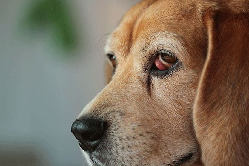
Dogs and puppies communicate through their eyes as much as they do with their voices. Whether it’s to express fatigue, pain or joy, a dog’s eyes tell their owners a great deal of information.
Behind these expressions are a number of moving parts that allow the eyes to function. But sometimes, due to genetics, the eyes don’t function as well as they should. What are we barking about? Inherited eye diseases in dogs.
Genetic eye conditions in dogs can occur at birth or appear later in life. They can range from mild to severe. Some can be treated. Others may eventually lead to blindness or require consistent monitoring and management.
Notable eye conditions in dogs that may be inherited can include:
- Achromatopsia (canine day blindness)
- Canine multifocal retinopathy
- Cataracts
- Collie eye anomaly (CEA)
- Congenital stationary night blindness (CSNB)
- Corneal dystrophy
- Dry eye curly coat syndrome (CCS)
- Glaucoma
- Goniodysgenesis
- Oculoskeletal dysplasia (OSD)
- Primary lens luxation
- Progressive retinal atrophy (PRA)
Many of these diseases are found in purebreds but can also occur in mixed breeds.
Could your dog be at risk? Read more below for an overview of each condition, which breeds they affect and how you can care for a dog that has been diagnosed.
1. Achromatopsia (canine day blindness)
Achromatopsia is an inherited, congenital (present at birth) disease caused by a mutation in the gene responsible for retinal development (the CNGA3 gene). The retina is the light sensitive tissue located in the back of the eye. In the retina are two types of light-sensitive cells, or photoreceptors, called cones and rods. Cones assist with vision in daylight, while rod cells are used to see in dim lights.
In a dog that has achromatopsia, the cone photoreceptor cells do not function normally. Since these are the cells that help a dog see in bright lights, a dog with the condition struggles to see in the daytime, hence the name “day blindness.”
The retinas finish developing between 8 and 12 weeks of age. By this time, a dog that has achromatopsia may begin to show signs of day blindness.
Can achromatopsia be treated? Gene therapy using an adeno-associated viral (AAV) vector restored cone function in some younger dogs but not in older ones. However, when some older dogs were first treated with ciliary neurotrophic factor (CNTF), vision was restored. Similar gene therapy trials in humans show that treatment may be generally safe and may improve vision, though full recovery could be limited.
Scientists performed the therapy on two dogs that showed promising results for treatment of the disease — a special imaging technique called electroretinography revealed that the gene therapy restored the function of the cone cells. But more research is needed before achromatopsia is considered appropriate for all pets.
Breeds that may be affected by achromatopsia include:
- German Shepherds
- Labrador Retrievers
- Alaskan Malamutes
- German Shorthaired Pointers
- Standard Poodles
Research has found that day blindness in standard poodles is caused by a different genetic mutation than in other breeds. The condition in Poodles is often associated with more advanced retinal degeneration, so scientists have named the mutation “DB/RD” (day blindness/retinal degeneration).
Because retinal degeneration may be more complete in these cases, it can eventually cause vision loss in both bright and dark environments.
2. Canine multifocal retinopathy (CRM)
Canine multifocal retinopathy is an inherited disease that causes areas of retinal detachment in the eye. Typically CRM typically does not cause vision defects or blindness — but there have been some reports of vision loss.
CRM has three different types of mutations:
- CRM1 — appears between 11 and 16 weeks of age
- CRM2 — appears at around 15 weeks of age
- CRM3 — appears between 9 and 24 months of age
Each mutation is associated with different breeds. While the CRM1 and CRM3 types are found in a variety of dogs, the CRM2 type is found in the Coton de Tuléar.
The condition can cause pink, orange, gray or tan blisters on the retina due to fluid that accumulates beneath the detached retina. Blisters heal in some cases, and the changes in the retina tend to occur slowly.
Breeds associated with canine multifocal retinopathy include:
- Australian Shepherds (CMR1)
- American Bulldogs (CMR1)
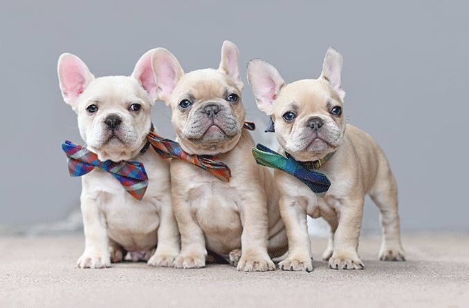
- French Bulldogs (CMR1)
- Mastiffs (CMR1)
- Cane Corsos (CMR1)
- Coton de Tuléar (CMR2)
- Finnish Lapphund (CMR3)
- Swedish Lapphund (CMR3)
- Lapponian Herder (CMR3)
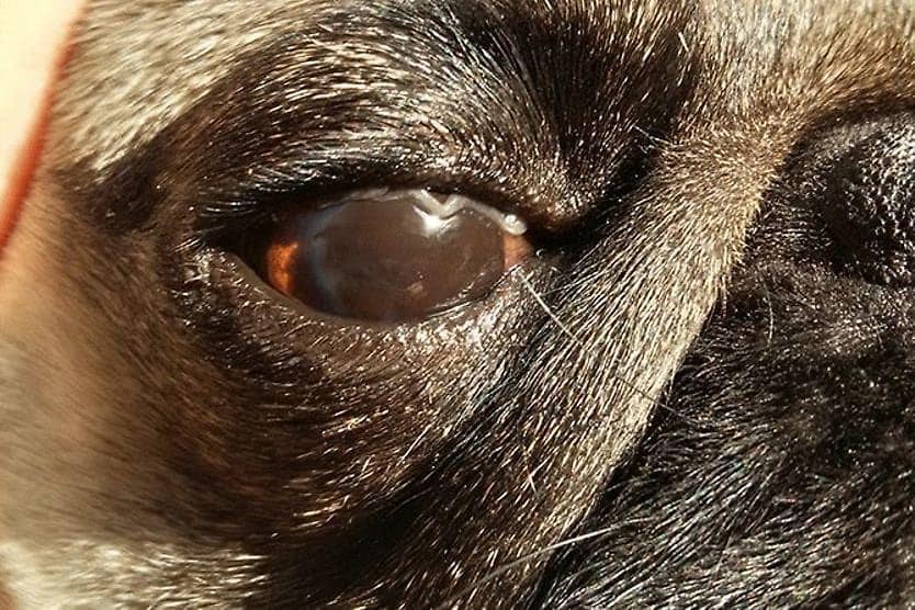
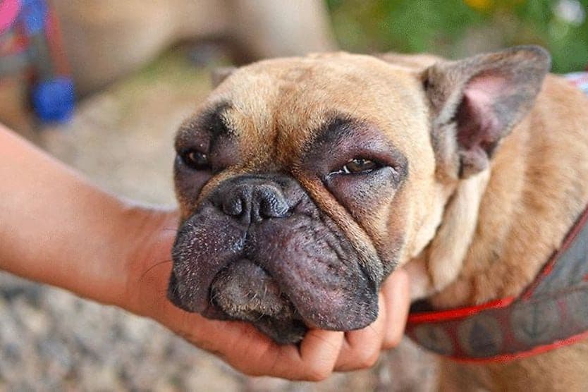
3. Cataracts
Just like humans, dogs can develop cataracts in one or both eyes. Cataracts are cloudy, thickened clumps of special proteins that cover the lens of the eye (the part that focuses light to create a crisp image).
As a cataract grows and matures, it blocks light from entering the eye through the lens and causes difficulty for a dog to see. Both eyes are often affected by cataracts, but they don’t always develop at the same time.
Cataracts can occur in dogs due to a number of factors, including aging or injury — but a common cause is genetics.
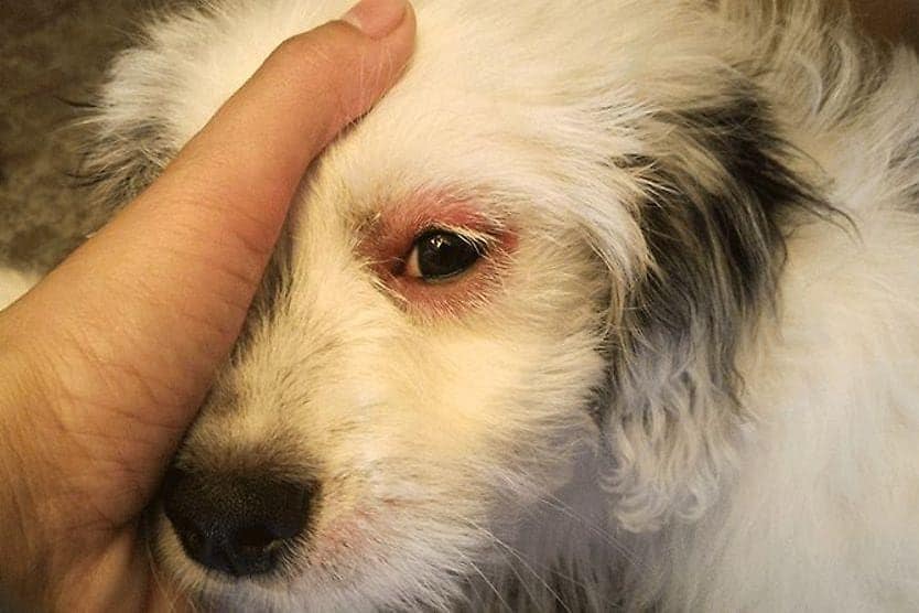
Symptoms associated with canine cataracts may include:
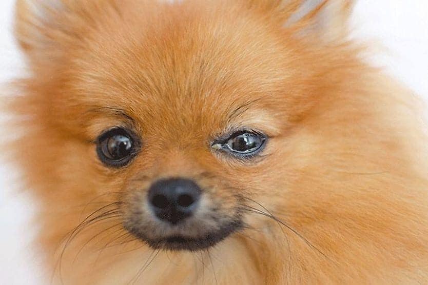
- Cloudy or bluish appearance in the pupil
- Clumsiness and confusion in unfamiliar environments
- New or sudden hesitancy to jump on furniture
- Eye discharge
- Redness in the eye or around the eyelids
- Constant blinking or squinting
- Rubbing or scratching the affected eye
Note: Cataracts, by definition, are a disease of the lens. Unless trauma or infection is involved, none of the listed symptoms may develop except the first three.
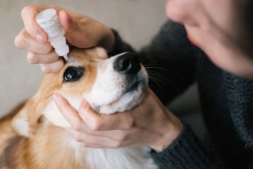
Treatment may be possible for cataracts, but the condition cannot be reversed. Some cases are managed with special eye drops and regular veterinary check-ups to monitor the cataract’s progression.
Cataracts can also be surgically removed if appropriate. But the procedure can be expensive, and some dogs may not be suited to be put under anesthesia to have it done.
Cataracts can eventually lead to glaucoma, a sight-threatening eye condition caused by high eye pressure. If this happens, your vet or veterinary ophthalmologist may prescribe eye drops to reduce the pressure.
Cataracts can also cause blindness if they are deemed untreatable. But dogs may adapt. And with the help of their owners, they may live a happy life despite vision impairment.
Breeds that may be associated with hereditary cataracts in dogs may include:
- Staffordshire Bull Terriers
- Boston Terriers
- Australian Shepherds
- Smooth Fox Terriers
- American Cocker Spaniels
- Miniature Schnauzers
- Siberian Huskies
4. Collie eye anomaly (CEA)
Collie eye anomaly, also called “collie eye defect” is a genetic eye condition in collies and similar breeds. CEA affects the development of the eye.
More specifically, it is caused by a mutation in the gene responsible for eye development. Because of this, the condition can cause a number of defects in each of the eye’s layers.
Sometimes “CEA” is used correspondingly with a condition called choroidal dysplasia (or bilateral choroidal hypoplasia [CH]). The choroid is the vascular layer of the eye sandwiched between the retina and the sclera.
A dog who has CEA also has choroidal dysplasia (which means abnormal growth). The condition causes the vascular tissue in the back of the eye to thin and does not affect vision significantly.
Signs, symptoms and abnormalities associated with CEA may include:
- Blindness – either partial or full, depending on the degree of CEA defects
- Retinal folds – when the retina folds in on itself during development
- Retinal detachment
- Coloboma – holes that occur within the structures of the eye, which can sometimes lead to retinal detachment
- Enophthalmia – eyes that are deeply sunken into the eye sockets
- Microphthalmia – eyes that appear smaller than expected
Treatment may not be available for CEA. Retinal detachment in a dog with CEA may be corrected with laser surgery to reattach the retina if it was caused by coloboma and caught early on. This procedure may not always be recommended.
Breeds that may be associated with CEA/choroidal hypoplasia may include:
- Collies
- Border Collies
- Australian Shepherds
- Shetland Sheepdogs
- Nova Scotia Duck Tolling Retrievers
5. Congenital stationary night blindness (CSNB)
Congenital stationary night blindness in dogs is a rare genetic eye disease caused by a mutation in the RPE65 gene. It is the same mutation that causes retinal dystrophy (abnormal growth of the retina).
With CSNB, dogs may display normal vision in daylight, but have trouble seeing objects and people during the night and in dim environments.
Night blindness can occur in dogs as young as 5 weeks, but major changes in the eye may not occur until a dog is at least 2 years old. As CSNB progresses, light brown patches may form on the retina's surface. Eventually, they may affect the entire retina.
Symptoms associated with CSNB may include:
- Normal vision during the day but loss of vision at night
- Reluctance to move or appearing to be lost in dark environments
- Hesitancy to enter dimly lit places
There currently may be no treatments for CSNB. Genetic testing can be done to determine the risk of your dog developing the disease.
Congenital stationary night blindness has been found in breeds including:
- Beagles
- Briards
SEE RELATED: A guide to caring for a blind or vision-impaired dog
6. Corneal dystrophy
Corneal dystrophy is a group of genetic eye disorders in dogs. There are three main types of corneal dystrophy: epithelial, stromal and endothelial. Each occurs in a different layer of the cornea.
All types of corneal dystrophy may cause clouding of the cornea. None of the types are typically associated with other medical conditions.
- Epithelial corneal dystrophy – occurs on the superficial layers of the cornea and may cause white or gray patches called opaque lesions. Lesions can cause erosions on the cornea and lead to eye pain. Many dogs with this type of dystrophy only experience opaque corneas.
- Stromal corneal dystrophy – occurs in the middle layer of the cornea and may cause silver, white or gray opacity in the center of the cornea. It can also cause a ring on the outer portion of the cornea or cover it completely. Stromal corneal dystrophy occurs when fatty deposits fall into the middle layer of the cornea.
- Endothelial corneal dystrophy – occurs in the deepest part of the cornea and causes fluid to slowly accumulate across the cornea. Endothelial corneal dystrophy can cause ulcers and eventual vision loss. This type of dystrophy typically affects older Chihuahuas, Boston Terriers and Dachshunds.
A condition called macular corneal dystrophy may also be inherited. It may cause similar symptoms of corneal clouding and is typically found in middle-aged Labrador Retrievers.
Many dogs who have corneal dystrophy may not experience vision problems aside from cloudiness in the cornea. But if corneal ulcers develop due to the condition, it is recommended that they be treated promptly.
Breeds that may experience corneal dystrophy may include:
- Cocker Spaniels
- Bearded Collies
- Samoyeds
- Weimaraners
- Airedales
- Cavalier King Charles Spaniels
- Dachshunds
- Chihuahuas
- Boston Terriers
7. Dry eye curly coat syndrome (CCS)
Dry eye curly coat syndrome is sometimes referred to as rough coat syndrome, congenital keratoconjunctivitis sicca or ichthyosiform dermatosis. CCS is a genetic condition that affects the skin and eyes.
Dogs with CCS are unable to produce tears, which causes inflammation of the cornea and conjunctiva. Additionally, the lack of tears prevents the dog’s eyes from filtering dust and other particles, which can cause infections, ulcerations and even blindness.
CCS is associated with the Cavalier King Charles Spaniel breed and is detectable in puppies. Aside from the eye-related symptoms, a dog with CCS also may have an exceptionally curly coat and may experience deformed toenails or teeth. The mouth and eyes also typically lack moisture.
Dogs affected by the disease are sometimes euthanized because it may be severely painful and treatment is limited. Pet owners may choose to treat dogs with CCS with very frequent medical bathing and topical eye medications.
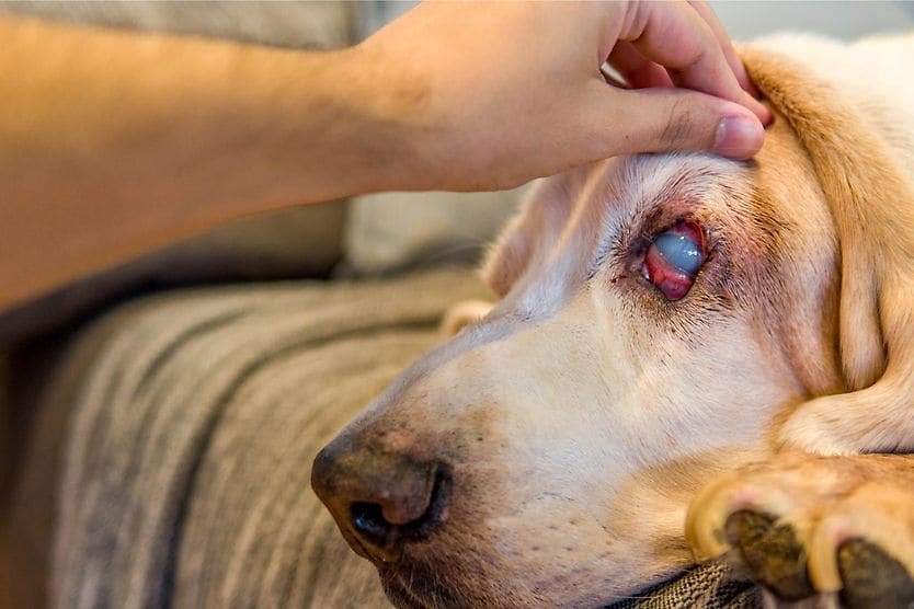
8. Glaucoma
Glaucoma is a disease that occurs due to a build-up of pressure inside the eye (called intraocular pressure or IOP). The increased pressure can cause complications in vision, such as optic nerve damage. It can also lead to blindness.
A dog can get glaucoma after an injury, surgery or other major eye problem — this is called secondary glaucoma. The condition can also be inherited. When genetics are to blame, it’s referred to as primary glaucoma.
Primary (inherited) glaucoma has two different forms:
- Primary closed-angle glaucoma (PCAG) – caused by goniodysgenesis, a genetic defect of the eye’s drainage angle. PCAG may be found in breeds such as Border Collies, Cocker Spaniels and Golden Retrievers.
- Primary open-angle glaucoma (POAG) – caused by a genetic mutation that is different from goniodysgenesis. POAG may be found in breeds such as Beagles, Basset Hounds and Shar-Peis.
All types of glaucoma in dogs can cause problems such as swelling, redness, eye discharge and dilated pupils. Eyes may also have a cloudy or hazy bluish appearance.
Genetic forms of glaucoma may be treated with eye drops, surgery and/or monitoring. Dogs may also be prescribed pain relievers for any discomfort.
In addition to the breeds mentioned above, glaucoma also may be associated with the following:
- Poodles
- Dalmatians
- Great Danes
- English and Welsh Springer Spaniels
- Many terrier breeds
9. Goniodysgenesis
Goniodysgenesis (gō’nē-ō-dis-jen’ĕ-sis) is an inherited condition that is associated with primary closed-angle glaucoma (PCAG). It is caused by a developmental defect in the anterior chamber of the eye.
A dog with goniodysgenesis isn’t guaranteed to develop PCAG, but the risk is greatly increased. Vets or veterinary ophthalmologist may recommend regular vision check-ups to make sure the eye pressure is normal in a dog with goniodysgenesis. Some may also prescribe eye drops as a method of prevention for glaucoma.
Goniodysgenesis can occur in any breed, but is primarily found in:
- Border Collies
- Basset Hounds
- Cocker Spaniels
- Golden Retrievers
- Siberian Huskies
10. Oculoskeletal dysplasia (OSD)
Oculoskeletal dysplasia is an inherited disease that affects a dog’s bones and eyes. OSD is caused by a specific genetic trait.
Dogs that have OSD may have shortened limbs (especially in the hips and elbows), as well as ocular defects such as cataracts, retinal folds, vitreous dysplasia and retinal detachment. Some dogs may also experience glaucoma and hyphema (blood in the anterior chamber of the eye) as a result of OSD.
Many dogs can experience mild forms of OSD, but breeds associated with stronger effects of the disease may include:
- Labrador Retriever
- Samoyeds
11. Primary lens luxation (PLL)
Primary lens luxation is an inherited disease that causes dislocation (luxation) of the lens of the eye. In a normal eye, the lens is located behind the pupil and iris, and is held in place by zonular ligaments (a group of fibers).
If these ligaments are damaged, it can push the lens out of place either partially or completely. When the lens is only partially out of place, it’s called lens subluxation.
PLL is considered a genetic disease, but it can also be caused by conditions such as cataracts, glaucoma, cancer, inflammation and trauma.
The condition can also cause secondary problems, including glaucoma and uveitis (eye inflammation). Both of these are recommended to be treated promptly to avoid long-term complications.
Surgical removal of the lens may be performed if PLL is identified early. Uveitis and glaucoma that occur due to PLL may be treated with oral or topical medication as well as monitoring vision.
Primary lens luxation can affect many breeds including:
- Australian Cattle Dogs (commonly known as Heelers)
- Jack Russell Terriers
- American Eskimo (also known as Spitz)
- Rat Terriers
- Yorkshire Terriers
- Welsh Corgis
12. Progressive retinal atrophy (PRA)
Progressive retinal atrophy is an inherited eye disorder in dogs, passed down by a genetic trait, that affects the retina and leads to blindness. Vision loss occurs gradually as the cells of the retina deteriorate over time. Blindness sets in within two years of the onset of the disease in many dogs.
The retina has light sensitive cells called photoreceptors. There are two subtypes of photoreceptors: cones and rods. Cone cells detect color and aid in daytime vision, while rods help dogs see in dim environments.
In PRA, both types of photoreceptor cells may deteriorate over time. Rods are usually affected before cones, which means dogs may initially lose their night vision. Cone cells deteriorate more slowly, but once they do, the dog experiences total blindness.
PRA can occur in the first few months of life, or later on, often between 3 and 9 years old. The early-onset type is sometimes called retinal dysplasia. Another type of the condition can also occur suddenly (sudden acquired retinal degeneration or SARD) and causes blindness within days or weeks.
No treatment is available for PRA. But, as mentioned, dogs are great at adapting to life changes, including blindness. Dogs with PRA are recommended to be monitored regularly, as the condition can lead to cataracts or cause inflammation in the eye.
Breeds associated with PRA may include:
- American Cocker Spaniels
- Bedlington Terriers
- Cavalier King Charles Spaniels
- English Springer Spaniels
- Golden Retrievers
- English Mastiffs
- Rottweilers
Can you test for genetic eye diseases in dogs?
Yes, you can test for most inherited canine eye diseases. But in order to do so, your breed typically must be at risk. In some cases, a diagnosis isn’t made until signs and symptoms develop.
If you got your dog from a breeder, it is recommended to ask for information about his or her medical history. (And consider doing outside research on the breed).
It’s also a good idea to talk to your veterinarian or veterinary ophthalmologist about your dog’s eye health and ask if you should have any concerns about disease. Your vet may recommend genetic testing or inform you of other concerns that you may not have expected.
The outlook and what to do if your dog has an eye disease
Genetic eye conditions can have mild to severe effects on your dog. But remember, just because your dog’s breed is at risk of developing a certain disease, it doesn’t mean that they absolutely will.
In any case, it’s important to monitor your dog’s general eye health and report any problems to your veterinarian or veterinary ophthalmologist. It is recommended to speak up if you notice any complications such as eye discharge, redness, swelling, light sensitivity or any behavioral problems that may be associated with an eye problem.
Depending on the condition, your veterinarian or veterinary ophthalmologist may prescribe eye drops or other topical medications as well as oral medications to treat it. More drastic measures, such as surgery or other procedures (euthanasia in very serious cases) may need to be performed at your vet or veterinary ophthalmologist’s recommendation.