Cornea of the eye

The cornea allows light to enter the eye for vision. Here is some basic information to know about this important part of the eye.
Cornea definition
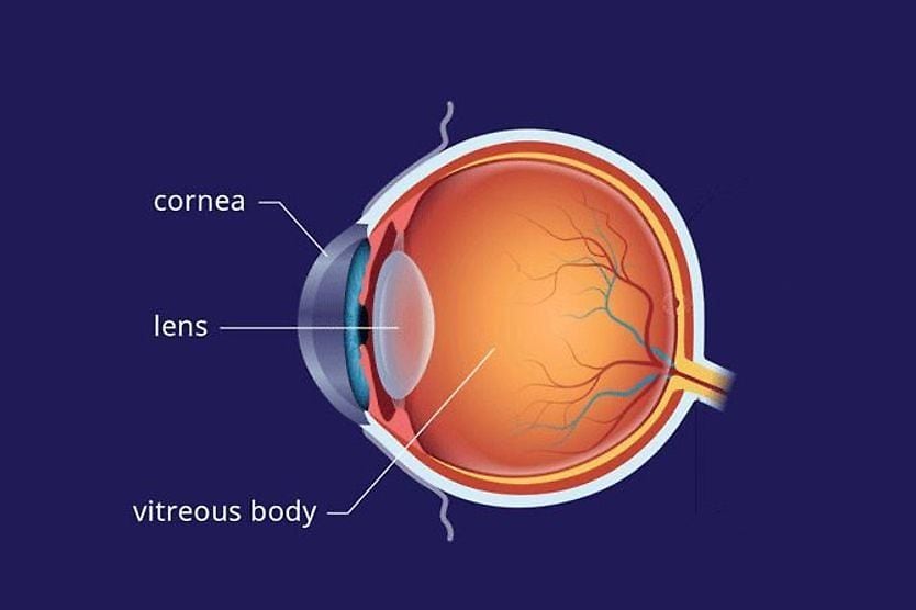
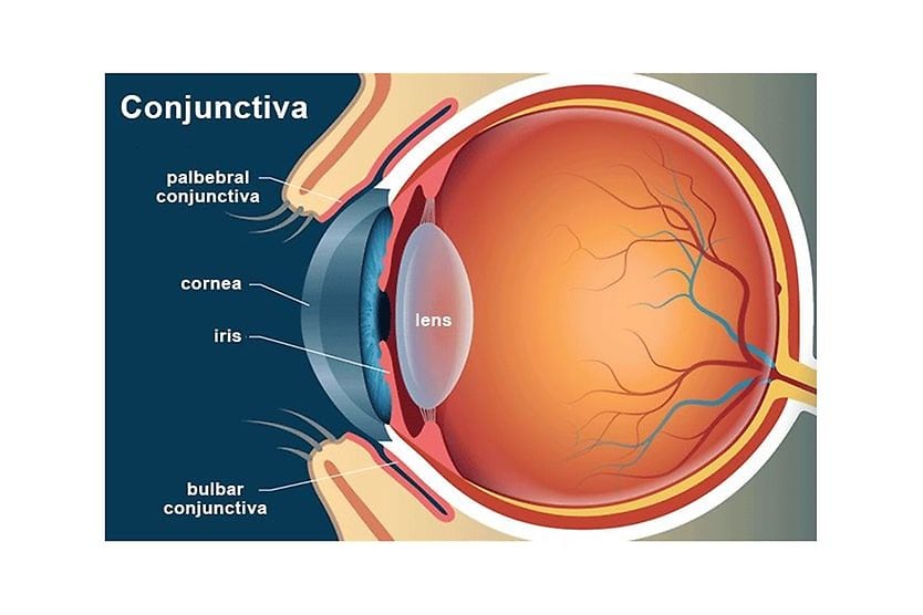
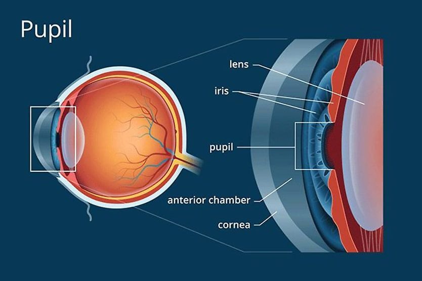
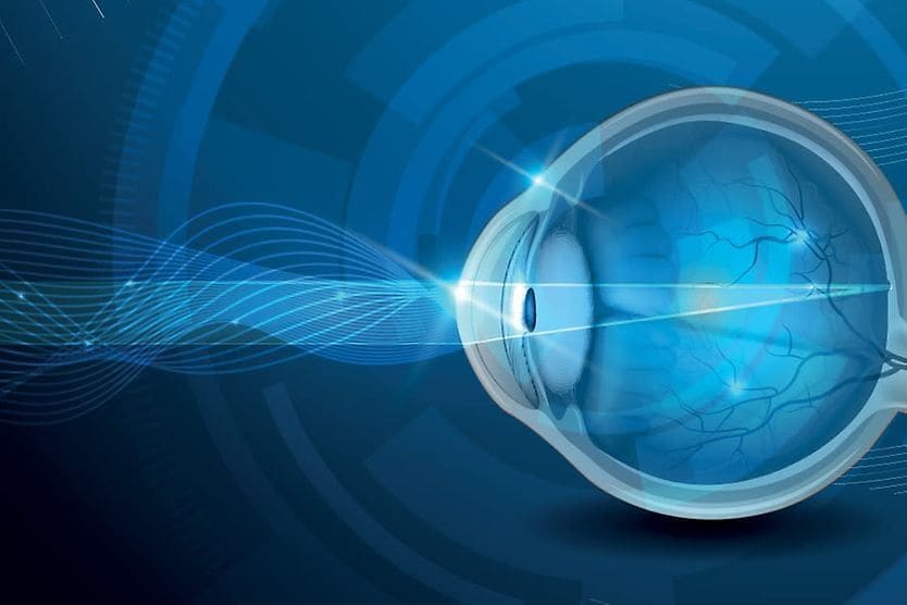
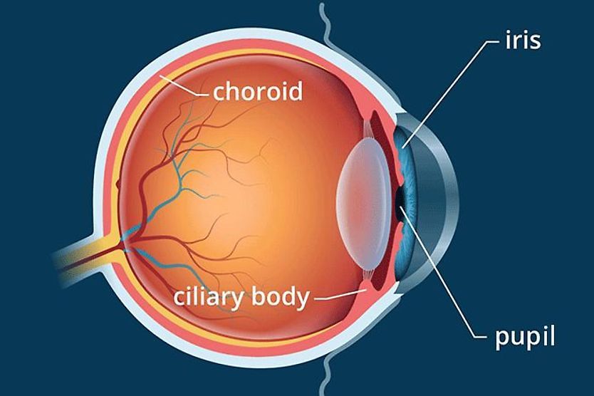
The cornea is the clear front surface of the eye. It lies directly in front of the iris and pupil, and it allows light to enter the eye.
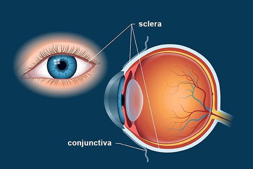
Viewed from the front of the eye, the cornea appears slightly wider than it is tall. This is because the sclera (the "white" of the eye) slightly overlaps the top and bottom of the anterior cornea.
The horizontal diameter of the cornea typically measures about 12 millimeters (mm), and the vertical diameter is 11 mm, when viewed from the front. But if viewed from behind, the cornea appears circular, with a uniform diameter of approximately 11.7 mm. This makes the cornea about two-thirds the size of a dime.
The center thickness of the average cornea is about 550 microns, or slightly more than half a millimeter.
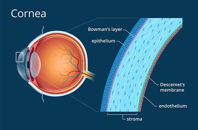
The cornea has five layers. From front to back, these layers are:
- The corneal epithelium. This outer layer of the cornea is five to seven cells thick and measures about 50 microns — making it slightly less than 10% of the thickness of the entire cornea. Epithelial cells are constantly being produced and sloughed off in the tear layer of the surface of the eye. The turnover time for the entire corneal epithelium is about one week.
- Bowman's layer. This is a very thin (8 to 14 microns) and dense fibrous sheet of connective tissue that forms the transition between the corneal epithelium and the underlying stroma.
- The corneal stroma. This middle layer of the cornea is approximately 500 microns thick, or about 90% of the thickness of the overall cornea. It is composed of strands of connective tissue called collagen fibrils. These fibrils are uniform in size and are arranged parallel to the cornea surface in 200 to 300 flat bundles called lamellae that extend across the entire cornea. The regular arrangement and uniform spacing of these lamellae is what enables the cornea to be clear.
- Descemet's membrane. This very thin layer separates the stroma from the underlying endothelial layer of the cornea. Descemet's (pronounced "DESS-eh-mays") membrane gradually thickens throughout life — it's about 5 microns thick in children and 15 microns thick in older adults. (Learn about corneal descemetocele)
- The corneal endothelium. This is the innermost layer of the cornea. The back of the endothelium is bathed in the clear aqueous humor that fills the space between the cornea and the iris and pupil. The corneal endothelium is only a single layer of cells thick and measures about 5 microns. Many of the endothelial cells are hexagonal (six-sided), but some may have five or seven sides. The regular arrangement of these cells is sometimes called the endothelial mosaic.
Cornea function
As already mentioned, the clear cornea allows light to enter the eye for vision. But it has another very important function as well — the cornea provides approximately 65% to 75% of the focusing power of the eye.
The remainder of the focusing power of the eye is provided by the crystalline lens, located directly behind the pupil.
The majority of refractive errors — nearsightedness, farsightedness and astigmatism — are due to a less-than-optimal curvature or symmetry of the cornea. Presbyopia, on the other hand, is due to an aging change in the crystalline lens.
In addition to allowing light to enter the eye and providing the majority of the focusing power of the eye, individual parts of the cornea have specialized functions:
Corneal epithelium. The corneal epithelium provides an optimal surface for the tear film to spread across the surface of the eye to help keep it moist and healthy and maintain clear, stable vision.
Bowman's layer. The dense nature of Bowman's layer helps prevent corneal scratches from penetrating into the corneal stroma. Corneal abrasions that are limited to the outer epithelial layer generally heal without scarring; but scratches that penetrate Bowman's layer and the corneal stroma typically leave permanent scars that can affect vision.
Corneal endothelium. The single layer of cells that forms the endothelium maintains the fluid content within the cornea. Damage to the corneal endothelium can cause swelling (edema) that can affect vision and corneal health.
Cornea problems
A number of conditions can affect the cornea. Among the more common corneal problems are:
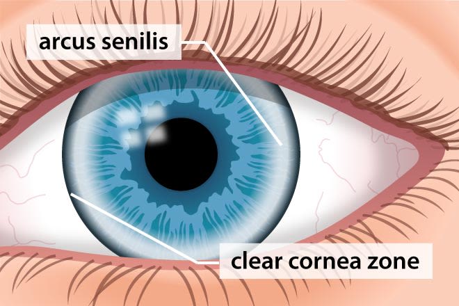
Appearance of arcus senilis (corneal arcus).
Arcus senilis. As people get older, a white ring often develops in the periphery of the cornea. This is called arcus senilis (also called corneal arcus), and it's the more common aging change in the cornea. Arcus senilis is typically separated from the limbus by an area of clear cornea. The white ring — which is composed of cholesterol and related compounds — can be barely noticeable or very prominent.
In older individuals, corneal arcus typically isn't related to blood cholesterol levels; but if it occurs in a person under age 40, blood tests should be performed to check for hyperlipidemia (abnormally high concentration of fats or lipids in the blood).
Corneal abrasion. A scratched cornea can be very painful and can lead to an eye infection.
LEARN MORE about corneal abrasions.
Pterygium. A pterygium is a fibrous growth that starts on the sclera but can grow into the peripheral cornea and cause irritation, vision problems and disfigurement of the front of the eye.
Dry eyes. Though the cause of dry eyes typically begins in the tear gland and eyelids, it can lead to damage of the corneal epithelium, which can cause eye discomfort and vision disturbances.
Corneal ulcer. A corneal ulcer is a serious abscess-like infection of the cornea that can lead to significant pain, scarring and vision loss.
Corneal dystrophy. A dystrophy is a weakening or degeneration of a tissue. The more common corneal dystrophy — called Fuch's dystrophy — affects the corneal endothelium, causing corneal swelling, foggy vision, light sensitivity and other problems.
Acanthamoeba keratitis. This is a very serious corneal infection that can cause significant pain and vision loss.
Fungal keratitis. Fungal keratitis is another dangerous corneal infection that (like Acanthamoeba keratitis) tends to affect contact lens wearers more often than people who wear glasses.
LEARN MORE about other types of keratitis.
Keratoconus. This is a thinning and deformation of the cornea that causes vision problems that can't be corrected with regular eyeglasses or contact lenses. In some cases, vision problems from keratoconus can be corrected with scleral contact lenses or hybrid contacts. But in severe cases, a cornea transplant may be required.
Corneal ectasia. Corneal ectasia is a thinning and deformation of the cornea that resembles keratoconus but can occur as a rare complication of LASIK or other corneal refractive surgery.
READ NEXT: Staphyloma and Tarsorrhaphy