Staphyloma

What is a staphyloma?
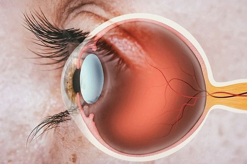
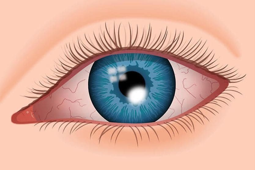
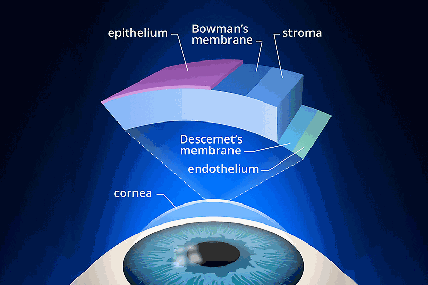
A staphyloma is a condition in which the sclera (white of the eye) or cornea (the clear layer in front of the pupil) stretches and thins, allowing tissue underneath to bulge through. This “outpouching” can cause vision problems. Associated vision problems include objects in the environment looking misshapen and abnormal.
Staphyloma stems from the Greek word staphyus, meaning bunches of grapes.
A staphyloma generally occurs in the front (anterior) or back (posterior) of the eye. Some people are born with a staphyloma (congenital staphyloma). Others develop it because of an infection or injury to the eye. It can also develop due to progressive, abnormal elongation of the eye.
Types of staphyloma
Eye doctors classify staphyloma according to where in the eye the weakening occurs and tissue pushes through:
- Anterior staphyloma involves the front of the eye, where the cornea is located.
- Intercalary staphyloma involves the area between the cornea and sclera.
- Ciliary staphyloma involves a structure called the ciliary body located behind the iris (colored part of the eye).
- Equatorial staphyloma occurs in the middle of the eye, about equal distance from the anterior (front) and posterior (back).
- Posterior staphyloma involves the back part of the eye. People with this type of staphyloma tend to have the most problems with their vision.
- Peripapillary staphyloma is a rare type of staphyloma some people are born with. The outpouching is seen around a normal-looking optic nerve. The optic nerve sends signals to your brain, helping you to see.
Posterior staphyloma in pathologic myopia
There are several classifications of staphyloma. An important classification, called posterior staphyloma, occurs in the back of the eye. It is associated with a vision-threatening form of nearsightedness.
Nearsightedness itself is a common eye condition. When you’re nearsighted, you can see near objects clearly. But things far away look fuzzy and blurred. Nearsightedness is usually connected with a risk factor called axial length elongation, when the eye grows too long.
High myopia, or severe nearsightedness, increases the risk of developing a type of nearsightedness called pathologic (degenerative) myopia.
Pathologic myopia occurs when degenerative changes, such as staphyloma, result from excessive elongation of the eye. These changes can lead to permanent vision impairment and blindness.
In degenerative myopia, stretching and thinning of the sclera and retina occurs in the back of the eye. This results in distortions in the overlying retina. Pathologic myopia is among the leading causes of staphyloma.
The presence of staphyloma is one of the signs that eye doctors use to determine whether someone has pathologic myopia.
LEARN MORE: Myopia (nearsightedness)
Signs and symptoms of staphyloma
Posterior staphylomas are seen in severely myopic patients. Markedly worsening vision, particularly in very nearsighted people, is one of the symptoms of posterior staphyloma.
A staphyloma can also have other symptoms, depending on its location, including:
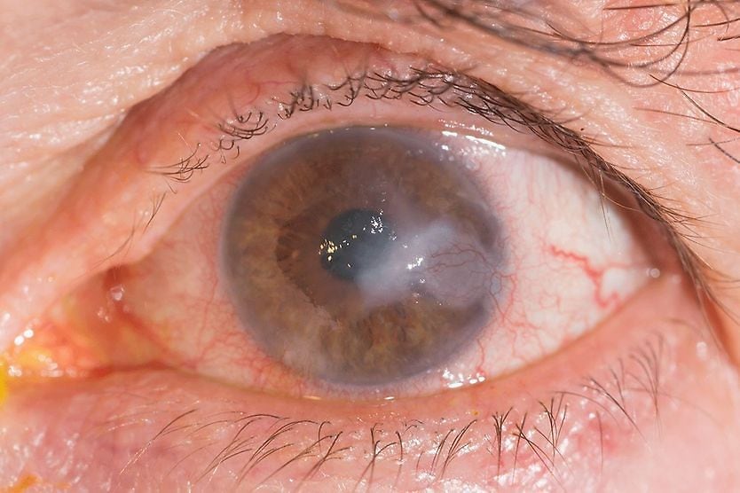
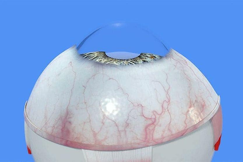
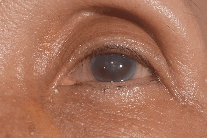
- An enlarged eye, especially when staphyloma is severe
- A bluish/blackish color to the white of the eye
Staphyloma causes
Anything that weakens the sclera can lead to eye staphyloma. Some of the more common causes include:
- Pathologic myopia. As discussed earlier, this is the most common cause. This is a type of nearsightedness that worsens (degenerates) over time. The sclera is stretched and weakened. About 3% of the people in the world have pathologic myopia, and 50% of those with the condition have staphyloma.
- Injury. Many eye injuries, including those that can produce staphylomas, are caused by sharp objects, like scissors or knives. Staphylomas due to injury can become punctured and require urgent repair to avoid vision loss.
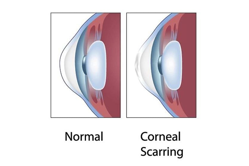
- Infection. Certain bacteria and fungi can cause infectious keratitis. This is an infection of the cornea that can lead to a staphyloma in the front of the eye. Even the measles, a common childhood viral disease, can lead to keratitis and, consequently, staphylomas.
- Certain diseases babies can be born with. For example, retinitis pigmentosa causes damage to the retina of the eye. Another is Alport’s syndrome, which can cause eye damage. These conditions can damage the innermost part of the middle layer of the wall of the eye, known as Bruch’s membrane.
- Surgery on the eye. Surgery can place stress on the eyes, and it is possible to develop a staphyloma due to eye surgery. But this is not common.
Staphyloma treatment
Your eye doctor will examine your eyes and order tests that take pictures of the inside of the eye. Optical coherence tomography (OCT) is a non-invasive test that allows a doctor to see cross-section images of the retina. This allows them to check for the presence of staphyloma.
In some cases, other imaging techniques may be used, including:
- B-scan ultrasonography – A type of ultrasound that uses sound waves to let a doctor see the back of the eye to diagnose a posterior staphyloma
- Orbital CT scan – Uses X-rays to see the eye and the bones around it
- Orbital MRI – A test that can give doctors a 3D image of your eye
Most types of staphylomas don’t need treatment. Your eye doctor may decide to take a wait-and-see approach, watching the staphyloma to see if it gets worse. Most do not.
If a staphyloma progresses, your eye doctor may recommend a scleral or corneal patch graft to reinforce the weakened area of the eye wall. The primary goal of this procedure is to restore structural integrity and prevent further thinning. While some patients may experience an improvement in vision, the main purpose is to protect the eye from complications, such as perforation.
Posterior staphylomas may require preventive treatment to avoid future complications. Treatments, such as collagen crosslinking and scleral reinforcement, may be considered for people with pathologic myopia with staphyloma.
When to see a doctor
Contact your eye doctor if you notice that you can’t see as well as you once did, have pain in your eye or see changes to the appearance or shape of your eyes.
People who have pathologic myopia with posterior staphyloma are at an increased risk for degenerative changes to the eye. These changes can include conditions, like retinal detachment. A retina that detaches, or pulls away, from the back of your eye can lead to vision loss in that eye.
Symptoms of a detached retina include seeing flashing lights (photopsia) and having specks (called floaters) or a gray shadowy area in your vision. A detached retina can be serious and requires immediate treatment.