PERRLA assessment test: What it is, how it’s done and what it stands for

What is the PERRLA eye test?
PERRLA is an acronym for “pupils are equal, round and reactive to light and accommodation.” The PERRLA eye test is used to check if your pupils look and function normally. It is essential that your eye doctor perform this test in any comprehensive eye exam.
The PERRLA eye test can indicate whether you have certain health issues, such as eye diseases and some visual and neurological conditions. Plus, a properly functioning pupil is critical for good vision.
Why you need the PERRLA eye test
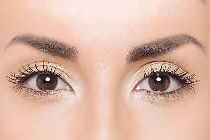
The pupil is the opening in the center of the eye. It allows light to enter the eye so that the light can be focused onto the retina. It dilates to let more light in and constricts to let less light in. Your pupil adjusts to different light levels to help you see clearly things that are up close and far away.
Your pupils also change size to give you the optimal vision depending on your current circumstances.
During a PERRLA test, an eye doctor checks to see if your pupils are:
- Equal – Both of your pupils should be the same size and shape.
- Round – Your pupils should be round.
- Reactive to light and accommodation – If your pupils are reacting to light as they should, they will get smaller when you’re looking at bright or direct light. They should also get smaller when you’re focusing on something very close to your eyes (accommodation).
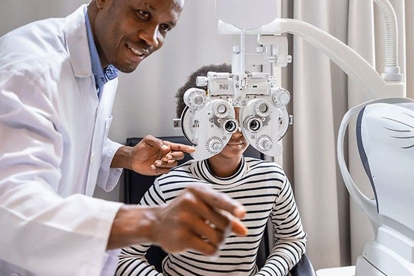
How the PERRLA eye test is administered
The PERRLA eye test includes the following steps:
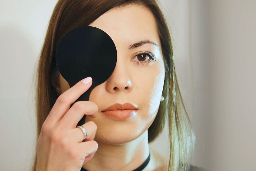
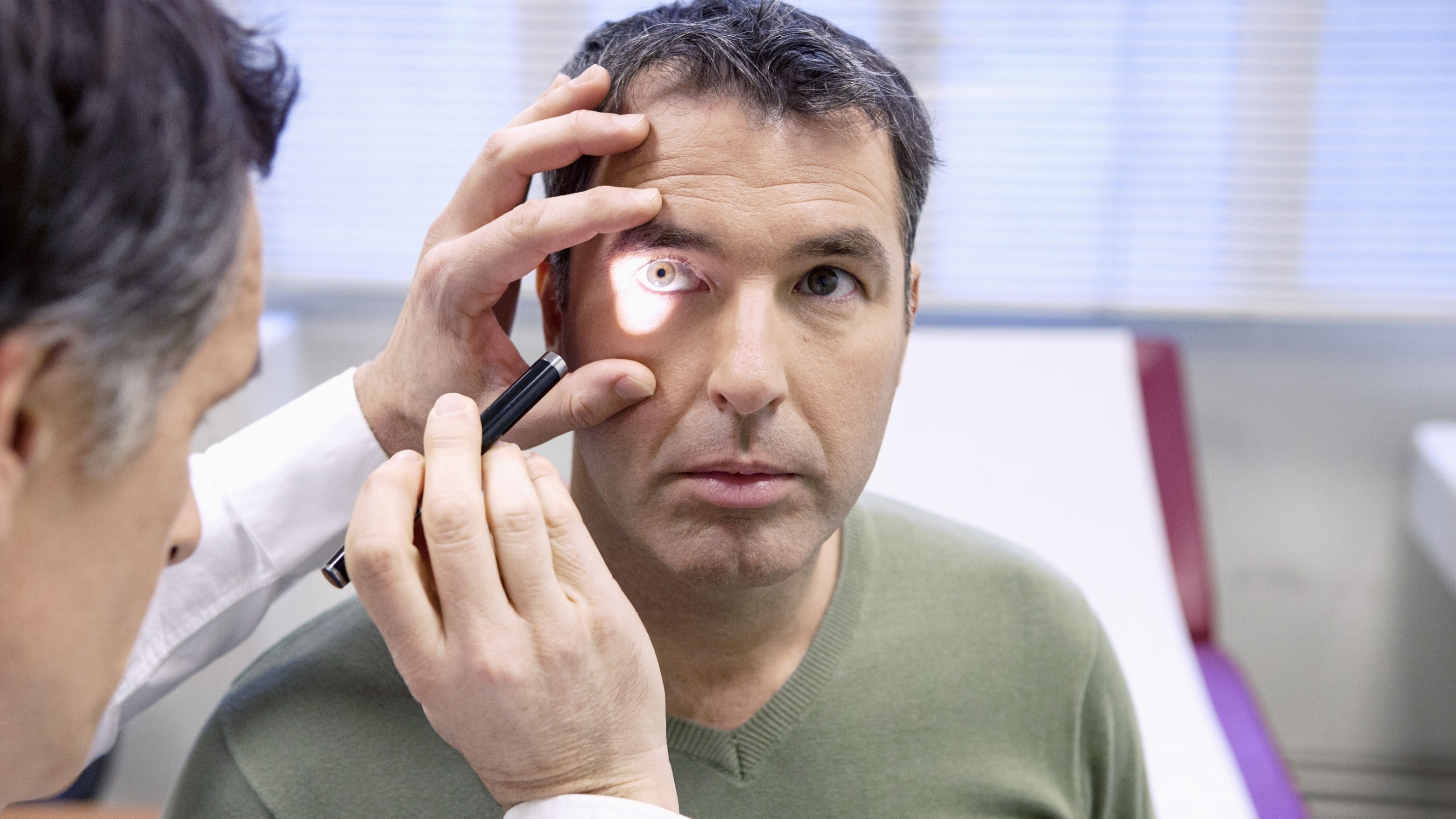
- First, your eye doctor will look at your pupils to see if they are round and the same size.
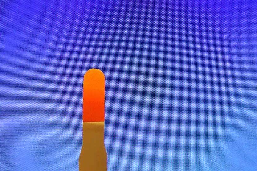
- Next, the doctor will perform the swinging light test to see how your pupils react to light. The doctor will dim the room lights and will shine a narrow beam of light into one eye for about three seconds and then into the other eye. If there is no optic nerve or retinal disease, both pupils should constrict (get smaller) equally at the same time in reaction to this bright light. And they should dilate equally (become larger) when the light is turned off.
- To test your pupils’ accommodation, the doctor will have you focus on an object they are holding in their hand while moving that object closer to your nose. Properly functioning pupils will get smaller as the object gets closer.
Common conditions the PERRLA test can detect
Some common conditions the PERRLA test can detect include:
Adie’s tonic pupil – This is a nervous system condition that may lead to unequal pupil sizes and cause a slow reaction to light. Adie’s tonic pupil usually affects only one eye but it can be present in both eyes. The cause is often unknown, but may occur as a result of viral infection, trauma, narrowing of a blood vessel due to migraine, eye surgery or tumors.
Anisocoria – This is when you have uneven pupil size that could be normal or could be a symptom of an underlying health issue. Normal anisocoria is called “physiologic anisocoria.” It affects up to 20% of the population and isn’t typically harmful. But this condition is a cause for concern when normal, even-sized pupils become uneven for reasons such as:
- A damaged blood vessel in the neck, often as a result of head or neck trauma.
- Damage to the third cranial nerve that manages a muscle that controls eye movement.
- A brain aneurysm, which is ballooning in a weakened area in the wall of a blood vessel in the brain.
- If certain topical medications like anti-nausea or motion sickness patches get into your eye.
Argyll Robertson pupil – Patients with this condition have small pupils that do not get smaller in response to bright light but do constrict when focusing on something close to their eyes. It is rare and is typically caused by late-stage syphilis.
Horner’s syndrome – This condition is characterized by a drooping eyelid on one side, and the pupil in the eye on that side is small. Horner’s syndrome is a result of damage to the part of the nervous system that increases pupil size. This damage can be caused by:
- A tear in the wall of the carotid artery that brings blood to the brain and head
- Pancoast tumors, which are related to rare cancer that starts at the top of the lung
- Nasopharyngeal tumors, which are rare tumors of the head and neck
- Lymphoproliferative disorders or uncontrolled growth of white blood cells
Relative Afferent Pupillary Defect (RAPD) – Also known as Marcus Gunn pupil, this occurs when there is an unequal pupillary response. One pupil will not constrict when a light is shone into that eye but will constrict when light is shone in the other eye. Some causes of an RAPD include:
- Inflammation of the optic nerve (optic neuritis)
- Optic nerve damage from trauma, radiation or a tumor
- When blood doesn’t flow properly to the optic nerve (ischemic optic neuropathy)
- Retinal disease
- Retinal detachment
- Severe glaucoma that causes trauma to optic nerve
- Severe age-related macular degeneration (AMD)
- Retinal infection caused by the CMV virus or herpes
PERRLA alone doesn’t diagnose a specific condition; it can only give your doctor clues about any disorders you may have. If you have any abnormal results from the PERRLA eye assessment, your doctor will do more tests to determine the potential causes and treatment.
When to see your eye doctor
If you look in the mirror and notice that your pupils look unusual, make an appointment with your eye doctor. Seek immediate medical treatment if you additionally experience severe head pain, confusion or dizziness.
SEE RELATED: What is a Dilated Eye Exam?