Diabetic eye exam: What to expect

What is a diabetic eye exam?
A diabetic eye exam is a detailed evaluation of the health of your retina and other parts of the eye that can be affected by diabetes.
Let’s go over what a diabetic eye exam is, what’s involved and why it’s important.
What does a diabetic eye exam include?
Diabetic eye exams can vary in length and scope, depending on what your eye doctor feels is necessary to manage your condition.
For example, if you have just been diagnosed with diabetes and you’ve recently had a comprehensive eye exam that showed no signs of diabetic retinopathy, your follow-up diabetic eye exam may require your doctor to simply recheck the condition of your retina.
But if you’ve had diabetes for a number of years and your doctor has already detected signs of retinopathy or other eye problems related to your disease, your diabetic eye exam may be more extensive and may even include some form of in-office treatment.
The following tests and procedures are commonly performed in many diabetic eye exams:
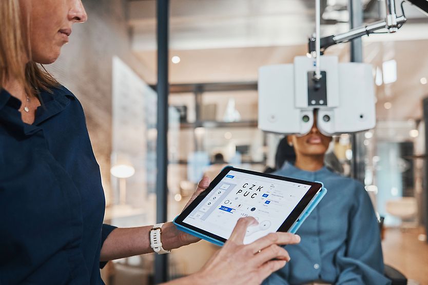
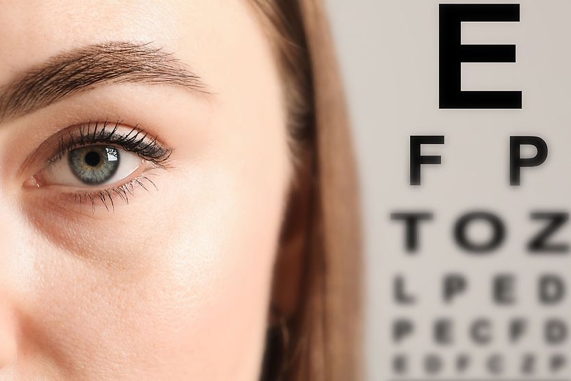
Visual acuity testing
Your eye doctor or an assistant will check your visual acuity with an eye chart.
Diabetes can cause several changes inside your eyes that can affect the clarity of your vision. So a visual acuity test is often included in a diabetic eye exam.
If your visual acuity has decreased since your last exam, your eye doctor may also perform a refraction. This is typically done for two reasons:
- To see if there’s been a significant change to your eyeglasses prescription
- To evaluate your ideal visual acuity with new lenses
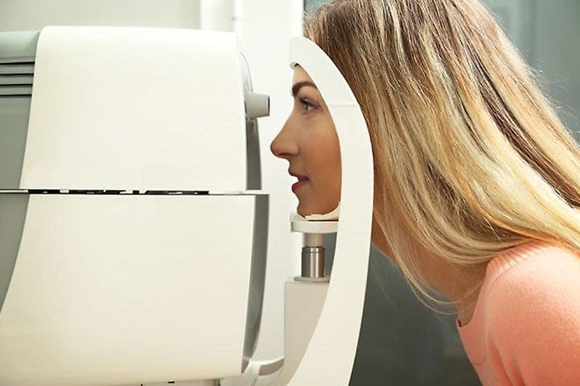
Pupil dilation
In many diabetic eye exams, your pupils will be dilated with eye drops.
This temporarily makes your pupils much larger and eliminates their normal reaction to light, allowing your eye doctor to get a better view of the back of your eye (fundus) to check for damage to the retina from diabetes.
Eye drops are applied to your eyes to cause pupil dilation. It usually takes about 20 minutes for your pupils to fully enlarge, and you typically will be escorted to a reception/waiting area to wait for the dilation to take effect.
Your pupils usually will remain dilated for about two to three hours (in other words, for a period of time that extends beyond your diabetic eye exam. For this reason, it can be a good idea to bring dark sunglasses with you to your exam and consider having someone drive you home).
If necessary, you may be given a pair of disposable sunglasses at your doctor’s office to wear home after your exam.
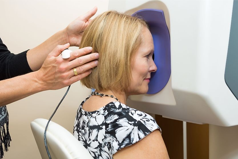
Fundoscopy
The term fundoscopy (or ophthalmoscopy) describes an examination of the back of the eyeball — where the retina, the blood vessels that feed the retina and the optic nerve are located.
Your eye doctor (typically after your pupil is dilated) will examine your fundus with one or more of the following instruments or procedures:
- Direct ophthalmoscope — This is a small, hand-held instrument with a bright light. It is positioned very close to your eye as your eye doctor looks through it to focus on the back of your eye.
- Indirect ophthalmoscope — This is a combination of a bright light your eye doctor wears on their head and a large hand-held lens that is held close to your eye. This device can provide your eye doctor a wider-angle view of your retina and the back of your eye.
- Slit lamp ophthalmoscopy — You and your eye doctor are seated at opposite ends of a table-mounted, illuminated binocular microscope (slit lamp), and your doctor will examine your eye with a small, hand-held lens. This can give your eye doctor a highly magnified, 3D view of specific parts of your retina, retinal blood vessels, and optic nerve.
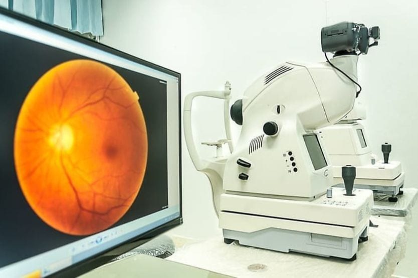
Fundus photography
In addition to (or sometimes, in place of) fundoscopy, your eye doctor may have an assistant take a wide-angle, high-resolution photograph of the back of your eye.
An advantage of this fundus photography is that it creates a permanent record of the appearance of your retina and retinal blood vessels on the day of your diabetic eye exam.
Another potential advantage is that your eye doctor can show you the image on a digital screen and point out any areas of concern.
This type of fundus photography is called ultra-widefield retinal imaging. It requires a large instrument and is typically performed by one of your eye doctor’s assistants in a separate testing room.
Glaucoma testing
Diabetes and diabetic retinopathy can increase your risk for glaucoma.
For this reason, a diabetic eye exam may also include a measurement of the pressure inside your eyes and a detailed examination of your optic nerve during the fundoscopy.
What do diabetic eye exams check for?
Diabetic eye exams focus on any possible eye health problems that can be caused by diabetes, including:
- Diabetic retinopathy
- Diabetic macular edema
- Cataracts
- Glaucoma
Diabetic retinopathy, a leading cause of blindness among American adults, occurs when blood vessels inside the eye start to leak. Left untreated, the leakages can worsen over time and lead to varying levels of blindness. Early detection and treatment can often save part or all of your eyesight.
Diabetic macular edema (DME) is a swelling of the macula, located in the back of the eye. It’s responsible for central vision and seeing color. When a patient with diabetic retinopathy experiences blurry vision, DME can often be the cause.
Is there an additional cost for a diabetic eye exam?
Diabetic eye exams may be billed as medical eye examinations. For this reason, costs will likely differ from a standard comprehensive eye exam.
Some or all of the fee for a diabetic eye exam may be covered by medical insurance. But be sure to ask your insurance carrier for details about the extent of your coverage.
When do I need a diabetic eye exam?
People with diabetes should usually follow strict precautions to ensure ongoing eye health:
- Adults with Type 1 diabetes should typically schedule an eye exam within five years of their diagnosis and every year thereafter.
- Adults with Type 2 diabetes should typically schedule an exam as soon after their diagnosis as possible.
- When possible, women with diabetes who are planning to get pregnant should typically consult with their eye doctor beforehand. An eye exam should also usually be scheduled within the first three months of pregnancy. Another exam should likely be scheduled a year after giving birth.
After an initial exam, the ADA recommends getting annual diabetic exams to monitor the health of your eyes. It’s important to specifically schedule a diabetic eye exam; a typical routine eye exam for people with no health problems may not focus enough on the specific needs of a diabetic patient.
Depending on the findings of your diabetic eye exam, your optometrist or ophthalmologist may recommend more frequent exams or may refer you to a retinal specialist.
The eyes can be among the first parts of your body affected by diabetes. But maintaining a regular routine of diabetic eye exams can help ensure any problems are diagnosed and treated early to protect your eyesight.
READ NEXT: Diabetic eye doctor and eye exam near me