Understanding proteomics and its impact on vision science

What is proteomics?
Proteomics is the study of proteins and how they impact individual cells, tissues and organisms. Proteins affect biological processes throughout the body, including the eyes and vision. By analyzing patterns in proteins, scientists can identify biomarkers that may help detect and manage diseases.
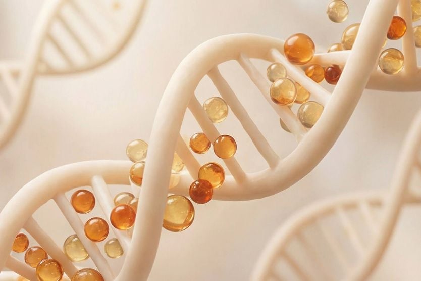
Evolving from genomics — the study of genes (deoxyribonucleic acid or DNA) — proteomics focuses on the proteins that genes help produce and how they function in the body.
Since genes provide the instructions for making proteins, inherited variations in a person's DNA can change how much of a given protein is produced, how it's structured or how well it functions. Proteomics can pick up these downstream effects even when a genetic test alone would not predict how a disease might actually behave in the body.
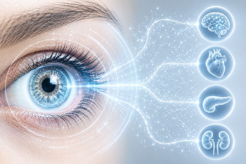
Some of these proteins may serve as biomarkers in oculomics — the study of how biomarkers found in the eye can provide information about systemic health.
Proteomics examines proteomes, or sets of proteins produced by a cell or organism. This includes their:
- Composition
- Structure
- Function
- Interactions within the body
- Expression profiling (changes in protein levels)
- Post-translational modifications (chemical changes occurring after proteins are produced)
Proteins serve an important role in the body, specifically in cellular function. They can detect changes in the body and help control biological processes in response to those changes. Changes in certain proteins can lead to the development and progression of disease.
The study of proteomics as it applies to health care is called “clinical proteomics.” It can offer scientists the opportunity to:
- Identify disease biomarkers
- Improve the ability to diagnose diseases
- Advance the development of disease treatments and therapies (such as drug discovery)
- Develop more effective medications
- Allow for more personalized treatment strategies
- Determine disease progression and prognosis
Types of proteomics
There are three main categories of proteomics:
- Expression proteomics – Examines which proteins are present in cells. It also studies how their levels change in response to factors, like diseases, medications or environmental circumstances.
- Structural proteomics (cell mapping) – Examines the structure and location of proteins within a cell or organism. It can aid scientists in understanding how proteins function and interact with each other.
- Functional proteomics – Focuses on what proteins do, including their role in various molecular processes. It also studies how they interact with other proteins to perform biological functions.
Proteomics and vision
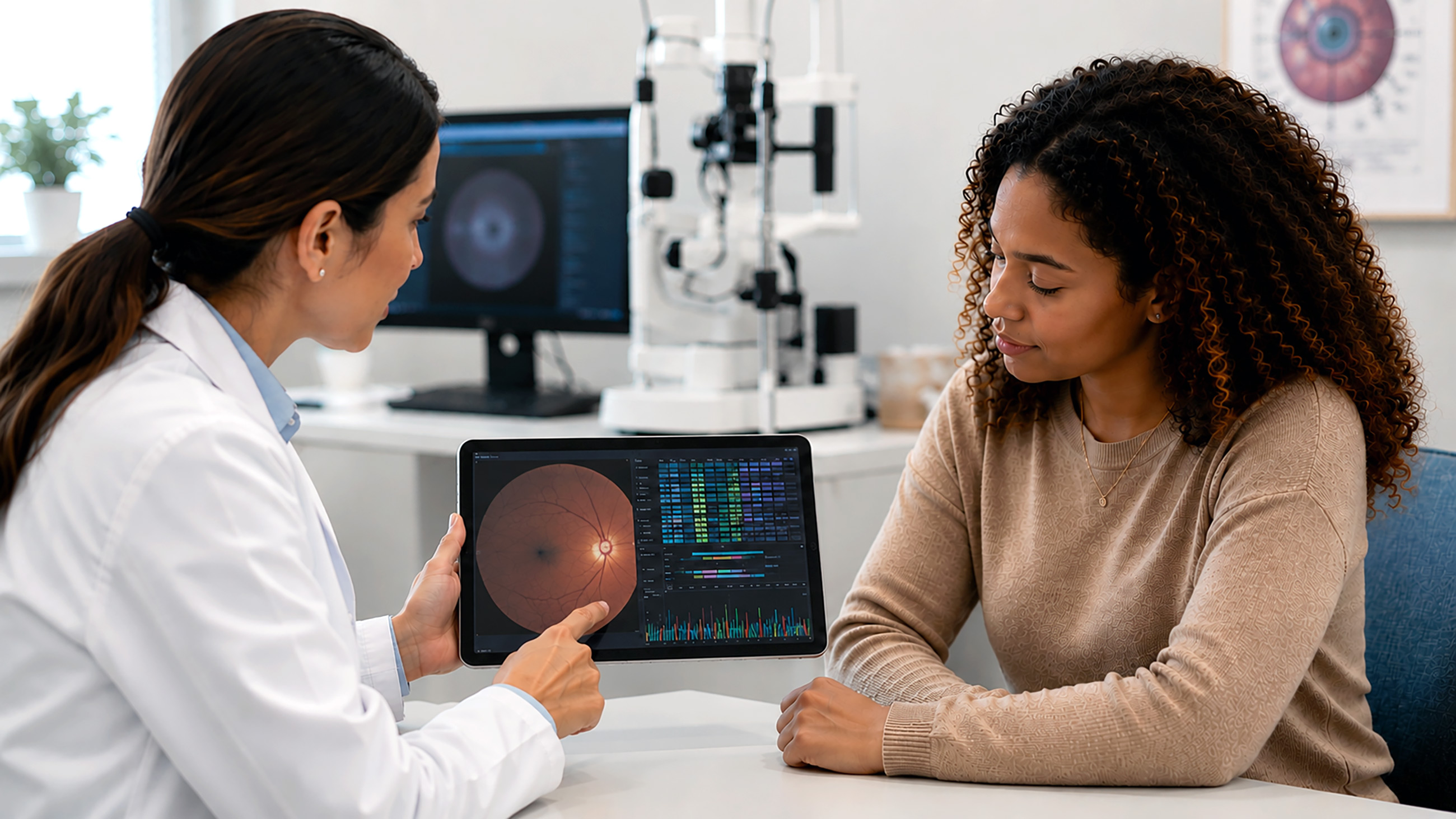
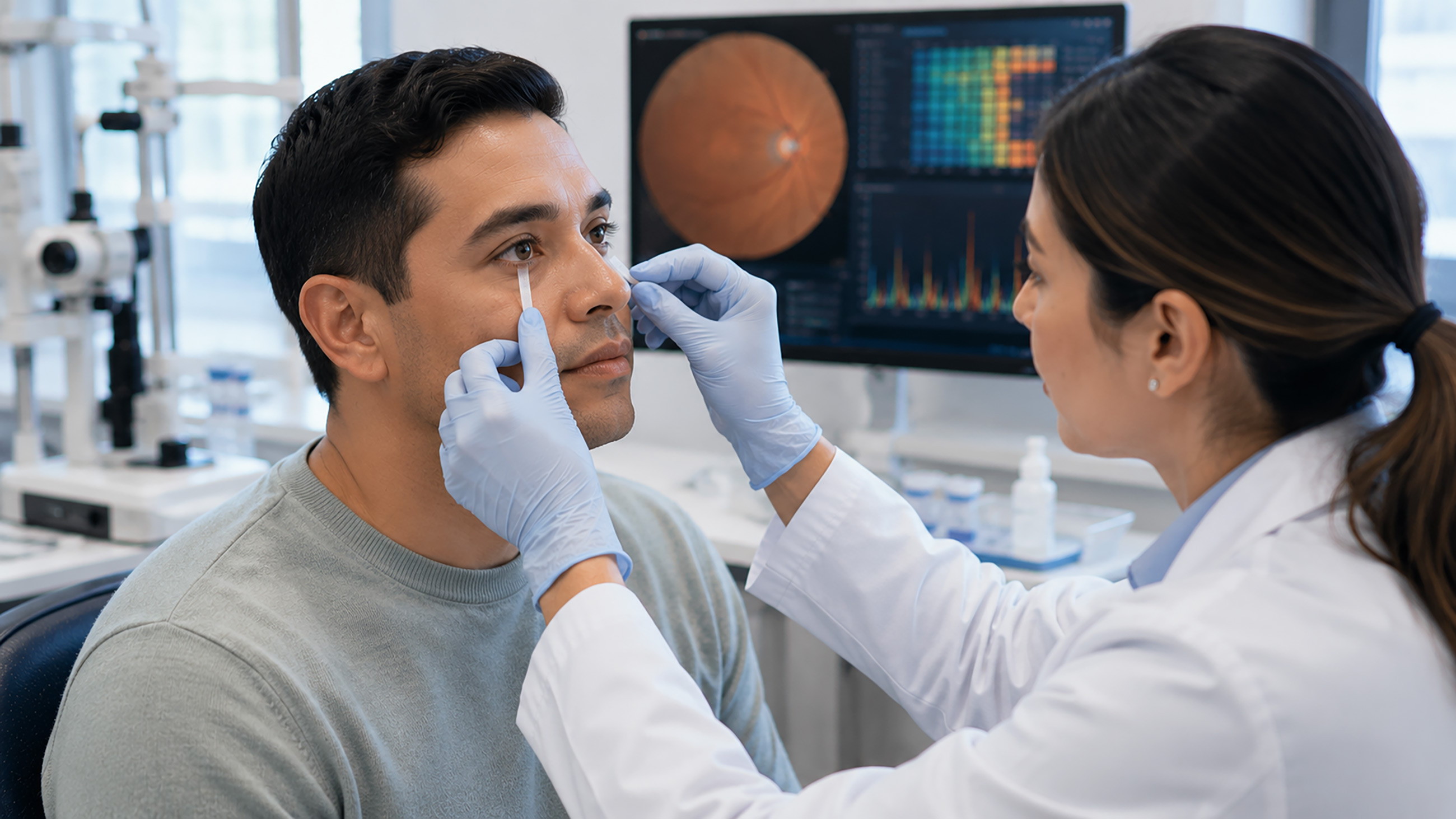
The eye contains thousands of proteins that help support eye health and vision. Various factors can affect protein levels and function in the eye, such as age, stress and other environmental conditions. Today, eye doctors primarily rely on tools like tonometry, optical coherence tomography (OCT) and dilated fundus exams to detect these changes. Proteomics is being studied as a way to complement, not replace these established methods.
Changes in these proteins can signal the development and progression of eye diseases. This makes proteomics a potentially effective method of detecting, diagnosing and treating ocular conditions.
In vision science, proteomics may be used to find protein biomarkers linked to eye conditions, such as glaucoma, diabetic retinopathy (DR) and age-related macular degeneration (AMD). These findings might help eye doctors detect diseases earlier, which could potentially improve how they manage, treat and predict outcomes for patients.
It's important to note that most of the protein biomarkers described are still investigational. They come from research studies and are not yet part of comprehensive eye exams or standard diagnostic testing. If you have or are at risk for glaucoma, diabetic retinopathy or AMD, it is recommended to talk with your eye doctor about how to appropriately manage your condition.
The Human Eye Proteome Project (EyeOME)
The Human Eye Proteome Project, or EyeOME, is a large-scale initiative that seeks to identify and catalog all proteins within the human eye. It’s a part of the broader effort led by the Human Proteome Organization (HUPO). They aim to understand how proteomics might help diagnose and treat diseases in different body systems.
In general, the goals of EyeOME are to:
- Develop tools to map proteins in the eye. This can help detect disease pathways and new biomarkers, which may ultimately lead to preventive and therapeutic measures for eye conditions.
- Create a collaborative network of scientists focused on eye proteomics.
- Characterize proteins within the human eye and how they change throughout the protein lifecycle.
- Establish standard methods for studying eye proteins. This includes collecting, processing, dissecting and storing protein samples.
- Establish reporting standards to create a human eye proteome database.
- Create shared repositories of human eye tissue samples. This can encourage collaboration, promote open access and facilitate the exchange of research.
- Secure funding for researcher exchanges and collaborative scientific meetings to expand proteomics in the field of ophthalmology.
EyeOME has helped advance vision science by identifying and mapping proteins in the eye. This work can provide researchers with a better understanding of eye diseases. It may also lead to the earlier detection and more effective management of eye and vision conditions.
Proteomics in eye diseases
Proteomics is increasingly being applied across a range of eye diseases to detect disease-associated protein patterns. Some of the more significant applications include:
Glaucoma and proteomics
Glaucoma is a group of eye diseases that can damage the optic nerve — the structure that connects the eye to the brain. It’s often associated with high intraocular pressure (IOP) or pressure within the eye. The condition can lead to vision loss and eventual blindness, especially if left untreated.
Many people don’t experience glaucoma symptoms until damage has occurred. Therefore, diagnosing and treating the condition early is important for preventing and slowing the progression of vision loss.
Proteomics is helping scientists identify proteins associated with optic nerve damage. It can also aid in glaucoma diagnosis, progression and treatment. Studies have found several protein biomarkers linked to certain types of glaucoma, such as:
- Serum ferritin – A type of protein associated with inflammation and oxidative stress.
- Brain-derived neurotrophic factor (BDNF) – A protein that helps support nerve health.
- Nerve growth factor (NGF) – A protein that helps support nerve health.
- Secreted protein acidic and rich in cysteine (SPARC) – A protein linked to higher eye pressure and trabecular meshwork (TM) remodeling.
- Thrombospondin-2 – A protein linked to higher eye pressure.
- Osteopontin – A protein linked to higher eye pressure.
Other proteins are also being studied as potential biomarkers of glaucoma.
Diabetic retinopathy (DR) and proteomics
Diabetic retinopathy (DR) is an eye disease that can affect people with diabetes. It can occur when:
- High blood sugar levels damage retinal blood vessels, ultimately causing them to swell, leak or become blocked
- New, abnormal blood vessels form in the retina
DR can damage the retina (the light-sensing structure at the back of the eye). It can lead to vision loss, especially without timely treatment and proper blood sugar control.
Researchers are using large blood protein datasets to pinpoint biomarkers associated with DR. Several stand out in terms of disease risk and progression, including:
- Plexin-B2 (PLXNB2) – A protein linked to inflammation and neurovascular stress in the retina. It may also signal disease activity.
- Growth differentiation factor-15 (GDF-15) – A stress-response protein associated with early cellular and vascular damage in the retina.
- Renin (REN) – A protein linked to inflammation and blood vessel dysfunction. It may also indicate potential causal effects of DR.
These blood proteins may aid scientists in diagnosing the condition. They may also help predict disease risk and identify effective treatment options.
Age-related macular degeneration (AMD) and proteomics
Age-related macular degeneration (AMD) is a progressive eye condition. It causes the deterioration of the macula — the portion of the retina that provides your central and sharpest vision. Over time, AMD can lead to the loss of central vision, or what you see directly in front of you. There are two main types, known as wet AMD and dry AMD.
Proteomics studies in AMD have detected proteins as biomarkers that can signal disease. Many of these proteins are found to be altered. This can show differences between healthy and diseased tissues. It can also reflect changes associated with disease progression.
Scientists have found several key biomarker proteins involved in AMD, including:
- Apolipoproteins – Linked to lipid metabolism.
- Complement proteins – Associated with immune, inflammatory and other defense responses in the body.
- Clusterin – Helps regulate the complement system. It is also linked to processes, such as cell death, tumor growth and nerve damage.
- Protease inhibitors – Help control processes, like inflammation, blood clotting, new blood vessel growth and tissue maintenance.
Anterior segment eye diseases and proteomics
Proteomics is also used to study anterior segment eye diseases, which affect the front part of the eye. This includes structures, such as the cornea (the clear, dome-shaped structure at the front of the eye) and crystalline lens (the flexible disc behind the pupil).
Proteomics has been used in vision research for several years, particularly for conditions like glaucoma, DR and AMD. However, its application to the anterior of the eye appears to be expanding.
One area includes tear proteomics, which examines proteins in the tear film. This helps identify biomarkers of conditions, such as dry eye disease (DED) and keratoconus.
Scientists are also applying proteomics to study other anterior segment diseases, including cataracts. Aqueous humor (the fluid that fills the anterior chamber) is often used for protein analysis in cataract studies.
Advances in proteomics technologies
Proteomics involves various techniques used to identify, measure and characterize proteins. It also explores how they impact health and disease. While the field continues to evolve, some of the current applications include:
- Mass spectrometry – A technique for finding the molecular weight and chemical makeup of biological structures. In proteomics, it helps scientists detect and quantify proteins for analysis.
- Gel electrophoresis – A technique for separating proteins based on their mass, charge and weight. This can allow for further characterization and analysis of individual proteins.
- Nuclear magnetic resonance (NMR) spectroscopy – An approach that helps scientists map the shape of proteins. This can further their understanding of protein function and applications, such as drug design.
- X-ray crystallography – A method that exposes crystallized protein samples to X-rays to determine their three-dimensional structure. This can aid in efforts, like drug design and studying protein interactions.
- Protein microarray – A technique that measures protein levels, functions and interactions. It can help identify disease-related protein changes.
- Edman sequencing – A protein sequencing technique that can help scientists better understand a protein’s composition. It can also aid in assessing the quality of protein-based medicinal therapies.
- Bioinformatics – An approach that uses proteomic algorithms and databases to analyze large protein datasets. It can help reveal biomarkers, protein pathways and protein interactions.
- Aptamer-based assays – A newer technique that uses DNA or RNA molecules to detect and measure a large number of proteins. It can potentially aid in finding new biomarkers, disease diagnosis and treatment planning.
Artificial intelligence (AI) and machine learning (ML) help synthesize large, complex proteomic datasets. Scientists can then use this information to pinpoint disease biomarkers. It can also aid in drug discovery and in monitoring the effectiveness of medications and other treatments.
Challenges and future directions
Proteomics has advanced the study of eye and systemic disease in numerous ways. It continues to grow as an important area of research. However, it is not without its limitations.
One of the primary challenges is ethical considerations. This includes privacy, data protection and the handling of sensitive health information, among other concerns. Other key challenges include turning lab-based proteomic information into useful real-world clinical data. The development of clear clinical guidelines is also needed on a global scale.
Some of the more technical challenges currently affecting proteomics involve:
- Managing, organizing and processing vast amounts of protein-related data
- Limited consistency between protein evaluation techniques
- Distinguishing technical from biological associations
- Validating findings across multiple platforms
- Standardizing data references when comparing results between studies
- Risk of false-positive findings in large-scale proteomic studies
Future advances in proteomics are expected to broaden the study of proteins across various biological tissues and diverse populations. They will also likely allow for more dynamic evaluation and tracking of proteins in the body.
Proteomics and the future of vision science
The eye contains many proteins that are essential to eye health and vision. Changes in proteins caused by factors, like aging and environmental conditions, can indicate disease development.
Proteomics helps identify these protein changes — called biomarkers — in eye conditions, such as glaucoma, diabetic retinopathy and AMD. This could allow for the earlier detection, diagnosis and management of eye diseases.
These tools are also feeding into precision medicine efforts. Proteomic profiles may eventually help match patients to specific therapies, including emerging gene therapies for inherited retinal disease, based on their individual protein signatures rather than a one-size-fits-all approach.
Ultimately, proteomics could have a significant impact on eye care and treatment in future.