Custom-built vision: What the next-generation eye exam can reveal about your health
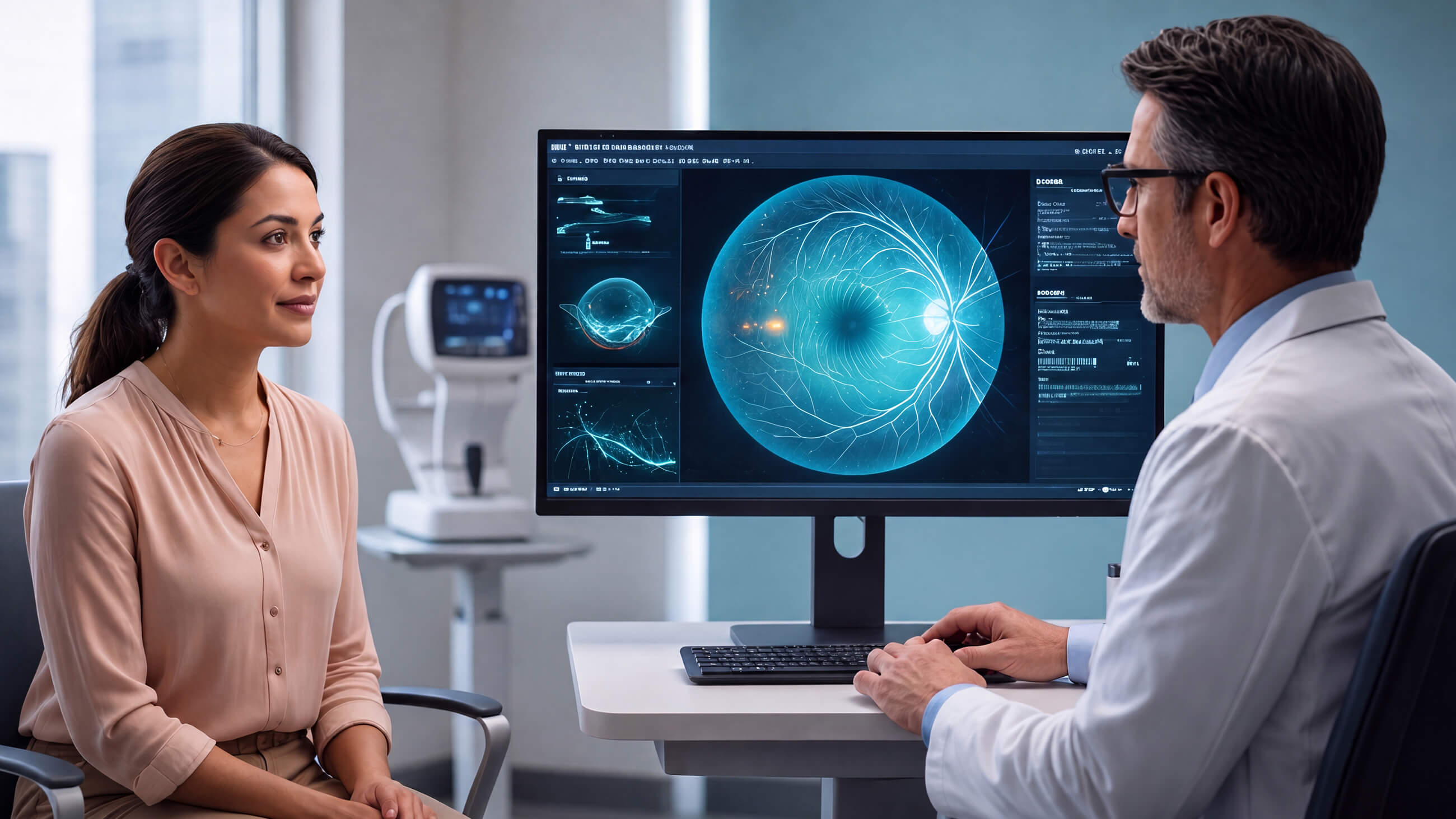

A person settles into the chair for their annual comprehensive eye exam. Things seem as they should. Their farsightedness hasn’t progressed, so no need for a stronger prescription. The eye pressure check was normal with no signs of glaucoma at the visit. Diabetic retinopathy isn’t a concern either.
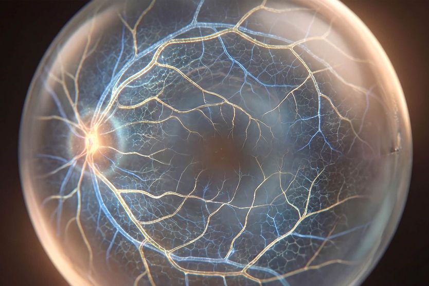
During the visit, the eye doctor takes a closer look at their retinal scan. The image shows subtle changes in the small blood vessels of the retina. The patient is referred to their primary care doctor, who catches undiagnosed high blood pressure.
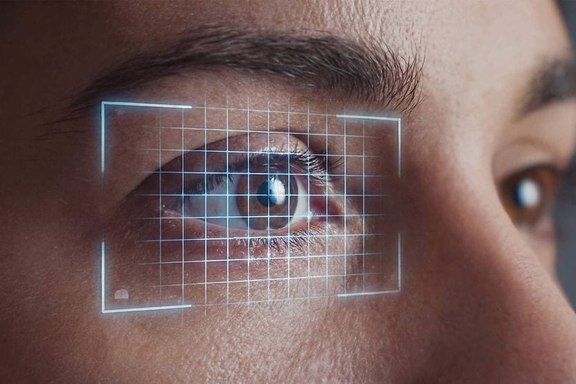
This scenario is one way the comprehensive eye exam is evolving to provide eye doctors with faster, data-backed analysis that unlocks other insights about a patient’s health.
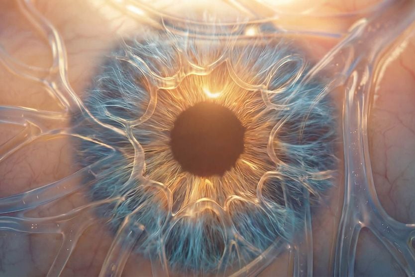
The eye has long been considered a unique window into one’s health. It’s the sole “accessible extension of the brain” that provides a non-invasive view of blood vessels and the nervous system.
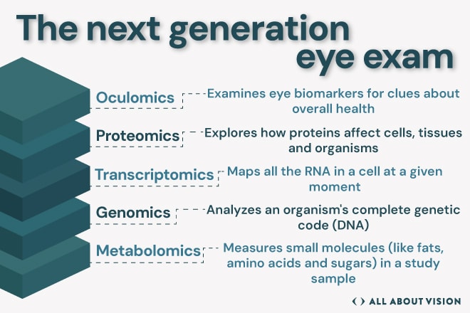
This thinking is represented by oculomics, a system that empowers physicians to make further sophisticated analysis and diagnosis of health conditions. Oculomics uses information known as biomarkers, the biological indicators of a person’s health, uncovered by image scans and analysis backed by artificial intelligence (AI) and machine learning.
The advances changing the eye exam
“The eye is an internal organ directly exposed to the outside world, and it is truly unique that by dilating your eye or looking inside your eyes, we literally have insight into what’s going on inside your body,” said Roya Attar, OD, MBA, DHA, FAAO, associate professor and director of optometric services in the department of ophthalmology at the University of Mississippi Medical Center.
Eye doctors hope to expand the range of diseases and health issues they can detect. The concept isn’t far-fetched, as AI-backed analysis is already used in FDA-cleared software to screen for diabetic retinopathy. These autonomous systems can produce a result on their own without an eye specialist interpreting the image in real time. They can detect whether more-than-mild diabetic retinopathy is present and if the patient should be referred to a specialist.
Health care professionals, such as primary care practitioners (PCPs), may use these devices to examine people who might not be getting their comprehensive eye exam. After the PCP takes a photo of the back of a patient’s eye, often with a compact or handheld portable device, the software scans and reads the image. Then, the PCP can decide if the patient needs to visit an eye doctor for a closer look.
“That is revolutionary because a lot of times, these patients were not able to get to an eye doctor, so at least there is some way they are being screened,” Dr. Attar said.
As with all technological advances, it’s important to understand limitations. Dr. Attar recalled a patient whose imaging screening report accurately identified the lack of diabetic retinopathy. The patient still required treatment for glaucoma, which was discovered by traditional screening methods.
“It does not take the place of a truly comprehensive eye exam,” said Dr. Attar.
Researchers also caution that AI screening tools are only as reliable as the images and data behind them. A portion of the images taken may be flagged as ungradable and require a repeat scan or a referral. Performance of AI-based technologies can vary across different patient populations. Experts recommend that these systems should be validated in diverse groups and more importantly monitored over time with the goal to enhance accuracy.
Imaging can also map blood flow in the retina without dye injection, a less invasive process. This method helps doctors identify vascular changes that can be linked to eye diseases, such as diabetic retinopathy or age-related macular degeneration.
Beyond what’s already in use, researchers are looking for more biomarkers that the eye might reveal. One active area of study is whether AI analysis of the retina’s blood vessels can flag a person’s risk of cardiovascular disease.
Other work is further into the future. There’s optimism that a retinal scan could uncover early signs of neurodegenerative disease. Scientists are studying links between changes in the retina and other conditions, such as Alzheimer’s disease.
“I believe that more and more biomarkers will be identified with AI to help us be on the precipice of diagnosing patients early, but also monitoring them, working much more collaboratively with other physicians and specialties,” Dr. Attar said.
The imaging tools behind the shift
Imaging is the foundational technology for this new layer of insights. By employing a non-invasive method to visualize the eye and analyze biomarkers, researchers aim to provide doctors with new ways to assist their patients.
A key player in this field is optical coherence tomography or OCT. The scan uses light waves to create a detailed picture of the retina. It’s already used to help evaluate and monitor glaucoma, diabetic retinopathy and age-related macular degeneration (AMD). This technology is increasingly being studied for detecting and monitoring neurodegenerative conditions such as Parkinson's disease.
“When we look at the clinical care of patients today, ophthalmology has become a very imaging-driven subspecialty,” said Justis P. Ehlers, MD, director of the Tony and Leona Campane Center for Excellence in Image-Guided Surgery and Advanced Imaging Research at the Cole Eye Institute at Cleveland Clinic. “When we think about the key pieces we need to make decisions, most of us would consider an OCT as perhaps the most critical component of optimal patient care, depending on the disease we’re looking at.”
An additional innovation is optical coherence tomography angiography or OCTA. It builds on OCT to map blood flow through the retina. The software acquires rapid OCT images sequentially to capture the body’s blood flow.
Fundus imaging is also widely used. It takes a wide-field photograph of the back of the eye and captures the retina, optic disc and blood vessels. It’s increasingly paired with AI-screening software to detect diabetic retinopathy.
There’s increased optimism about adaptive optics. The technology comes from astronomy, where telescopes cancel the blur introduced by Earth’s atmosphere, bringing distant objects into sharper focus. Applied to vision, adaptive optics seeks to remove blur and sharpen images for more precise viewing of the tiniest blood vessels and other parts of the eye, giving doctors more targeted data than before.
How the science of omics technologies is shaping eye care
Imaging is only part of how a next-generation eye exam may help eye doctors develop new treatment and diagnostic pathways for patients.
The innovations hoped for in personalized medicine draw on a number of omics technologies, with each focused on a particular layer of human biology.
Genomics is the closest to clinical use. Researchers are using a person’s genetic data to estimate the risk of conditions, such as AMD or glaucoma. Genetic risk scores for such conditions are still being tested in studies and aren’t yet part of a comprehensive eye exam.
This work focuses on common, yet complex conditions, where many genes each add a small amount of risk. That’s different from inherited retinal diseases (IRDs) caused by a single gene where genetic testing has started to become a part of clinical care and can help guide newer, gene-based treatments.
Other “omics” fields are in various stages of development.
Sources:
Oculomics – The eyes talk a great deal. Indian Journal of Ophthalmology. March 2022. Multi-omics profiling for health. Molecular & Cellular Proteomics. June 2023.
Proteomics focuses on the proteins the body makes. Researchers are digging into proteins in tear fluid as a non-invasive approach to look for disease markers.
Metabolomics examines metabolites, the small molecules produced as the body breaks down food, medication and other substances. Observing such changes can work as a critical signal for eye disorders like macular degeneration, glaucoma, diabetic retinopathy and myopia.
Transcriptomics studies are what’s known as “gene expression,” which looks at how genes are switched on or off and the implications for disease. For vision, researchers are examining these patterns in retinal tissue to spot the molecular changes that may signal disease.
According to Dr. Ehlers, oculomics holds the most promise to interpret information gleaned from the eyes and apply it to other areas of one’s health.
“Oculomics, in my mind, is taking it to the next level using high-level computational technologies, like artificial intelligence, to interpret different signatures in the images obtained from the back of the eye. Studies have demonstrated that using these signatures’ unique associations can be identified with systemic conditions, from cardiovascular disease to Alzheimer’s,” he said.
What’s available near you
Depending on where you live or the capabilities of a particular health system, some approaches may be more readily available than others.
However, it’s becoming more common to see robotics in the exam room or have an eye doctor employ machine learning with analysis to screen and diagnose conditions.
Dr. Ehlers said advanced algorithms are already capable of making accurate diagnoses and analyses.
“We have been able to deploy what are near-automated cameras that have AI-enabled image analysis for diabetic retinopathy screening. In this case, a patient with diabetes can come in, get a picture taken and the AI analysis determines whether or not they need to be referred for further evaluation,” he said.
Note: A positive screening result is a prompt to see an eye doctor — not a final diagnosis. Confirming the disease, staging it and deciding on treatment still require a comprehensive, in-person exam.
Today’s comprehensive eye exam is a fuller, deeper analysis than was available in the past. Your eye doctor has more tools to check for a wider scope of diseases and may one day alert you to other health problems.
For now, eye doctors generally agree that everyone needs an annual comprehensive eye exam, even if there are no present vision problems. That line of thinking could take on new meaning as imaging technology and analysis software get even more powerful.
Dr. Attar sees future advances in AI, helping fill the gaps that can occur in human diagnosis, especially with diseases like glaucoma. The diagnosis has relied on judging the nuances of conditions, like eye pressure and damage to the optic nerve. AI can reduce the risk of error and improve objectivity in critical exams.
“Three doctors could look at the same optic nerve and judge it differently, size it differently,” she said. “The same doctor could look at the same eye, the same nerve and call it one size at one exam and another size at another exam.”
Catching disease earlier matters because several sight-threatening conditions like glaucoma and diabetic retinopathy often cause few or no symptoms until vision is already affected. Early detection and monitoring, with the help of AI technology, can mean proactive treatment and a better chance of protecting everyday vision for reading, driving and work.
Dr. Attar said that while the clinician role will always be essential, AI technology will be an integral assistant to enhance what an eye doctor can do for their patients. Its role in examining retinal scans and analyzing large data sets will become a core part of the eye exam and its post-visit analysis.
“AI doesn’t have those biases,” she said. “It looks at everything.”