How transcriptomics can advance vision science and eye health
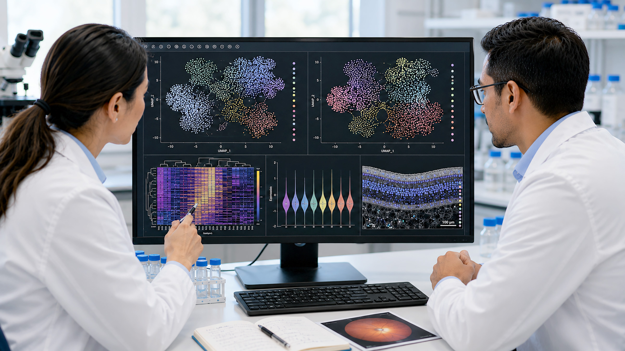

What is transcriptomics?
Transcriptomics is the study of all the RNA molecules in a cell or tissue at a given moment. This collection of RNA is called a transcriptome. You can think of a transcriptome as an always-changing cookbook of RNA recipes for your cells and tissues to follow.
Your body constantly makes new RNA so cells can use the information stored in your genes (DNA). This happens because your genes are always:
- Being switched on and off
- Being made more or less active
Unusual changes in a transcriptome can disrupt a cell’s normal routine. This can lead to diseases or other problems. Studying RNA can help researchers see how and where the changes are happening.
Transcriptomics research can help scientists:
- Learn which genes are active in different types of cells and how active they are
- Compare healthy cells with diseased ones to see how they’re different
- Discover information about gene behavior to help support new treatments
Understanding transcriptomics and its role in vision
In transcriptomics, researchers examine RNA to see which genes are active and how active they are. This is a constantly changing process called gene expression.
Transcription and messenger RNA (mRNA)
There are different types of RNA, but the main type used to study gene expression is mRNA, which stands for messenger RNA.
mRNA molecules are like temporary instruction manuals that tell your cells how to make other molecules called proteins. They can also affect how many proteins a cell makes. Proteins help your body grow, repair itself and stay alive.
The original instructions for making proteins are in the genes in your DNA. But DNA doesn’t leave the cell’s control center (nucleus), so it has to be copied into a messenger.
The instructions are transcribed (copied) from your genes into mRNA molecules and carried to the protein-making parts of the cell. This is how gene expression starts.
Gene expression in the eye
Scientists use gene expression to understand how cells work in different parts of the eye and how eye diseases can disrupt the cells’ routines and change their behavior.
Transcriptomics can be used to study many different parts of the eye, since most are made of living cells with RNA.
Eye structures like these are vital to healthy vision:
- Retina – Detects light and sends visual signals to the brain.
- Optic nerve – Carries visual signals from the retina to the brain.
- Cornea – Helps focus light and protects the inside of the eye.
By looking at RNA in the eye, scientists can try to find out how cells react to things, like age, lifestyle and other factors.
This information helps them understand vision-threatening conditions, like glaucoma and age-related macular degeneration (AMD). It can also be an important step toward developing new ways of diagnosing and managing those diseases.
Key applications of transcriptomics in vision research
Transcriptomics helps scientists learn more about different parts of the eye and their cells. Eventually, this can help other researchers protect people’s vision.
Studying retinal health through transcriptomics
The retina isn’t just part of the eye; it’s an extension of the brain. It’s the only part of the brain system that can be seen from the outside, so it’s very useful in scientific research.
The retina contains millions of special cells called photoreceptors. The cells react to light and send visual signals to the “processing” parts of your brain so you can see.
There are two types of photoreceptors:
- Rod cells (rods) – Let you see along the sides of your view (peripheral vision) and in dark or low-light situations.
- Cone cells (cones) – Let you see colors and sharp details.
The neural retina is a major part of eye-related transcriptomics research.
All rods and cones live here, along with other important types of retinal cells. Each kind of retinal cell has a different job, so they each rely on a different gene for instructions.
Scientists can study RNA to see which genes are active in different retinal cells and how those cells are working.
Related areas of research
Transcriptomics is an important piece of the retina puzzle, but there are other pieces, too.
Researchers can combine transcriptomics research with genomics (the study of full sets of genes), proteomics (the study of proteins) and tissue samples to help create a cell “atlas” of the retina.
Together, these help them get a more complete picture of the eye’s inner workings.
Identifying disease mechanisms
Researchers use transcriptomics to understand how both common and rare eye diseases affect the eye at the cellular level. It's important to note that transcriptomics is a research tool used to study disease at the molecular level. It is not itself a diagnostic test or treatment, and any therapies that emerge from this research must go through their own clinical trials and regulatory approval before they reach patients.
They often look for harmful patterns in gene expression. These can act like “red flags” called disease biomarkers.
Learning how diseases develop like this can be an early step toward new diagnoses and treatment options.
Glaucoma
Glaucoma is a group of eye diseases that damage the optic nerve. It can lead to permanent vision loss, especially when it’s diagnosed too late.
People with primary open-angle glaucoma (the most common type) tend to lose parts of their peripheral vision first. This can be hard to notice until the disease is already advanced.
Peripheral vision loss can develop gradually and often outside a person's central focus. It can affect everyday tasks like driving and workplace safety well before it's consciously noticed. This is why it is recommended to schedule a comprehensive eye exam even without symptoms. An early diagnosis from an eye doctor can be important to potentially help preserve your vision.
However, genetics alone usually doesn’t decide someone’s risk for a more common eye disease, like glaucoma. This is because other factors, like aging and lifestyle, can also affect how the disease develops.
Since different eye cells use genes in different ways, researchers use transcriptomics to see which genes are active in the disease-related eye cells.
One group of researchers used transcriptomics to identify 37 different types of cells in the eye. Then, they looked at more than 180 genes already linked with glaucoma to see which types of cells had those genes switched on and how active they were.
By matching these genes to specific cell types, they built a clearer picture of glaucoma’s effects at a microscopic level. This kind of research helps refine, rather than replace, the treatments glaucoma patients already receive today, which can include:
- Intraocular pressure (IOP)-lowering eye drops (preserved and preservative-free)
- Selective laser trabeculoplasty (SLT/DSLT)
- Minimally invasive glaucoma surgery (MIGS)
- Minimally invasive bleb surgery (MIBS)
- Procedural pharmaceuticals
- Glaucoma filtering surgeries
Macular degeneration
Transcriptomics has helped map different types of retinal cells and see how they change when people have AMD. This is when cells in a small part of the retina (the macula) are damaged over time, which can cause vision loss.
Age-related macular degeneration comes in one of two forms, dry or wet. Dry AMD is more common and develops gradually in stages when tiny protein deposits, called drusen, form in your macula. On the other hand, wet AMD is less common and happens when your body creates abnormal blood vessels under your retina in the foveal to macular areas. These blood vessels can then begin to leak, causing fluid accumulation and bleeding in the macula.
By comparing RNA in healthy retinas with RNA from retinas affected by AMD, studies have found that certain immune-related genes become more active. This helps researchers understand what role someone’s immune system might play in AMD.
The standard treatment for wet AMD today is a class of drugs called anti-vascular endothelial growth factor (anti-VEGF) injections, which transcriptomic research may help refine further rather than replace.
Diabetic retinopathy
Diabetic retinopathy (DR) happens when diabetes causes harmful changes to blood vessels in the retina. This can cause visual symptoms and vision loss.
Transcriptomic data (boosted by AI and machine learning) helped researchers in one study find early changes in gene and cell activity before there was serious damage.
In another study, researchers used RNA data to examine individual retinal cells from patients who had advanced diabetic retinopathy. This helped them pinpoint which types of cells were involved in the late-stage damage. Diabetic retinopathy is currently managed with anti-VEGF injections, laser treatment and blood sugar management. Transcriptomic findings like these are aimed at sharpening that care, not replacing it.
Inherited retinal diseases
Rare eye conditions called inherited retinal diseases (IRDs) can be caused by a mutation (change) in one or more genes. These conditions can follow several inheritance patterns which determine how likely a mutation is to be passed on and who in a family may be affected:
- Autosomal dominant
- Autosomal recessive
- X-linked
- Mitochondrial (less common)
They can affect the retina in ways that often lead to vision loss or blindness.
Some of these diseases are:
By studying RNA, scientists can see how mutations change the way genes act in certain retinal cells. They can also look for mutations that affect RNA splicing, the actual process of turning a gene on or off.
When RNA splicing goes wrong, the cell can make faulty proteins that cause diseases.
Some research shows that around 11% to 15% of mutations that cause an inherited retinal disease are linked to RNA splicing problems.
Findings like these could eventually support new treatments for conditions that are very hard to treat right now. In fact, this kind of research has already led to an approved gene therapy. Voretigene neparvovec received approval from the U.S. Food and Drug Administration (FDA) and is used to treat RPE65 mutation-associated retinal dystrophies such as Leber congenital amaurosis. This illustrates how transcriptomic and genetic research can translate into real therapies for people.
Advancing personalized medicine
While transcriptomics research alone isn’t enough to make a new treatment, it can help scientists look for unusual changes and decide which cells to target.
This could contribute to more personalized treatments, where eye doctors can choose therapeutics based on a patient’s specific disease.
Emerging technologies in transcriptomics
RNA isn’t very stable on its own, so scientists don’t always study it directly. Instead, they can use different tools and techniques to study snapshots of RNA.
A common way to get these snapshots is with a technique called RNA sequencing, or RNA-seq for short.
With RNA sequencing, an enzyme (a type of protein) called reverse transcriptase is used to make a DNA copy of the mRNA instructions. This DNA copy, called complementary DNA (cDNA), is easier to measure in a lab.
The standard method of doing this is bulk RNA sequencing. It’s used to look at the average gene activity in a sample of tissue from the body.
For example, a researcher might take a small tissue sample from a glaucoma-affected retina to see how genes are generally affected. Then, they’ll compare this with the average gene activity in a healthy eye.
Bulk RNA sequencing is still widely used today, along with other techniques, such as:
- Reverse transcription polymerase chain reaction (RT-PCR) – Also involves copying mRNA to cDNA, but the genes are amplified many times to examine them in more detail.
- Microarrays – mRNA attaches to pieces of DNA on a slide or “chip” to measure the gene expression across many genes at once.
- Nanopore sequencing – Reads the sequence of individual RNA molecules as they pass through microscopic openings (pores).
Newer transcriptomics technologies have led to even more ways of studying RNA. Two important methods are:
Single-cell RNA sequencing
Single-cell RNA sequencing (scRNA-seq) is when researchers look at the RNA in individual cells. This lets them see how cells act on their own instead of getting an average from an entire tissue sample.
There are different variations of single-cell RNA sequencing. One example is single-nucleus RNA sequencing (snRNA-seq). It’s used to examine the RNA only in a cell’s nucleus instead of the entire cell.
By comparing thousands of cells or nuclei, researchers can learn more about different cells and see which ones express genes related to eye diseases, like glaucoma. In some cases, single-cell RNA sequencing can also discover new cell types that weren’t clearly known before.
When researchers can figure out which specific cells are affected by a disease, it gets easier to understand how the disease develops and which cells future treatments might need to target.
Spatial transcriptomics
While single-cell RNA sequencing provides a lot of information about individual cells, it doesn’t show where the cells are. This is where spatial transcriptomics can help map out the gene activity in a tissue.
Spatial transcriptomics not only measures how active genes are but also where the gene signals are coming from. This method can also be useful for cells that are more sensitive and harder to separate because it works right on the tissue while the cells are in place.
One example is retinal detachment, an emergency in which the retina pulls away from the back wall of the eye.
Posterior vitreous detachment (PVD) is a common issue where the gel inside the eye separates from the retina. It’s usually age-related and harmless, but some cases can lead to a torn or detached retina.
Researchers don’t always know why that happens. But using spatial transcriptomics together with other techniques could help them figure out where the problems might start. That way, people can help protect themselves in advance.
Comparing these technologies
To understand these three methods, you can think of them like looking at traffic on a highway:
- Bulk RNA sequencing tells you how many vehicles are on the highway without any specific details.
- Single-cell RNA sequencing lets you group the individual vehicles by their type. Think sedans, SUVs, minivans, buses, semi-trucks, and so on. It tells you how many of each is on the highway, but it doesn’t show you their location.
- Spatial transcriptomics plots the vehicle types onto a map of the highway so you can see where each type is.
Challenges and ethical considerations
Like any scientific technology, transcriptomics has limitations and other important issues to consider. Some of the current concerns include:
- Analyzing complex data – Transcriptomics involves processing huge amounts of information. The data can often be hard to interpret accurately, even when it’s mostly done by computers.
- The need for more research – Many transcriptomic findings still need more research before they can be directly used in tests and treatments available to the public.
- Privacy and security – Transcriptomics is linked to someone’s genes, so results can include (or imply) sensitive health information. This needs to be protected and used responsibly.
- Accessibility – Transcriptomic equipment and testing are often expensive, especially newer techniques, like single-cell sequencing and spatial transcriptomics. Although some costs are falling, many research facilities may not have access to them. Some technologies also aren’t widely available yet, both in terms of the equipment itself and the experts trained to use it.
- Information bias – Studies that don’t include diverse groups of people could end up with results that don’t apply equally to everyone. The same could apply to transcriptomic techniques that detect certain genes or patterns better than others.
Future directions in transcriptomics and vision science
Transcriptomics technologies keep improving every year. In the future, they may help researchers learn even more about how eye diseases develop and how new therapies could help patients protect their vision:
- Artificial intelligence – AI is already being used to analyze large sets of data and look for things that would be very hard for humans to find on their own. AI-assisted discoveries like these could become faster, more reliable and work in new ways as time goes on.
- Better disease biomarkers – Researchers will likely keep finding new biomarkers related to eye diseases and other conditions that can affect your vision, such as diabetes. These could help detect and monitor eye problems earlier on.
- More personalized treatment options – Scientists will continue to learn more about the ways different eye diseases affect different people. Over time, this data may be used together with other research to create treatments targeted for individual patients.
The transformative power of transcriptomics in vision
Transcriptomics has already started to make an impact on eye care, but the future is even more promising.
By studying gene activity in the eye’s cells and tissues, researchers are starting to understand eye diseases in much more detail. This includes finding new types of cells, new disease biomarkers and mapping where different cell types are involved in the eye.
Transcriptomic technology will continue to improve in the coming years. Alongside traditional eye exams, advances in transcriptomics could help eye doctors catch problems earlier and personalize their care for each patient’s unique condition.