Peripheral vision: Tests, issues and FAQs


What is peripheral vision?
Peripheral vision is what many refer to as “seeing out of the corner of your eye.” It is your ability to see objects outside of your direct line of sight without turning your head or shifting your eyes. This allows you to do things like walk without bumping into things, drive and play sports.
Peripheral vision is also sometimes referred to as “side vision” or “indirect vision.” The peripheral is the edge of your visual field, or the full range you can see at a given moment.
The typical visual field for humans is 170 degrees: 70 degrees for central vision and 100 degrees for peripheral vision. Peripheral vision can be further broken into three sections:
- Far-peripheral – your vision furthest to the side
- Mid-peripheral – your vision in the middle of the peripheral
- Near-peripheral – your vision nearest to the center
The further from the center (or the more peripheral) vision gets, the lower the acuity or sharpness of what you see.
Peripheral vision is made possible by an area of the retina called the peripheral retina. The retina is the layer of tissue at the back of the eye that converts light into nerve signals. These nerve signals are what the brain uses to interpret what you see.
The peripheral retina contains cells called photoreceptors, which can be broken into two types: rods and cones. Rods handle vision in dim light, while cones handle color vision.
The center of the retina is an area called the macula. The macula is what allows you to process objects directly in front of you. The outside area of the retina is the peripheral retina, which deals with objects on the edge of the visual field.
While the macula is made up of cones, the peripheral retina is made up of rods. This means that the macula is sensitive to color and can focus on smaller details. The peripheral retina is better able to see in lower light but won’t interpret much color or detail.
How is peripheral vision tested?
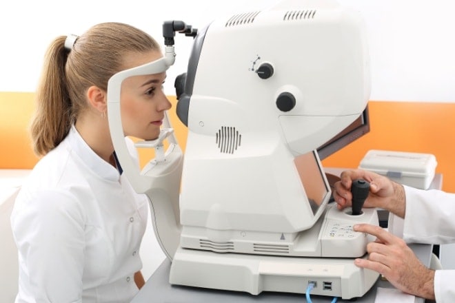
An eye doctor can check your peripheral vision by performing a visual field test. These tests measure how far outside the center of vision your eyes can see without moving. They also help determine how sensitive your vision is throughout your visual field.
Visual field tests are an important part of comprehensive eye exams because they help detect blind spots, or scotoma. These blind spots can point to an eye disease like glaucoma or a brain disorder.
There are six main types of visual field tests. Some are routine, like the confrontation field test and frequency doubling technology. These only test part of the visual field. If the patient shows signs of particular conditions, like glaucoma, they may then conduct a full field test. They may also do a full field test if the patient is on certain medications that can impact vision.
Pro tip: Remember to get your eyes checked annually to protect your vision.
Confrontation visual field test
The confrontation visual field test allows the eye doctor to test each eye’s peripheral vision on its own. The test requires the patient to look straight ahead while covering one eye. The doctor may then hold up a certain number of fingers outside the patient’s central line of vision and ask them how many fingers they see.
By doing this in different areas, the eye doctor can test each of the sections of the patient’s peripheral vision.
Automated static perimetry test
The automated static perimetry test allows for a more detailed view of a patient’s peripheral vision. The test involves a perimeter — a bowl-shaped tool that flashes light in different areas at varying levels of brightness.
Each time the patient sees a flash of light, they are asked to push a button. The machine keeps track of which flashes they didn’t see. This helps the eye doctor spot potential loss of peripheral vision, as well as where the loss has occurred. As with the confrontation visual field test, one eye is covered for this test.
Kinetic visual field test
The kinetic visual field test is similar to the automated static perimetry test. However, while the lights in the static test blink off and on, the lights in the kinetic test move around.
Frequency doubling perimetry
The frequency doubling perimetry test involves an optical illusion to check for any vision loss. The optical illusion uses vertical bars that flicker at different rates. If the patient is unable to see certain bars at certain times, it can indicate damaged vision.
Electroretinography
Electroretinography (ERG) measures the electrical signals of photoreceptors. During this test, the eye doctor dilates the patient’s eyes. Dilation is a process in which eye drops trigger the pupils to widen. This allows more light to enter the eye, giving the eye doctor a clearer view of the back of the eye.
The eye care provider will also likely numb the patient’s eyes with eye drops during the ERG.
Once the eyes are dilated and numbed, the eye doctor will use a tool called a speculum to hold the eyes open. An electrode is then placed on the cornea while the patient looks at flashing lights, similar to those in the static or kinetic visual field tests. The electrode keeps track of the photoreceptors’ electric responses to the light.
Amsler grid
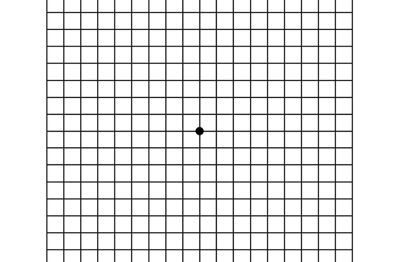
An Amsler grid consists of straight lines, with a reference dot in the center.
The Amsler grid is a simple test that consists of a square with a grid pattern and a dot at the center. Patients are asked to look at the central dot and tell the eye doctor if any areas of the grid look distorted.
This test is mostly used to measure the center of the visual field. It can be used to evaluate vision 10 degrees outside the patient’s point of focus. The Amsler grid can provide your eye doctor with information about your overall vision but doesn’t offer much on peripheral vision. It is most commonly used by patients with age-related macular degeneration (AMD).
What symptoms can impact peripheral vision?
If you are having difficulties with your peripheral vision, it can mean a number of things. Different symptoms can be linked to different issues.
Eye floaters
Eye floaters come in many shapes. They may appear as strings or cobwebs at the edge of your vision, or black spots that move around like bugs. Floaters often disappear when you try to look directly at them.
Eye floaters are caused by shadows cast on the retina. The shadows are typically cast by small lumps of collagen — a protein that helps build skin, muscle, bone and other tissues in the body. The collagen lumps form as the vitreous — a jelly-like substance inside the eye — liquifies and shrinks. This is a common process as you age.
Floaters are typically harmless. However, if you notice a sudden increase in floaters, seek immediate medical attention.
Seeing stars
Some people may experience a sensation of sparkles or flashing lights that seem like fireworks or bolts of lightning. Some describe it as “ seeing stars.” These symptoms are often caused by an issue in the retina or the brain.
If the retina becomes inflamed or the vitreous moves and tugs on the retina, it doesn’t cause pain. Instead, the brain interprets these signals as flashes of light. This is true even if no light has entered the eye.
On the brain’s end, if something disrupts its electrical activity, it can send false signals to the eyes. Some of the most common causes of these disruptions are:
- Trauma to the head
- Migraine
- Movement of the vitreous
- Retinal detachment
- Torn retina
Kaleidoscope vision
Kaleidoscope vision refers to the experience of seeing sudden bright, fractured colors. These may look like prisms or the inside of a kaleidoscope. These visual auras can last anywhere from five minutes to one hour. A visual aura is the eye’s response to a sudden uptick in nerve cell activity.
Migraines are the most common cause of kaleidoscope vision. Strokes and brain damage may also trigger this visual aura.
Blind spots
Blind spots, or scotomas, are areas of vision loss in a person’s visual field. These may appear as dark, blurry spots; arcs of light or a single, flickering spot of light. On occasion, those with scotomas may struggle to see certain colors or to see in dim light.
Depending on their size and location, scotomas can make daily activities like driving and reading more difficult. There are many conditions linked with scotomas, including:
- Retinal detachment
- Diabetic retinopathy
- Glaucoma
- Macular degeneration
- High blood pressure
- Stroke
- Head injury
- Brain tumor
- Optic nerve damage or inflammation
- Multiple sclerosis
- Migraine
- Stress
- Hormonal shifts
Visual shadows
Visual shadows, which may look like dark curtains at the edge of your vision, can indicate a medical emergency. These shadows can block vision from above, below or to the sides of the eyes. They may appear in one or both eyes.
Potential causes of visual shadows may include:
- Impending stroke
- Retinal infection
- Retinal detachment or damage
- Blocked blood vessels in the eye
SEE RELATED: Eye strokes: CRAO, BRVO and other retinal artery and vein occlusions
Visual auras
Visual or migraine auras are warning signs that a migraine is about to hit. Potential auras may include:
- Flashing lights
- Colorful spots
- Zig zags
- Foggy vision
- Blind spots
- Haze, similar to seeing an object through water or heat waves
These visual disturbances may also be joined by ringing ears, dizziness or difficulty speaking.
READ MORE: Tunnel vision (peripheral vision loss)
FAQs
What is the normal range of peripheral vision?
The typical field of peripheral vision is:
- 100 degrees away from the body’s center (laterally)
- 60 degrees toward the body’s center (medially)
- 60 degrees up
- 75 degrees down
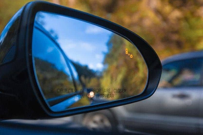
How does peripheral vision impact driving?
Peripheral vision is what helps you see pedestrians, cyclists and other cars alongside you as you drive. Diminished peripheral vision can lead to difficulties with:
- Driving at night
- Gauging the speed of oncoming traffic
- Recognizing unexpected events on the sides of the road
- Difficulty seeing objects clearly
All of these factors can increase the chances of a collision. If you have peripheral vision loss, you may consider:
- Using public transportation
- Asking for rides from friends or family
- Only driving during the day
- Limiting night driving to familiar routes
- Avoiding lane changes
Do women have better peripheral vision than men?
While there is no peer-reviewed medical evidence to support the assumption, women are often thought to have better peripheral vision than men. Some have linked this to humanity’s hunter-gatherer days.
The male’s traditional role as a hunter may have required stronger focused, central vision. The female’s traditional role as a gatherer may have relied more heavily on wider peripheral vision.
What is known is that women generally have more cones in their peripheral retina than men do. This may also feed into the idea that women have better peripheral vision.
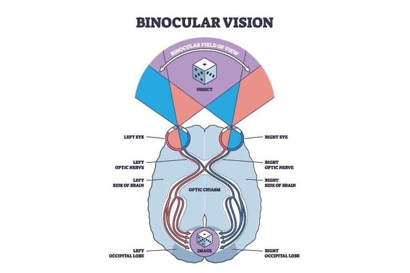
What is “120 degrees peripheral vision”?
The visual overlap between an individual’s eyes is about 120 degrees. This is called binocular vision and is what allows you to have depth perception. Visual field requirements for driving vary by state, and some states may not have these requirements.
What is the difference between focal vision and ambient vision?
Humans have two modes of processing vision: focal and ambient. Ambient mode, which helps you understand your space and surroundings, relies more on peripheral vision. Focal mode, which helps you see details, relies more on central vision.
Do cats have peripheral vision?
Cats don’t just have peripheral vision — they have a wider visual field than humans! They can see 200 degrees horizontally, compared to humans’ 180 degrees.
When to see an eye doctor
Routine comprehensive exams can help your eye doctor monitor the health of your eyes, including your peripheral vision. If you notice any sudden changes like floaters, visual shadows or blind spots, it is important to seek medical help to check for potential underlying conditions.