Amsler Grid: Macular Degeneration Test (Screening)
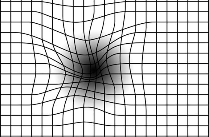
The Amsler grid is a tool that eye doctors use to detect vision problems resulting from damage to the macula (the central part of the retina) or the optic nerve.
The damage may be caused by macular degeneration or other eye diseases, so the Amsler grid is useful in detecting these problems.
An early diagnosis means early treatment, so it may help to limit or at least slow the vision loss you experience.
If you are at risk for macular degeneration or other eye diseases, you can use this chart at home to monitor your vision. But using the chart doesn't mean you should skip regular visits to your eye doctor, because you can easily miss signs that only a trained eye care practitioner will find.
The chart below is an approximation of the printed chart used by eye doctors. For more accurate detection of macular damage, you will need a complete eye exam.
Click here to download a copy of an Amsler grid and instructions for use.
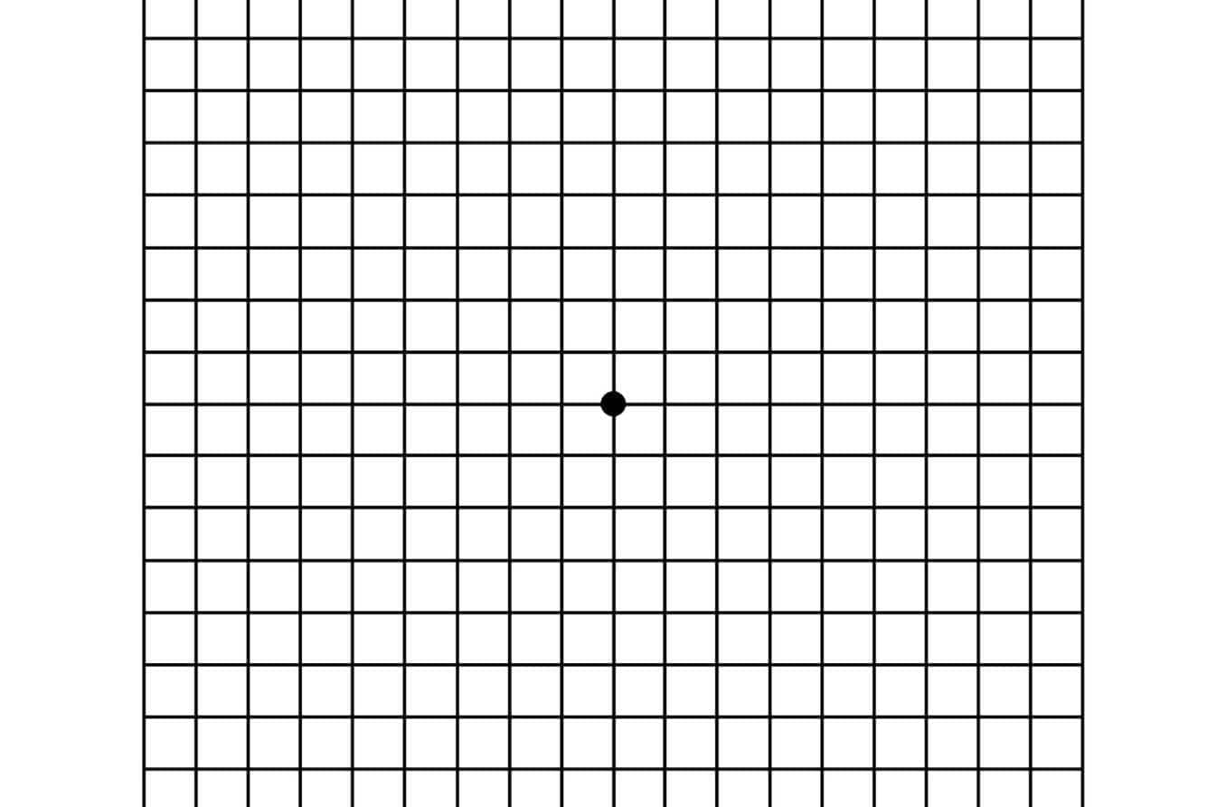
An Amsler grid consists of straight lines, with a reference dot in the center.
How to use the Amsler grid test
Testing your eyes with an Amsler grid is easy and takes only a few minutes. Here are the basic steps:
- Test your eyes under normal room lighting used for reading.
- Wear eyeglasses you normally wear for reading (even if you wear only store-bought reading glasses.
- Hold the Amsler grid approximately 14 to 16 inches from your eyes.
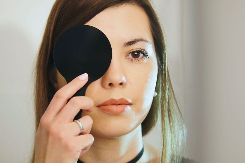
- Test each eye separately: Cup your hand over one eye while testing the other eye.
- Keep your eye focused on the dot in the center of the grid and answer these questions:Do any of the lines in the grid appear wavy, blurred or distorted?
- Do all the boxes in the grid look square and the same size?
- Are there any "holes" (missing areas) or dark areas in the grid?
- Can you see all corners and sides of the grid (while keeping your eye on the central dot)?
- Switch to the other eye and repeat.
IMPORTANT — Report any irregularities to your eye doctor immediately. Mark areas of the Amsler grid that you're not seeing properly (print two grids if you notice problems in each eye), and bring the grid(s) with you when you visit your eye doctor.
Check your eyes with the Amsler grid as frequently as your doctor recommends, or whenever you notice a significant change in your eyesight.
Please note: If you have an urgent question about your eye health, contact your eye doctor immediately. This page is designed to provide general information about vision, vision care and vision correction. It is not intended to provide medical advice. If you suspect that you have a vision problem or a condition that requires attention, consult with an eye care professional for advice on the treatment of your own specific condition and for your own particular needs. For more information, read our Terms of Use.