Optomap retinal exam: Pros and cons
What is an optomap eye exam?
During a comprehensive eye exam, your eye doctor evaluates the retinas, the light-sensitive tissue at the back of your eyes. To view the full retina, your doctor may dilate your pupils or use optomap technology.
Traditionally, to view the entire retina, patients are given eye drops that dilate the pupils. But these drops can be irritating to some patients, and can make the process a bit nerve-racking. Dilating drops can also cause blurry vision and light sensitivity.
Fortunately, advanced technology now offers a way to perform the retinal exam without the drop and dilation — all through something called an optomap retinal exam.
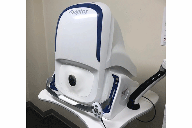
Over 20 years ago, a company known as Optos introduced optomap ultra-widefield retinal imaging. Optomap imaging provides a view of up to 80% (200 degrees) of the retina at one time. The exam is performed quickly and does not require pupil dilation. Given the wide and detailed view, optomap imaging has the potential to provide a more advanced look into the retina than other exams.
How does optomap imaging work?
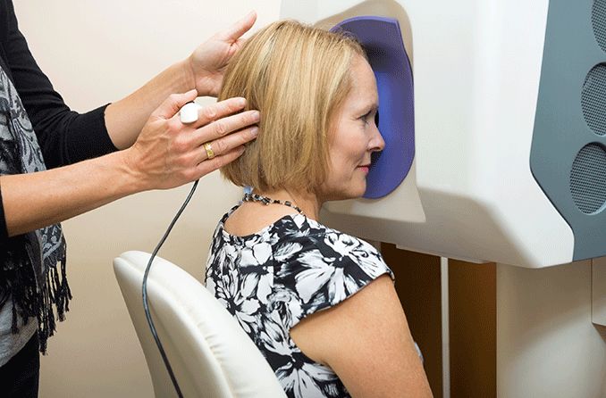
To capture an image of the retina using optomap technology, the following steps are taken:
First, the patient looks into the optomap instrument and looks for an LED target.
This action is displayed on a touch screen that the doctor or operator sees. The operator touches the monitor to capture an image of the retina, much like taking a photograph.
A set of green and red lasers is projected and a scanning process begins. The various wavelengths of laser light capture multiple images.
The final product provides a wide, high-resolution view of the retina.
The entire process takes about one minute per eye.
There are numerous Optos machines that provide many of the same imaging capabilities. With this efficient instrumentation, your eye doctor is able to see up to six different views of the retina.
Optomap retinal exam vs. dilation
One of the main differences between optomap retinal imaging and dilation? Optomap imaging provides a detailed look of the retina without the use of eye drops. Dilation, on the other hand, requires eye drops, which can have irritating side effects like blurry vision and light sensitivity.
That being said, a dilated eye exam is considered the standard among many eye doctors, as it provides a look at the retina and a more magnified view of the optic nerve. Because Optomap imaging is focused on the retinas, it provides less magnification of the optic nerve.
Some claim that optomap exams can give doctors more insight into the health of the retina, and help identify things that may not have been visible through dilation alone. In addition, the optomap images can be stored and compared for changes over time. Others argue that optomap imaging can miss important details, like signs of glaucoma.
For more details about what optomap imaging may provide or lack, be sure to continue reading the next section that highlights the advantages and disadvantages of the technology.
READ MORE: Frequently asked questions about eye dilation: What is it, how is it done and why?
Pros and cons of an optomap exam
Like many special imaging techniques, the optomap retinal examination comes with both pros and cons:
Advantages of optomap retinal imaging
It can give more in-depth imaging and information than traditional pupil dilation.
It often provides early detection of several eye diseases.
The imaging acts as a useful tool to detect retinal lesions before an eye surgery.
Exams are quick and painless.
It can be especially useful in pediatric ophthalmology.
Images can be saved and used to check for changes at later exams.
Disadvantages of optomap retinal imaging
The cost is often not covered by vision insurance.
Availability can be limited among vision care providers.
If a problem is detected through the imaging, dilation is often still needed to investigate the issue further.
Retinal detachment has been falsely identified through some optomap imaging.
Dilation is still important for conditions that involve the optic nerve, such as glaucoma.
READ MORE: Retinal imaging and scans
Disorders managed through optomap imaging
Optomap exams can be a helpful tool in diagnosing, monitoring and treating a variety of eye conditions, including:
Diabetes/diabetic retinopathy
Peripheral vitreoretinal disease
Tumors and lesions
Retinal detachment
Retinal dystrophies
Pediatric retinal disorders such as Coats disease
Although optomap exams are a helpful tool in the management of these conditions, your eye doctor may choose alternative (or additional) methods. The best decision depends on each individual patient and their needs.
SEE RELATED: Photos may help detect eye diseases in your children
Cost of an optomap exam
Is optomap covered by insurance? Optomap exams may be covered by your vision insurance if there is a problem with your retinas that needs to be further evaluated.
But in most cases of healthy retinas, optomap is not covered by insurance, and is considered an optional test. That being said, many optometry practices offer the testing at an affordable rate that is often $40 or less. Cost varies among practices, so be sure to ask your eye doctor’s office about their rates. Many patients feel the cost of an optomap is warranted in order to forgo the blur and light sensitivity that comes with dilation and last several hours.
In some cases, you may be able to pay for an optomap exam with your flexible spending account (FSA) or health savings account (HSA) if you have one.
Before you make a decision, you can always talk to your doctor to find out if optomap is right for you. And be sure to bring up pricing questions if cost is a concern.
Does your eye doctor offer optomap exams?
Retinal exams are a very important part of any eye exam, as they help to ensure the health of the retinas and detect any problems that may be present — both in children and adults.
Optomap retinal exams can offer a more advanced look at the state of the retinas and help establish a successful plan of action if one is needed. They also help monitor the status of any eye diseases or underlying conditions that affect the retinas.
Some eye care practices do not offer optomap imaging, so it’s best to ask your eye doctor if they offer optomap exams and if you may be a good candidate.
Whether or not you decide to have optomap imaging performed, remember to schedule your comprehensive eye exam on a yearly basis. This is the best way to ensure that your eyes and vision are both healthy and functional.
Wide field retinal imaging systems. American Academy of Ophthalmology. May 2022.
About Optos. Optos. Accessed July 2022.
Products. Optos. Accessed July 2022.
How an optomap image is captured. Optos. Accessed July 2022.
Point-counterpoint: Ultra-widefield imaging vs. dilated fundoscopy. Review of Optometry. March 2017.
Ultra-widefield imaging boosts retinopathy Dx. American Academy of Ophthalmology. May 2016.
Why optomap ultra-widefield retinal imaging? Optos. Accessed July 2022.
Application of mydriasis and eye steering in ultrawide field imaging for detecting peripheral retinal lesions in myopic patients. British Journal of Ophthalmology. March 2022.
Ultra-widefield imaging adding depth to pediatric ophthalmology. Ophthalmology Times. May 2020.
Ultra-wide field retinal imaging: A wider clinical perspective. Indian Journal of Ophthalmology. April 2021.
The dilated exam in the age of ultra-widefield imaging. Review of Optometry. November 2018.
Page published on Wednesday, August 10, 2022
Medically reviewed on Tuesday, July 19, 2022


