Contrast sensitivity testing

What is a contrast sensitivity test?
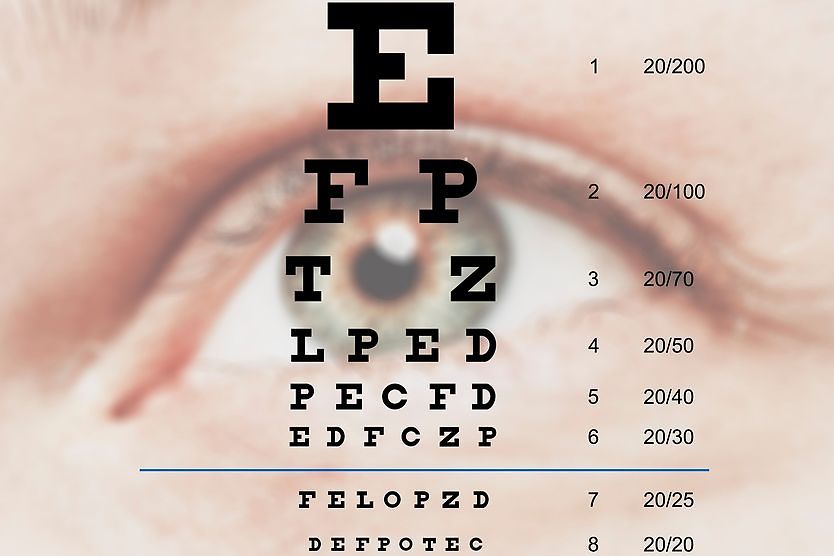
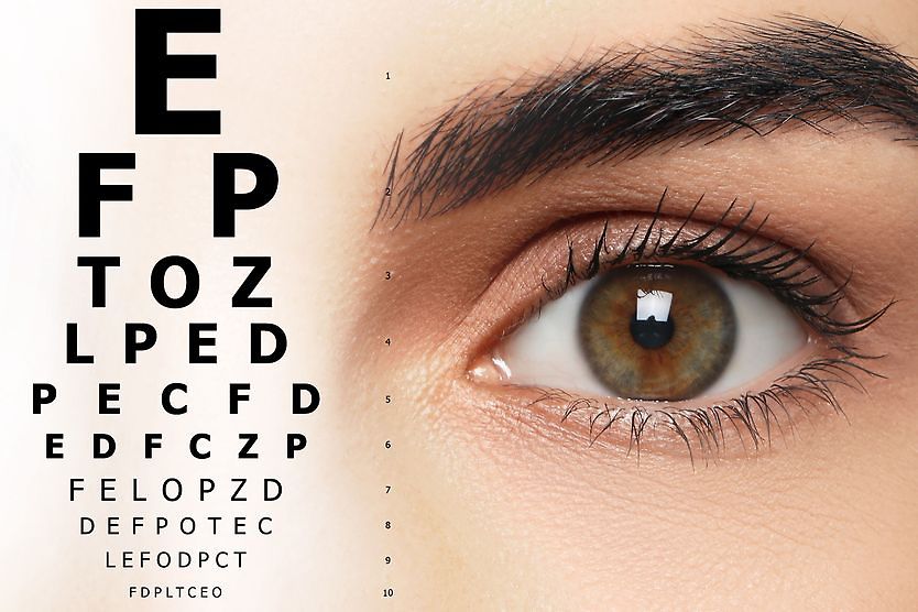
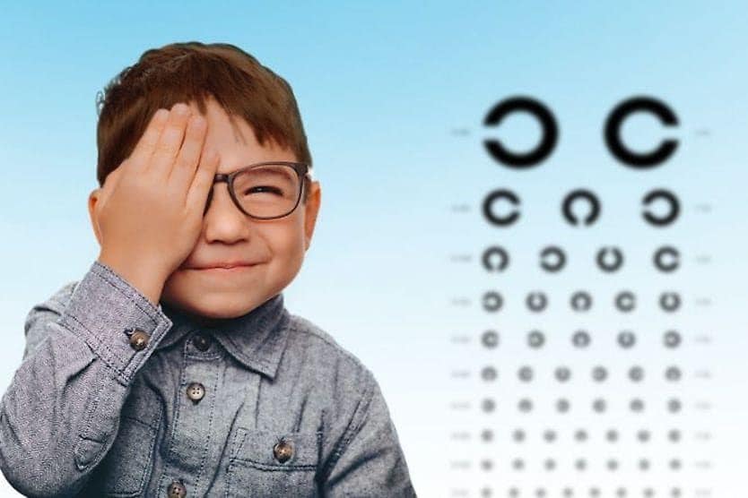
A contrast sensitivity test measures your ability to distinguish between finer and finer increments of light versus dark (contrast). This differs from common visual acuity testing in a comprehensive eye exam, which measures your ability to recognize smaller and smaller letters on a standard eye chart.
Contrast sensitivity is a very important measure of visual function, especially in situations of low light, fog or glare, when the contrast between objects and their background is often reduced. Driving at night is an example of an activity that requires good contrast sensitivity for safety.
Even if you have 20/20 visual acuity, you can have eye or health conditions that may diminish your contrast sensitivity and make you feel that you are not seeing well.
Symptoms of reduced contrast sensitivity
If you have low contrast sensitivity, you may have problems with night driving, including difficulty seeing pedestrians walking alongside poorly lit streets. Or you might notice that your eyes tire more easily while reading or watching television.

A person with normal visual acuity but poor contrast sensitivity might see the trees in the foreground clearly (high contrast), but have trouble seeing the contours of the mountains against the sky in the background (low contrast).
Poor contrast sensitivity can also increase your risk of a fall if you fail to see that you need to step down from a curb onto similarly colored pavement.
LEARN MORE about tactile paving.
Low contrast sensitivity can be a symptom of certain eye conditions or diseases, such as age-related macular degeneration (AMD), cataracts, glaucoma or diabetic retinopathy.
Changes in contrast sensitivity can also occur after LASIK, PRK and other types of refractive surgery.
For example, sometimes a person who has LASIK may be able to see 20/20 after the procedure but complains of poor night vision. This could be caused by a loss of contrast sensitivity from the surgery.
In the majority of cases, people with cataracts notice a significant improvement in both visual acuity and contrast sensitivity after cataract surgery.
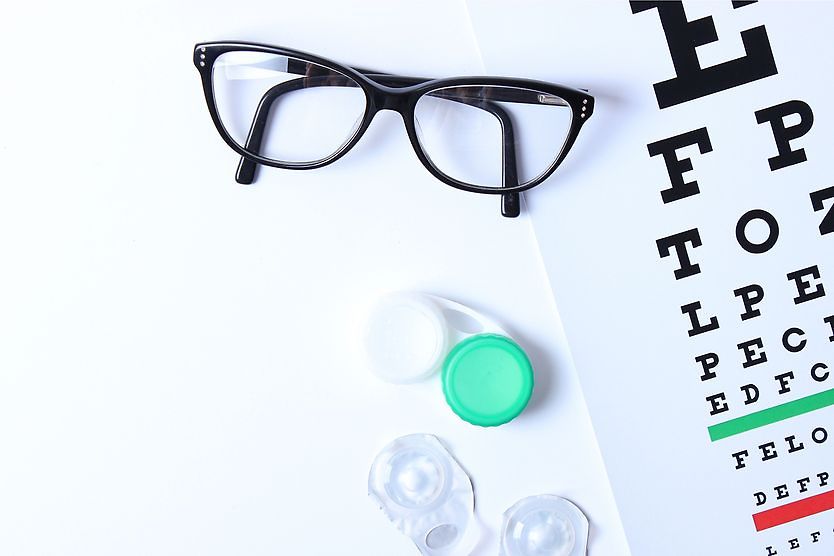
Contrast sensitivity testing
Contrast sensitivity testing often isn't included in a comprehensive eye exam. Your eye doctor might perform a visual contrast sensitivity test because of a specific visual complaint you have or because he or she suspects you have a condition that is affecting your ability to discern contrast.
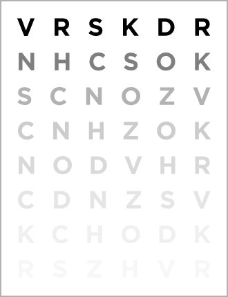
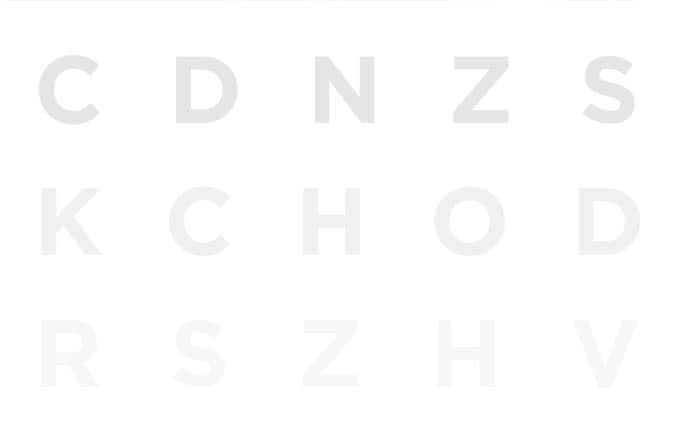
The Pelli-Robson chart is widely used to test contrast sensitivity. The letters on the chart gradually blend into the background as you go further down the chart, making them progressively harder to detect.
Like a standard Snellen visual acuity chart, the Pelli-Robson chart has horizontal lines of capital letters. But instead of letters getting smaller as you move down, the letters stay the same size — they just blend more into the background on each line.
The Bailey-Lovie chart is another test that can help assess contrast sensitivity issues. It evaluates how many letters can be read against a high-contrast background compared to a low-contrast background.
More sophisticated devices are also available to test contrast sensitivity. One such device uses targets called sine-wave gratings — fuzzy, parallel bars in shades of light and dark gray. Because these bars can vary in width and contrast, they provide a thorough picture of your overall contrast sensitivity.
Some sine-wave grating tests include a bright light source that can be directed toward your eyes during the test to simulate glare situations, such as oncoming headlights during night driving.
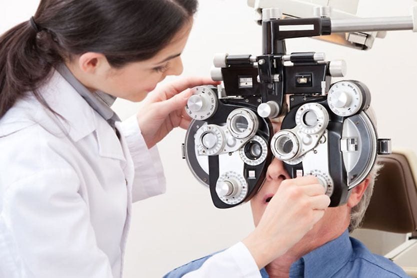
If your eye doctor determines that you need a contrast sensitivity test, it will likely be administered after a standard visual acuity test and before your pupils are dilated.
The testing is typically done while you wear your eyeglasses or contact lenses if you require vision correction.
For the evaluation of eye disease, contrast sensitivity is usually tested on each eye individually.
For other reasons, such as sports vision testing or to evaluate vision after a contact lens fitting, LASIK or cataract surgery, the testing might be done with both eyes open.
Contrast sensitivity function (CSF)
Your contrast sensitivity function is a detailed measurement of how well you see contrast. It's created by testing you with targets called sine-wave gratings. These are patterns made of parallel bars that vary in two ways:
- Their width (also called “spatial frequency”) from thick to thin
- Their contrast (how clearly they stand out against the background)
Thicker bars represent low spatial frequencies, while thinner bars represent high spatial frequencies, much like how hearing tests use different pitches and volumes.
The testing creates a map showing the minimum contrast you need to see each bar width. This helps your eye doctor understand your complete contrast sensitivity profile.
What can be done about poor contrast sensitivity?
Your contrast sensitivity test results can help your eye doctor determine if you have vision errors known as higher-order aberrations or some other problem that could be corrected with special eyewear or eye surgery.
If you are diagnosed with low contrast sensitivity, your eye doctor might advise you to wear corrective lenses with a yellow filter to improve your ability to discern contrast.
If you need prescription eyeglasses, many people find that they see better in low-light conditions when wearing lenses that include an anti-reflective (AR) coating, compared with wearing the same prescription lenses without AR coating.
Some vision correction options incorporate wavefront technology and may reduce higher-order aberrations. This can potentially enhance vision quality, though individual results vary. These options may include:
- Wavefront lenses (glasses and contacts)
- Wavefront LASIK
- Premium intraocular lenses (IOLs) for cataract surgery
Ask your eye doctor if wavefront technology might benefit your vision.
See an eye doctor
The only way to know for sure that you have normal contrast sensitivity is to see an eye doctor.
Ready to have your eyes checked? Find an eye doctor near you.