Iris/uvea of the eye

What is the uvea?
No, the uvea is not that little lobe of tissue that dangles from the back of your mouth when your doctor tells you to open wide and say "Ahh!" That's your uvula.
Here are the key things you should know about the uvea of your eye.
Iris and uvea definition
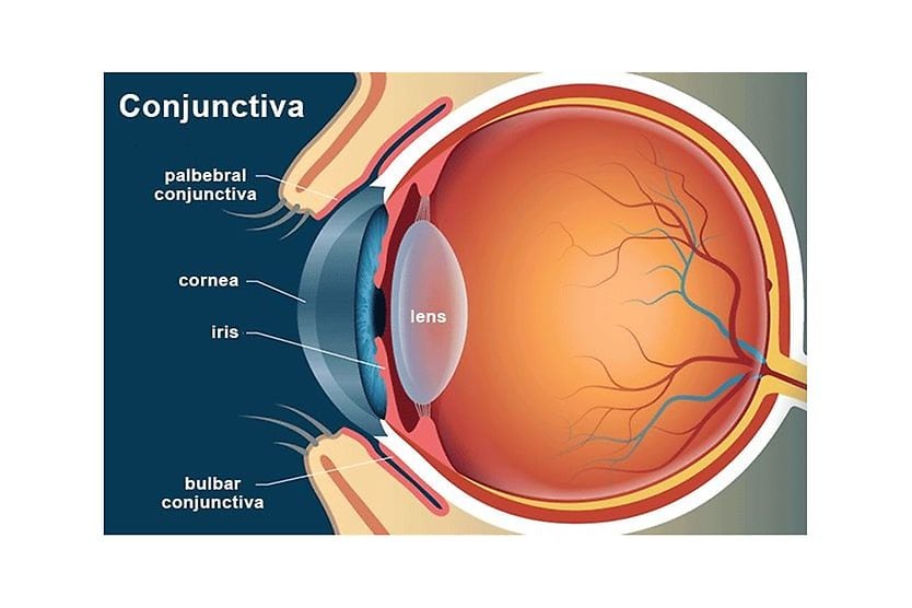
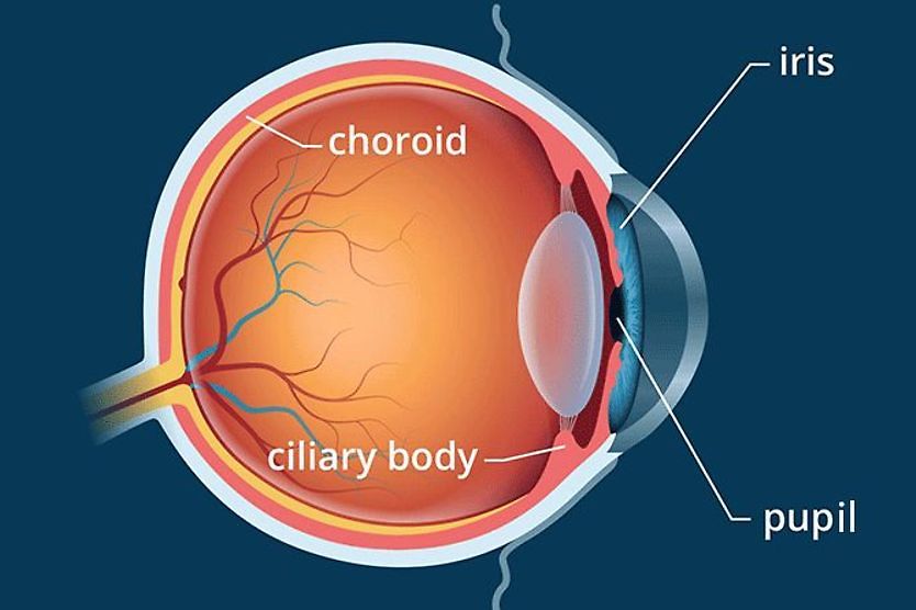
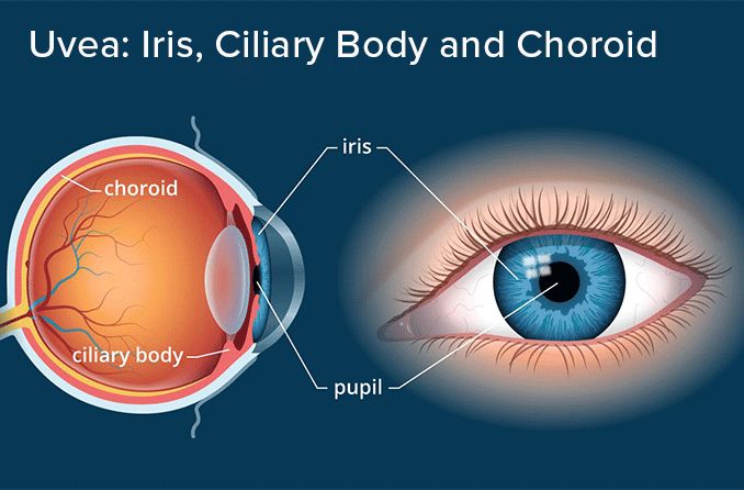
The uvea is the pigmented middle layer of the eyeball. It has three segments: the iris, the ciliary body and the choroid.
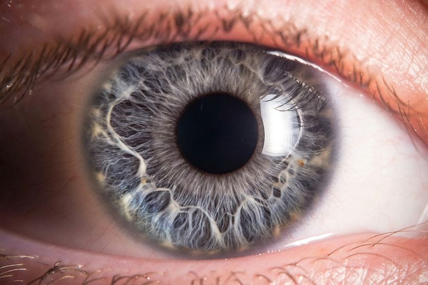
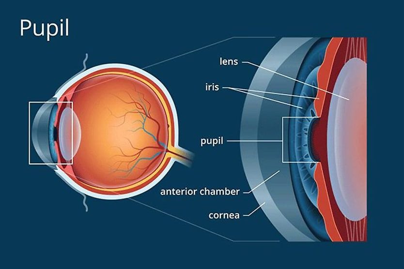
Iris: The iris of the eye is the thin, circular structure made of connective tissue and muscle that surrounds the pupil. The color of our eyes is determined by the amount of pigment in the iris.
Ciliary body: The second part of the uvea is the ciliary body. It surrounds the iris and cannot be seen because it's located behind the opaque sclera (white of the eye).
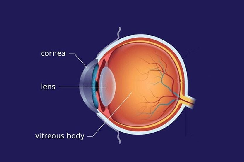
Choroid: The posterior portion of the uvea is the choroid, which is sandwiched between the tough outer sclera of the eye and the retina in the back of the eye.
LEARN MORE about eye color
Iris, ciliary body and choroid functions
Each component of the uvea has a specific function:
Iris: In addition to giving the eye its color, the iris acts like the diaphragm of a camera and controls the size of the pupil. One muscle within the iris constricts the pupil in bright light (full sunlight, for example), and another iris muscle dilates (enlarges) the pupil in dim lighting and in the dark.
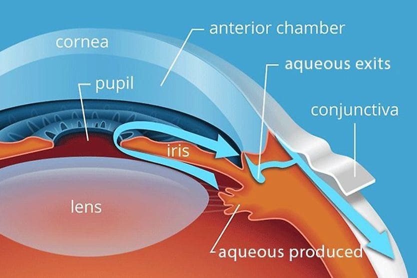
Ciliary body: The ciliary body holds the lens of the eye in place. It is connected to the lens with a network of many tiny ligaments (called ciliary zonules or zonules of Zinn) that suspend the lens in place behind the pupil. The ciliary body also secretes the clear aqueous fluid that fills the space in the anterior segment of the eye between the cornea and the iris and lens, and it contains the muscle that controls accommodation of the eye.
Choroid: The posterior portion of the uvea — the choroid — contains many tiny blood vessels and has the vital role of nourishing the retina. [See eye anatomy illustration.]
SEE RELATED: What is Hypopyon?
Uvea problems
A number of things can go wrong with the uvea. Some uvea problems are genetic, while others are age-related conditions or are associated with other health problems.
Here's a short list of conditions and diseases of the uvea:
Uveitis: This is inflammation of the uvea. When confined to the iris, it's called iritis. If the inflammation affects the iris and the ciliary body, it's called anterior uveitis or iridocyclitis. There are many potential causes of uveitis, but often the cause cannot be determined. Symptoms include a painful red eye, sensitivity to light and decreased visual acuity.
READ MORE about uveitis and iritis
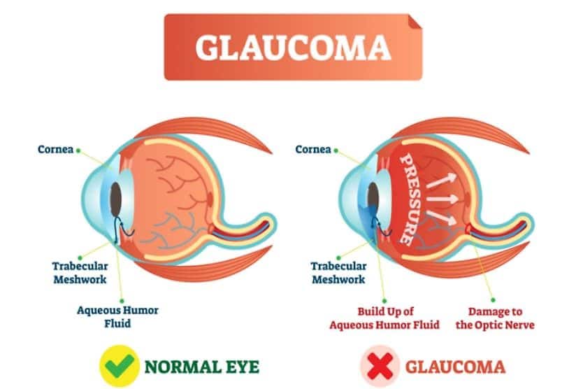
Synechia: Pronounced "si-NECK-ee-ah," this is when parts of the iris adhere to the back surface of the cornea or the front of the lens. Synechiae (plural; pronounced "si-NECK-ee-ee") may occur because of trauma to the eye, iritis or other causes. Synechiae are dangerous because they could lead to certain types of glaucoma.
Iris coloboma: This is a congenital disorder where a portion of normal iris tissue is missing, causing a misshapen "keyhole" or "cat-eye" appearance to the pupil. In some cases, iris colobomas can cause blurred vision, decreased visual acuity, double vision and ghost images. Often, people with iris colobomas choose to wear prosthetic contact lenses to mask the appearance of the eye and decrease any visual symptoms.
Uveal melanoma: This is a cancerous growth within the iris, ciliary body or choroid. If the growth occurs in the posterior uvea, it's called a choroidal melanoma; if in the iris, it's called an iris melanoma. The tumors are called melanomas because they develop in pigment cells (melanocytes) located within the uvea. Uveal melanomas are a common type of cancer inside the eye, and the tumors can metastasize to other parts of the body. There are treatment options, but in some cases, removal of the affected eye (enucleation) may be necessary.
READ MORE about ocular melanoma
Choroidal nevus: A choroidal nevus is a flat, benign pigmented freckle in the choroid. If a choroidal nevus is detected during a comprehensive eye exam, your eye doctor typically will take a photograph or other type of image of the interior of your eye to document the shape and size of the freckle and monitor it over time to ensure no changes occur.
Choroideremia: This is a hereditary, progressive degeneration of the choroid that primarily affects men. Choroideremia ("ko-roy-duh-REE-me-ah") is characterized by night blindness, decreased visual field and (eventually) blindness.
Iris nevus: This is a freckle (localized concentration of pigment) in the iris of the eye. Like freckles on your skin, iris nevi (plural of nevus) almost always are stable and harmless. However, if you have an eye freckle that appears to be getting larger, see your eye doctor immediately. In rare cases, an iris nevus can transform into a malignant growth.
SEE ALSO: Chorioretinitis (a type of uveitis)
Importance of routine comprehensive eye exams
Many uvea problems can be detected by your eye doctor before you notice any significant symptoms.
Routine comprehensive eye exams are essential to examine your eyes for signs of uveal melanoma and other serious eye conditions so treatment can begin as soon as possible to protect your eyesight.
SEE RELATED: Brushfield Spots