Choroidal nevus: A hidden “freckle” in the eye


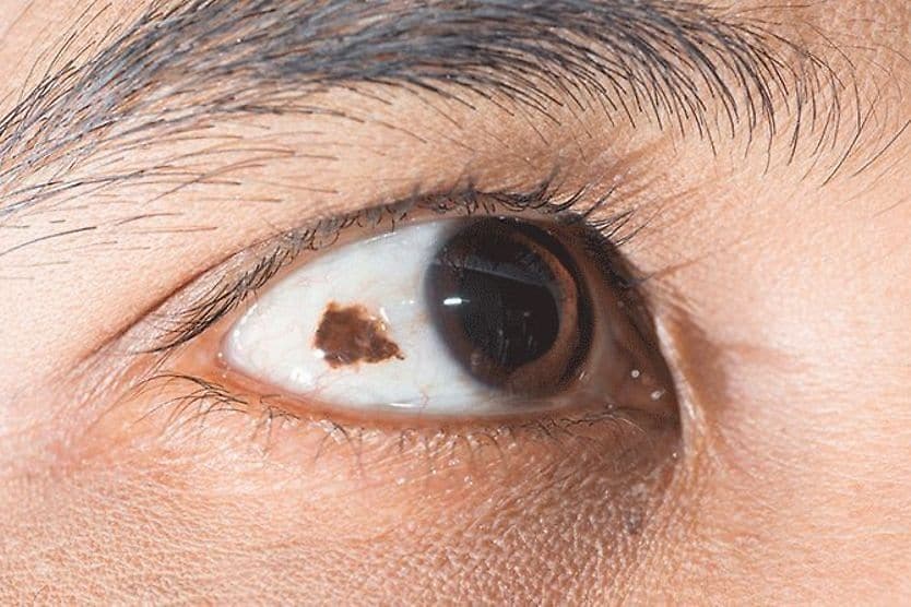
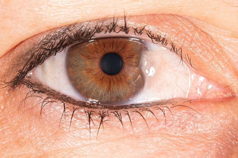
Just as you can get a freckle or mole on your cheek or nose, you can develop a spot known as a nevus on (or in) your eye. A choroidal nevus (plural: nevi) is usually a harmless spot inside of your eye that can be flat and gray in color. However, it is not visible in the mirror.
It’s unlikely you will know you have a choroidal nevus unless your eye doctor spots it during an exam.
Choroidal nevi are common (about 10% of the population has them). It’s a good idea to know what a choroidal nevus is, how likely it is to turn into cancer — just like a mole on your skin can — and why it’s important to get it checked by your eye doctor regularly.
Anatomy of a choroidal nevus
A choroidal nevus gets its name from its location in the choroid. This is a layer of tissue rich in blood vessels that is located between the white of the eye (sclera) and the retina (the sensory tissue lining the back of your eye).
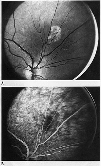
Choroidal nevus. (A) Red-free photograph shows small nevus of choroid with mottled pigmentation and no changes in overlying retina. (B) Arteriovenous phase shows poorly defined margins of nevus. Lesion blocks choroidal fluorescence poorly and does not leak dye. [Image credit: Permission granted by © 2022 American Academy of Ophthalmology]
A choroidal nevus is a type of growth sometimes known as an eye freckle. Other types of eye freckles are also named for their location in the eye. These are the three main types of nevi in the eye:
- Choroidal nevus – A spot in the choroid, a part of the eye that has many important functions, including supplying the outer retina with oxygen and nutrients.
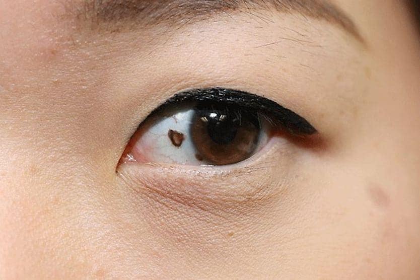
- Iris nevus – A spot on or in the iris, the colored part of the eye that surrounds the pupil.
- Conjunctival nevus – A spot on the conjunctiva, a clear membrane that covers the surface of the eye and the inside of the eyelids.
A choroidal nevus can be a variety of different colors, including gray, brown, yellow or multicolored.
Choroidal nevus symptoms
A patient with a choroidal nevus typically will have no symptoms at all. A choroidal nevus is typically found during a comprehensive eye exam.
In some very rare cases, a choroidal nevus may cause symptoms such as:
- Eye floaters – A choroidal nevus may cause eye floaters, which “float” in your field of vision and may look like specks or dots.
- Vision changes – A choroidal nevus may cause central or peripheral vision loss in some cases.
These symptoms are also linked with many other eye conditions, some of which are serious. See your eye doctor right away if you have any eye symptoms that affect your vision.
Choroidal nevus causes
A choroidal nevus occurs when pigmented cells called melanocytes clump together to form a growth on the inside of the eye.
Doctors don’t know exactly what causes a choroidal nevus. But it’s possible that exposure to the sun’s ultraviolet (UV) rays may play a role in the development of an eye freckle.
The risk of developing a choroidal nevus increases with age and is linked to race. In people aged 40 and over, almost 6% of white adults, almost 3% of Hispanic adults and less than 1% of African American adults have a choroidal nevus.
Choroidal nevus vs. melanoma
A choroidal nevus is typically a benign condition that may look similar to a melanoma, a type of cancer in melanin-producing cells. In some cases, a choroidal nevus can turn into a melanoma over time.
Some common warning signs that may alert your eye doctor that a choroidal nevus is or has turned into a melanoma include:
- The nevus is not flat.
- The nevus is orange in color.
- The nevus is leaking fluid.
- The nevus is growing quickly.
Fortunately, only about 1 in 8,000 choroidal nevi turn into cancer.
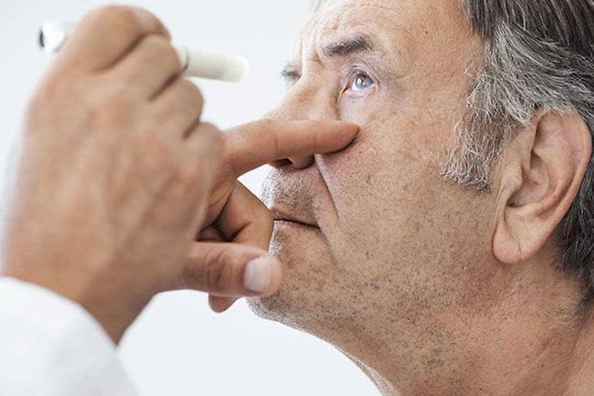
Choroidal nevus diagnosis
As part of a comprehensive eye exam, your eye care professional may do a fundoscopic exam, also known as ophthalmoscopy.
Your eye doctor may use lights and special lenses to look deep into the eye to see structures, including blood vessels, the optic nerve and the retina. A fundoscopic exam can help an eye doctor diagnose a choroidal nevus and many other conditions, including glaucoma, retinal detachment and macular degeneration.
Choroidal nevus treatment
You typically don’t need treatment for a choroidal nevus. Your eye doctor may do optical coherence tomography (OCT) imaging to get a more detailed look at the choroidal nevus.
Your doctor may take a picture of the lesion to see if it grows or changes over time. You may need to see your eye doctor for a recheck in about six months, and you’ll need to continue getting comprehensive eye exams.
In rare cases, a choroidal nevus can turn into ocular melanoma. If a choroidal nevus does turn into cancer, there are a number of treatment options. These include:
- Surgery
- Laser therapy
- Radiation
- Cryotherapy
Your eye doctor can refer you to an ocular oncologist, a doctor who specializes in diagnosing and treating eye cancer. An ocular oncologist can look for signs of eye cancer and can recheck your eye regularly.
Is a choroidal nevus dangerous?
In the vast majority of cases, a choroidal nevus is a benign condition and is not considered dangerous. But it’s important to have your eye doctor monitor a choroidal nevus closely.
That’s because it’s not always possible for an eye doctor to tell the difference between a choroidal nevus and an ocular melanoma. A melanoma can resemble a choroidal nevus in size, shape, color, etc.
See your eye doctor regularly
It’s important to see your eye doctor regularly. They will usually be able to spot a choroidal nevus during a comprehensive eye exam. Your eye doctor can monitor it over time with regular checkups to see if it grows or changes.
It’s also a good idea to practice prevention by protecting your eyes with sunglasses that block 100% of UV rays. If you don’t wear sunglasses all the time, get a quality pair and remember to put them on before heading outdoors.
SEE RELATED: Linear nevus sebaceous syndrome