Your eye exam may be one of your most valuable preventive health care visits

Your eye doctor saw it first
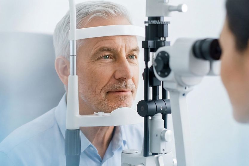
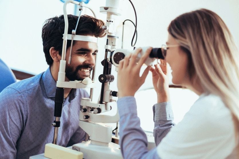
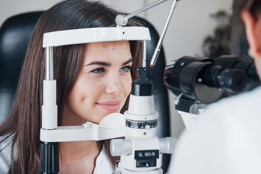
Medical tests often require something: a needle, a scan or a sample. A comprehensive eye exam requires almost none of that. You sit in a darkened room. A light passes across the retina (the light-sensitive tissue at the back of your eye), and within a few minutes, an eye doctor is looking at your blood vessels and nerves, visible without a single incision.
Almost everywhere else in the body, the blood vessels and nerves are hidden beneath skin and tissue, accessible only through imaging or invasive procedures. In the retina, both systems come to the surface. What an eye doctor sees looking at the retina is like a map, not only of the eye but of the continuous networks running through the brain, the heart and the rest of the body.
A brief history of looking inside the eye
For centuries, scientists suspected the eye held clues to the rest of the body’s health, but they lacked the tools to confirm. That changed in 1850, when Hermann von Helmholtz, both a physicist and physician, invented the ophthalmoscope, a small handheld device that used a mirror and light source to illuminate the inside of a living eye. A doctor could now look directly into a person’s eyes and detect not just eye disease but changes occurring in the blood vessels and nerves throughout the body.
What Helmholtz could only imagine
The number of available professional tools has grown considerably since Helmholtz’s time. OCT, or optical coherence tomography, generates high-resolution cross-sectional images of the retinal layers. OCT angiography (OCTA) maps blood flow through retinal vessels in three dimensions without injected dye. Digital fundus photography captures detailed color images of the retina, the head of the optic nerve (which connects the eye and brain) and blood vessels.
While the technology has changed substantially, the core principle remains the same. The retina can be examined and analyzed, offering a glimpse into both your eye health and whole health.
According to the American Optometric Association (AOA), eye care professionals can identify early warning signs of more than 270 systemic (affecting the whole body) and chronic diseases through a comprehensive eye exam, including diabetes, hypertension, cancers and some autoimmune conditions. Many of these signs appear before a patient notices anything’s wrong.
Millicent Knight, OD, FAAO, FAARM, is an integrative optometrist (combining traditional with holistic care) who has spent her career studying this. “The eye reflects patterns related to inflammation, metabolic health, stress and aging,” she said. “The technology utilized during a comprehensive eye examination can be insightful, and when combined with lifestyle and nutrition insights, it becomes a powerful guide for proactive wellness.”
Studying the patterns with oculomics
Oculomics is the study of what retinal imaging can reveal about systemic health. Its organizing question is simple. If the retina shares biology with the brain, the heart and the organs beyond it, what can a close look tell us?
Artificial intelligence (AI) is central to how this research is advancing. At the foundation of oculomics are the same imaging tools already in clinical use. Data from digital fundus photography, OCT and OCTA is analyzed by AI algorithms trained to detect patterns that are too subtle or too early for the human eye to catch.
Researchers are finding that systems trained on large datasets of retinal images may detect early disease. Signs of cardiovascular disease, chronic kidney disease, metabolic conditions and neurological changes could potentially be found before symptoms appear.
In most cases, however, the research is still early and can’t be used in routine clinical practice. More validation and real-world testing are needed.
Your eye doctor could be the first to detect high blood sugar
One area in which AI retinal tools have already begun to be implemented is diabetic retinopathy screening. Several FDA-cleared autonomous AI systems analyze retinal photographs for signs of diabetes-related damage, flagging images that need a specialist’s review. These tools can screen; however, they do not diagnose.
Diabetes is a disease that damages blood vessels throughout the body. This includes the retina, with its dense network of tiny vessels, which is often where damage can be evident. For some patients, the first indication that their blood sugar may be high comes not from a blood test or a primary care visit but from an eye exam. While the exam itself cannot officially confirm diabetes, it can give the eye doctor a reason to send you for blood work that will.
Diabetic retinopathy is the condition that results from uncontrolled blood sugar in the eye. It is the leading cause of blindness in working-age American adults, according to the CDC.
Signs of diabetic retinopathy can be visible in the retina before they become symptomatic elsewhere. An eye doctor can see vessel damage, abnormal vessel growth and fluid buildup — markers that blood sugar is elevated. The eye exam allows a patient to begin managing the disease before it progresses and causes further damage.
Blood vessels under pressure
When blood pressure stays high over time, the vessels in the retina begin to change. They narrow. They cross each other at abnormal angles. They bleed. Doctors call this pattern hypertensive retinopathy, a term for the cumulative damage that elevated pressure inflicts on the eye’s smallest vessels. Research has linked it to a higher risk of stroke and cardiovascular disease.
When the retina shows signs of high blood pressure, an eye doctor becomes the first link in a chain of care. The referral to other specialists has become increasingly well-documented and precise. Retinal photography and OCT angiography can be used to map and measure changes over time.
Telemedicine has made it possible to share those records across care teams without requiring an additional in-person visit. These tools have widened who can be reached. Retinal images can now be captured in a primary care office, a community health center or some pharmacy clinics and read remotely. This matters because cost, lack of insurance and lack of transportation remain the most common reasons adults may skip eye care services altogether.
Immune signals in the eyes
The eye’s relationship to disease extends to the immune system as well. Two eye conditions in particular have been linked to autoimmune disease. The first is uveitis, which is inflammation inside the eye. It’s a group of conditions rather than one, varying by which layer is inflamed and by how urgently they need attention. The other is scleritis, which is inflammation of the sclera (the white part of the eye).
Both have been documented in people with rheumatoid arthritis (RA), systemic lupus erythematosus (SLE), ankylosing spondylitis and inflammatory bowel disease (IBD). In some cases, the eye condition appears before the underlying disease is diagnosed, making the eye doctor the first to make the connection.
Dr. Knight sees this pattern regularly. “I find most often that they don’t realize the potential connection to systemic conditions, like autoimmune diseases, which are on the rise, especially in women,” she said. “The red eye the patient presents with may occur concurrently with a sudden onset of stiff joints. The patient does not connect them, and the doctor does not ask probing questions that might unmask the root cause.” If a person experiences these symptoms, particularly with pain, light sensitivity or blurred vision, it is recommended to call your eye doctor for an emergency appointment.
Early research into what the eyes can tell us about our brain
The eye’s relationship to the rest of the body runs even deeper. The retina develops from the same tissue as the brain and shares its blood supply. That connection has led researchers to ask whether changes in the retina might provide early signs of conditions like Alzheimer’s and Parkinson’s disease.
The primary tools are OCT and OCT angiography, which measure the thickness of retinal nerve fiber layers and map changes in the smallest blood vessels, tracking shifts that may correspond to what is happening in the brain.
The research is still early, and results haven’t been consistent across studies. Retinal imaging for neurological conditions is not yet available as a clinical screening tool. However, it is an active area of study and researchers are cautious but optimistic.
You (and your eyes) are what you eat
Not every connection between the eye and the rest of the body involves disease. Many people who spend significant time on screens experience eye strain, blurred vision, dry eyes or headaches, a cluster of symptoms that clinicians call digital eye strain. An eye doctor can identify what is contributing to the strain and suggest ways to reduce it, from small adjustments in screen use to corrective lenses that ease the demand on the visual system.
Nutrition is another place where patients are often surprised by the eye-body connection, says Dr. Knight. “They understand that nutrition is important for their body but don’t naturally equate it with eye health.”
Research supports the link. Lutein and zeaxanthin (carotenoids found in leafy greens) have been shown to slow the progression of age-related macular degeneration (AMD), according to the National Eye Institute. Leafy greens carry well-documented benefits for the heart and brain, a reminder that what nourishes the eye tends to nourish the rest of the body, too.
Dr. Knight has noticed a shift in what patients want from their care. “Today’s patients are looking for a partner in their health care and want to know what they can do in addition to the conventional treatments they are prescribed,” she said.
One example has stuck with her. “A 50-year-old patient I diagnosed with DED (dry eye disease) opted for a holistic approach, which included inflammatory detoxification and gut cleanse,” she said. “Afterward, she reported that her eyes felt more comfortable but raved about her joints being pain-free for the first time in years. Inflammation in the eyes is inflammation throughout the body.”
In her practice, she extends the idea further. “Nutrition, more broadly, is more than what you consume with food. It extends to what you consume in your environment, including mental, physical movement, sleep quality, spiritual connections, as well as environmental toxins and even negative news.”
Taking a GLP-1 medication? Tell your eye doctor
About 12% of U.S. adults are currently taking a GLP-1 receptor agonist, the class of medications that includes semaglutide and tirzepatide. These medications have well-established benefits for blood sugar control, weight management and cardiovascular health, but research is ongoing about their effects on the eye.
Some studies have found a possible link between GLP-1 medications and a slightly higher rate of a condition called non-arteritic anterior ischemic optic neuropathy (NAION). While rare, this optic nerve condition can cause sudden, painless vision loss.
Another association has come up often and is frequently confused. Diabetic retinopathy can worsen temporarily when blood sugar falls quickly. It is a long-recognized effect of rapid glycemic improvement coupled with weight loss rather than something specific to these drugs, and a reason to have the retina watched more closely during the first months of treatment if you already have retinopathy.
The American Optometric Association (AOA) recommends a dilated eye exam either within the 12 months before starting GLP-1 therapy or as a baseline within one month of starting it, with regular follow-up after.
One eye exam offers a glimpse into the whole body
Among the tools available for preventive care and long-term health monitoring, the eye exam is gaining recognition. “The eye functions as an important biomarker for systemic wellness,” said Dr. Knight. For this reason, those with diabetes, hypertension or an autoimmune condition should get their eyes examined annually or as often as their care team recommends. Guidelines on exam frequency vary for healthy adults, but the AOA recommends every two years for those without known risk factors.
Not everything an eye exam turns up is something you acquired. Some findings are inherited or present from birth. Family history of eye disease itself is one of the reasons to be examined more frequently, which is why your eye doctor asks the question.
Advances in health and technology have expanded what a comprehensive eye exam can detect. Understanding what your eyes say about your health and taking a whole-picture approach to health and vision means recognizing the eye exam as a tool to assess the entire body, not just eye health and vision.
A light, a lens and a few minutes in a darkened room can now open a conversation about the heart, the brain, the immune system and the body’s long-term health that no other routine clinical encounter quite provides. “Optometrists and ophthalmologists have the tools and skill sets for earlier identification of potential eye or systemic conditions, which can then be properly triaged and better managed, often with more optimal outcomes,” said Dr. Knight. “Vision is foundational. Who wants to live to be over 100 without quality vision?"
Editors’ disclosure note: Dr. Knight was senior vice president of medical and professional affairs for North America at EssilorLuxottica. Her opinions are her own and do not reflect those of the company.