How many vision conditions are identified through your comprehensive eye exam?

Clear vision isn’t the whole story
It's an appointment that's easy to postpone. Vision still feels sharp. Nothing hurts. The phone screen is readable and night driving is fine. When nothing seems wrong, it's natural to assume all is well and put off getting an eye exam a little longer.
That assumption is common and it's exactly why many serious eye conditions go undetected until they've already caused damage.
Consider what can happen at a single routine visit. A person arrives with no complaints, symptoms or sense that anything is wrong. They leave with a diagnosis they didn't expect: the beginning stages of macular degeneration, early signs of undiagnosed diabetes or dangerously high eye pressure. They had no idea.
Being able to see clearly is not the same as having healthy eyes. That’s why the American Optometric Association (AOA) identifies a comprehensive dilated eye exam as the primary tool for early detection of sight-threatening conditions. In fact, according to the AOA, that same visit could uncover early warning signs of more than 250 systemic and chronic conditions — without a single invasive procedure.
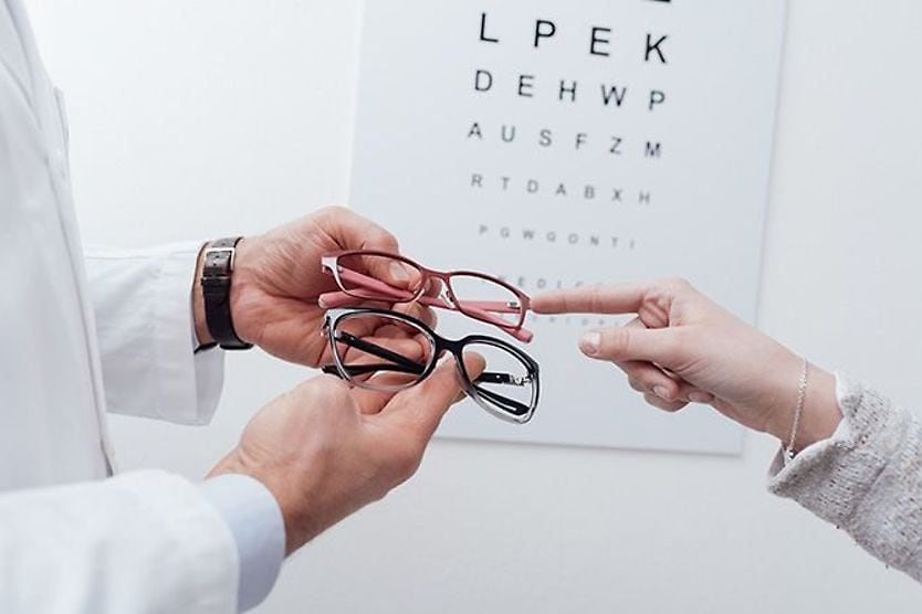
A screening checks your sight; an exam checks your eyes
Having clear vision may give the sense that the eyes themselves are healthy and passing a vision screening often reinforces that impression. Eye doctors hear this reasoning all the time. But a screening and an exam accomplish very different things.
“Both are so important, but do not mean the same thing,” notes Sumitra Khandelwal, MD, a professor of ophthalmology at Baylor College of Medicine. “There are many conditions where the vision is still clear, but the eyes have problems that need to be addressed.”
Vision screenings, such as those at schools, the DMV or pediatrician offices, aren't typically designed to diagnose or treat disease. In some settings, screenings simply check whether someone meets minimum vision criteria. For example, they determine how clearly someone can read letters on a chart from a set distance. Screenings are basic assessments meant to flag people who might need further evaluation.
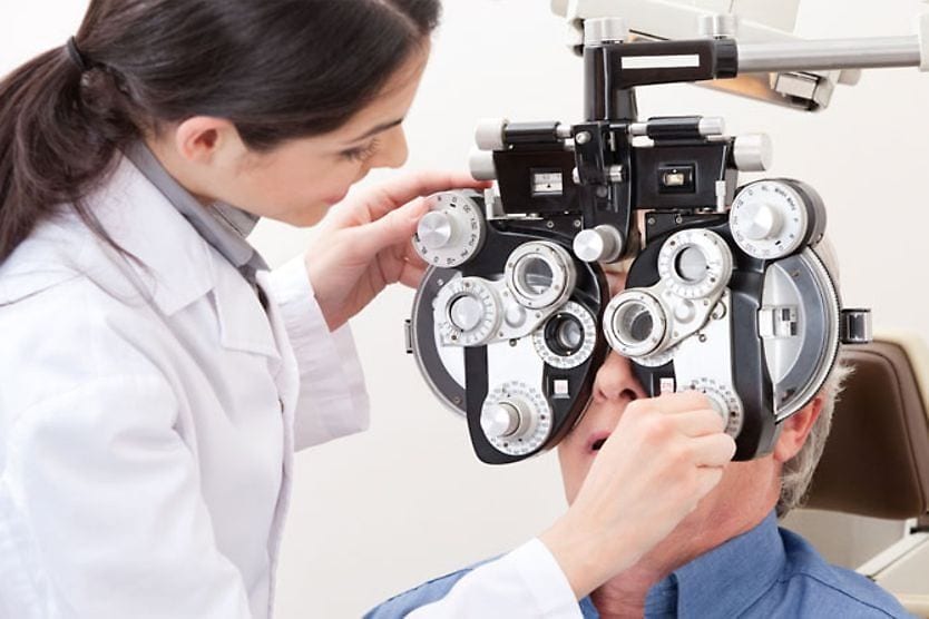
A comprehensive eye exam evaluates how the eyes function as a whole. It detects whether a disease may be developing inside structures you can't see or feel. These distinctions matter because several common eye conditions may develop slowly without causing noticeable symptoms. The difference between a vision screening and a comprehensive eye exam is not a matter of degree. It’s structural.
What your eye doctor sees that you can’t
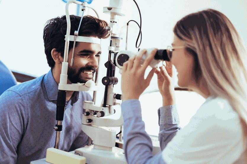
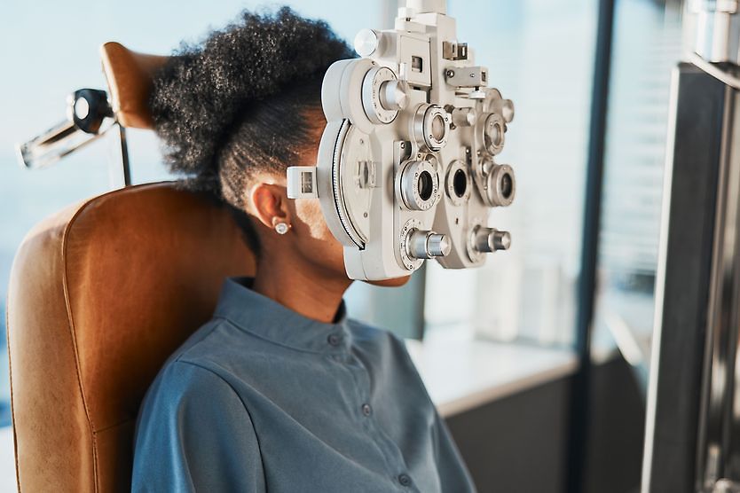
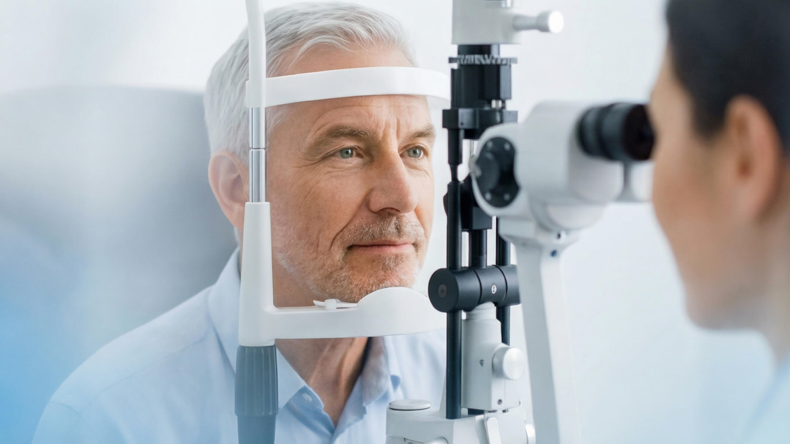
A comprehensive eye exam typically starts with a detailed health and family history, visual acuity testing and refraction to determine your prescription. From there, your doctor examines the front of the eye using a slit lamp. This high-powered microscope provides a close-up view of the eye's structures, such as the cornea, iris and crystalline lens. They'll also check your intraocular pressure, the fluid pressure inside the eye.
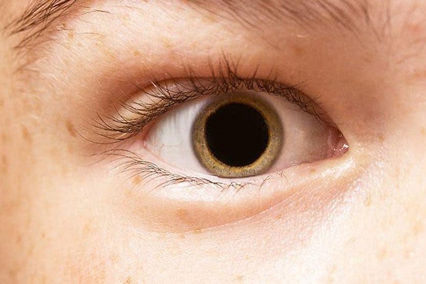
Dilating drops temporarily widen the pupil, giving your doctor a clear view of structures such as the light-sensitive retina and the optic nerve that connects the eye to the brain. Without dilation, important areas of the eye's interior simply can't be seen as well. The drops cause temporary light sensitivity and blurred near vision for several hours, but that small inconvenience makes it possible to spot conditions that may otherwise stay undetected.
Since the retina is the only place in the body where blood vessels and nerve tissue can be directly observed, your eyes can also reveal clues about your overall health. Signs of heart disease, high cholesterol, hypertension, diabetes, autoimmune diseases, some cancers and neurological problems may be detected during an eye exam, sometimes before the patient is even aware of them.
What diseases can an eye exam detect?
Several of the most vision-threatening eye diseases may produce no early symptoms. Detecting them depends on a comprehensive examination or technology such as retinal imaging.
“Common examples are glaucoma, retina changes from diabetes or even early cataracts,” says Dr. Khandelwal. “Vision may be stable, but the vision loss is occurring and won't affect the central vision until very late.”
Glaucoma
Glaucoma damages the optic nerve — and elevated intraocular pressure (IOP) is a major risk factor for developing the disease. Peripheral vision can gradually decrease, so a person may not notice a problem until the disease is advanced. An estimated 4.22 million U.S. adults are living with glaucoma, including 1.49 million with vision-affecting disease. Roughly half don't know they have it.
The National Eye Institute (NEI) says a comprehensive dilated exam is the most effective method for detecting glaucoma in high-risk groups, including Black adults over 40, adults over 60 and anyone with a family history of the disease.
Diabetic retinopathy
High blood sugar can damage the retina’s blood vessels, and in the early stages, you're unlikely to feel a thing. When caught early, retinal damage may respond better to treatment than when the disease is discovered after your vision has changed. Without an eye exam, these initial diabetic retinopathy changes may go undetected.
About a quarter of those with diabetes in the United States — an estimated 9.6 million people — have diabetic retinopathy. About 1.84 million people have vision-threatening disease.
Hypertensive retinopathy
High blood pressure can leave its mark. While there is no specific eye exam for high blood pressure, changes in the retina's blood vessels could indicate uncontrolled hypertension. This is called hypertensive retinopathy. It can cause narrowing of the small arteries. It can also cause compression of the arteries where they cross veins, which may appear before a formal hypertension diagnosis. These findings don't confirm the condition on their own, but they may be an early sign worth investigating.
Cholesterol-related changes
In some cases, an eye doctor may notice cholesterol-related signs, such as corneal arcus, a visible ring of fat deposits around the edge of the cornea. Less commonly, cholesterol emboli (small masses), known as Hollenhorst plaques, may be visible in retinal blood vessels and more rarely in the iris. These findings may point to high cholesterol or possible underlying cardiovascular disease. These may prompt a referral for further evaluation.
Age-related macular degeneration
Age-related macular degeneration (AMD) affects central vision and is the leading cause of vision loss in Americans over 60. Dry AMD, the more common form, often progresses in its early stages without any warning signs. A dilated exam can reveal drusen, small deposits beneath the retina, before visual distortion ever develops, opening a window for earlier management.
An estimated 20 million Americans age 40 and older are living with some form of AMD, with prevalence increasing sharply after age 75.
Retinal tears/detachments
Breaks in the retina, such as retinal tears and retinal detachments, may develop with little warning. Finding them typically requires a dilated peripheral retinal exam. The risk is elevated for highly nearsighted people. In fact, research suggests the likelihood of retinal detachment may be five to six times higher in people with severe myopia. Routine eye exams are critical for higher-risk individuals.
Cataracts
Cataracts usually develop gradually as the eye's natural lens becomes cloudy; they're among the most common age-related eye conditions. One study tracking U.S. adults over 65 found prevalence was about 37% in 2021, with nearly 20 million cases at that time. The good news: Early lens changes are often discovered during a comprehensive exam well before vision noticeably declines, giving you and your doctor time to plan.
Uveitis
Uveitis, inflammation inside the eye, can be linked to autoimmune conditions such as rheumatoid arthritis (RA), inflammatory bowel disease (IBD), systemic lupus erythematosus (SLE), sarcoidosis, psoriasis and ankylosing spondylitis. Infections, including shingles and Lyme disease, may also be a cause. In some cases, uveitis is the first clinical sign of an underlying condition a person didn't know they had and it's detectable during a routine eye exam.
Conditions at the eye surface and beyond
A slit lamp examination can reveal problems at the eye's surface. These conditions can affect comfort, vision and long-term eye health.
Dry eye disease
Dry eye disease disrupts tear film stability, leading to chronic irritation, fluctuating vision and/or difficulty wearing contact lenses. About 16 million Americans are diagnosed with this condition, though the actual number experiencing symptoms is likely much higher.
The slit lamp exam often finds two contributors to dry eye disease. Meibomian gland dysfunction (MGD), a blockage of the oil-producing glands along the eyelid margins, is present in an estimated 70% to 90% of dry eye cases. Blepharitis, an inflammation of those same eyelid margins, frequently occurs alongside MGD. Both can be identified during an eye exam.
Keratoconus
Keratoconus can cause the cornea to thin and change shape, distorting vision in ways glasses can't fully correct. It sometimes goes undiagnosed until young adulthood to the early 20s. However, when caught early, doctors can appropriately manage it to help optimize vision.
Thyroid eye disease
Thyroid eye disease (TED), associated with thyroid dysfunction such as Graves' disease, can produce changes around the eyes and eyelids (e.g., bulging eye appearance) that are visible on examination. In each case, what the doctor sees in the eyes doesn't confirm a systemic diagnosis. However, it can be the first clinical sign that something else may be going on that warrants further evaluation.
Neurological conditions
The same is true for conditions affecting the brain. Optic neuritis, inflammation of the optic nerve, is often the first sign in up to 30% of multiple sclerosis cases and can be identified during a comprehensive eye exam.
Swelling of the optic disc in both eyes, known as papilledema, may signal increased pressure inside the skull from causes that include brain tumors or aneurysms. Confrontational visual field testing, a standard component of many comprehensive eye exams, can reveal patterns of vision loss associated with neurological issues such as stroke.
Other tests that may be used to detect neurological diseases:
- Automated Perimetry: a computerized eye test that maps your peripheral (side) and central vision to detect blind spots
- Optical coherence tomography angiography (OCT / OCTA): non-invasive eye scan that creates 3D images of blood vessels of the optic nerve and retina without requiring dye injections.
Children's eye conditions
Amblyopia, or reduced vision in one eye caused by abnormal visual development, is estimated to affect 2% to 3% of the general population. It is the leading cause of visual impairment in children and young adults.
Children born prematurely or with a family history of amblyopia or strabismus (eye misalignment) are at higher risk of developing amblyopia. So are children with uncorrected refractive errors (nearsightedness or farsightedness). Conditions that obstruct vision in one eye, such as a droopy eyelid or a childhood cataract, can also increase the risk of amblyopia.
The brain's visual system is most adaptable before age 6 or 7, and treatment works best within that window. Once the critical developmental period closes, the condition may not be fully correctable. The challenge is that amblyopia causes no pain or visible symptoms. A child who has it usually doesn't know anything is wrong. That's what makes the eye exam so important.
In children with juvenile idiopathic arthritis (JIA), uveitis may be present without symptoms. This requires regular monitoring by an eye care provider.
In rare cases, a dilated eye exam may reveal signs of inherited retinal diseases such as Stargardt disease or retinitis pigmentosa (RP). Connective tissue conditions, such as Marfan syndrome, can affect the lens and may be identified during examination. Pediatric glaucoma is another finding that a dilated exam can uncover. In each of these cases, early identification can meaningfully change the course of care.
The technology behind the exam
In many practices, the comprehensive eye exam extends well beyond what a doctor can observe directly. Specialized diagnostic tools enable eye care providers to examine the eye's internal structures in extraordinary detail, identifying changes that would otherwise remain invisible until symptoms appear.
Scheduling an eye exam
Professional guidelines generally agree that comprehensive eye exams should be conducted regularly, though how often depends on your age, health history and risk factors. People with conditions such as diabetes or high myopia or a family history of eye diseases should have more frequent exams. So should people with prior eye surgery, trauma or medications affecting the eyes.
There are several options to access eye exams. InfantSEE provides a no-cost comprehensive eye assessment for infants aged 6 to 12 months, regardless of income or insurance. For older children and adults, options include vision plans, health insurance and nonprofit programs. The NEI has resources available on its website.
Bottom line: Don’t skip your eye exam
What diseases can be detected in an eye exam? More than most people expect. The eye diseases that cause the most lasting damage share something in common: They rarely give you a warning. Glaucoma takes your side vision slowly. Early diabetic retinopathy is silent. A child with amblyopia doesn't know that one eye isn't developing normally.
Dr. Khandelwal notes that many providers recommend scheduling your next exam before you leave the clinic. Plan ahead for dilation, which can blur near vision for several hours.
The reason is simple: "Conditions like glaucoma or retina changes are often 'silent' in that they don't have symptoms until very late," she says. "The vision loss is irreversible, so following up as directed is important even if you feel your eyes are healthy."
If it's been a while since your last eye exam, or you've never had one, this is your nudge to schedule an appointment.
And if you’re over 40, have diabetes, or have a family history of glaucoma or AMD, you may need that appointment more than you realize. By the time symptoms show up, the window for early intervention has already narrowed.