Diabetic retinopathy

What is diabetic retinopathy?
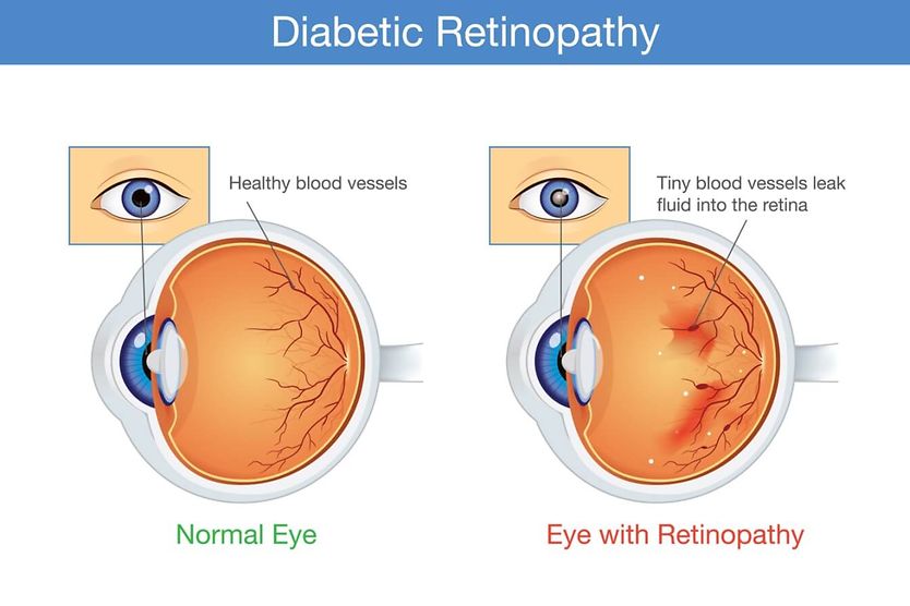
Diabetic retinopathy (DR) is a serious eye disease that can affect people with diabetes mellitus. It occurs when high levels of glucose (sugar) in the blood cause changes to the network of blood vessels located in the retina. Not everyone with diabetes develops diabetic retinopathy, and it can occur even when blood sugar is well controlled. These changes can damage the retina and, if untreated, may lead to vision loss. Diabetic retinopathy does not always cause vision loss or blindness, especially when it is detected and managed early.
Diabetes is a condition characterized by the chronic elevation of glucose levels in the blood. In the body, blood sugar levels are controlled by insulin. When there is too little insulin produced or the body is resistant to insulin, diabetes may result. Diabetes can impact many organs and systems of the body, including the eyes.
The retina is the light-sensing membrane located in the back of the eye. It converts light and visual images into special signals. These signals are then sent to the brain to process what you see.
Changes to the retina, such as those that occur from diabetes, can impact its ability to function properly. These changes can result in issues that can lead to visual impairment.
Diabetic retinopathy is a common eye condition among individuals with diabetes. It is also the leading cause of vision loss among working-age adults in the U.S., while age-related macular degeneration is the more common cause of vision loss in older adults. In 2020, diabetes-related retinopathy affected an estimated 103 million people worldwide. However, by 2045, that number is expected to rise to almost 161 million people.
SEE RELATED: Other types of retinopathy
Click image to enlarge.
Signs and symptoms of diabetic retinopathy
The early stage of diabetic retinopathy may not cause any noticeable eye- or vision-related effects. In fact, the most common early experience is no symptoms at all. This is why diabetic retinopathy is often found in a comprehensive dilated eye exam before any vision change is noticed. However, as the condition advances, signs and symptoms may develop. Some of the more common include:
- Metamorphopsia (distorted vision)
- Blurred vision
- Diplopia (double vision)
- Eye floaters
- Flashes of light
- Scotomas (blind spots)
- Dark spots
- Eye pain or pressure
- Eye redness
- Fluctuating vision
- Difficulty seeing in dim lighting
- Changes in color vision
- Partial or total vision loss
When these signs and symptoms occur, they typically affect both eyes.
READ MORE: Blurry vision and diabetes
What is the first sign of diabetic retinopathy?
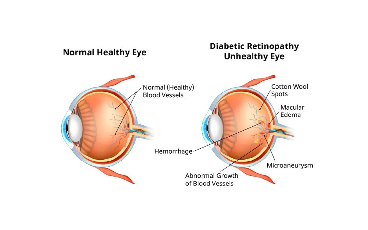
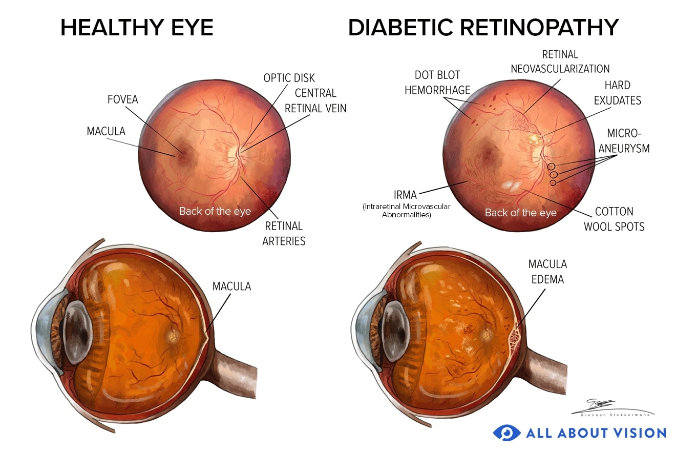
One of the first signs of diabetic retinopathy can be the presence of microaneurysms in the blood vessels of the retina. These are bulges within the vessels that leak fluid or blood. They often look like small red dots sprinkled across the retina when viewed through diagnostic equipment.
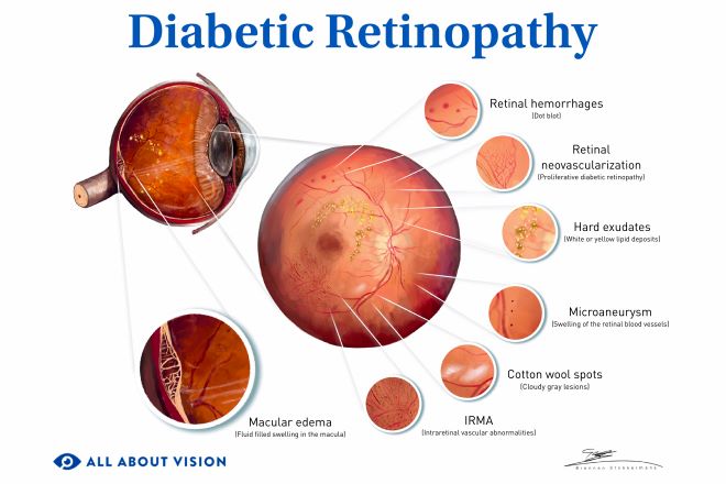
Microaneurysms are a characteristic feature of diabetic retinopathy. Other signs may include:
- Blood in the retina (retinal hemorrhage)
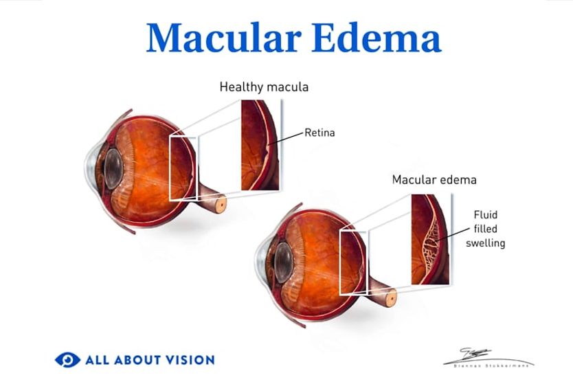
- Thickening of the macula (macular edema)
- Fatty deposits (hard exudates) in or under the retina
- Cotton wool spots (soft exudates) in the retina
As the condition progresses, further changes to the retina may develop, such as:
- Abnormal vessels and vascular changes within the retina
- New vessels growing within the retina or iris
- Blood in the vitreous (vitreous hemorrhage)
- Scarring of the retina
- The blockage or loss of macular blood vessels, depriving the macula of nutrients and oxygen (macular ischemia)
READ MORE: Types of diabetic eye problems
Causes of diabetic retinopathy
Several factors may contribute to the development of diabetic retinopathy. However, prolonged hyperglycemia (high blood sugar levels) is thought to be a leading reason behind its onset.
High blood sugar can cause the blood vessels in the retina to become enlarged and leak fluid. In other cases, abnormal new vessels may develop through a process known as neovascularization. These new vessels are fragile and prone to leaking.
Blood vessels in the retina may also become blocked. This can prevent blood flow through this part of the eye, depriving it of oxygen and nutrients.
Each of these factors can damage retinal blood vessels, causing diabetic retinopathy. Diabetic retinopathy is an acquired complication of diabetes rather than an inherited or congenital eye disease. Even so, genetic factors may help to explain why some people with longstanding — even poorly controlled — diabetes never develop significant retinopathy, while others do.
Risk factors
Diabetic retinopathy can affect people with type 1 or type 2 diabetes. Women who develop pregnancy-related diabetes (gestational diabetes) may also be at risk. Diabetic retinopathy may occur in those with both diagnosed and undiagnosed diabetic conditions. Around 1 in 8 globally, and higher in some populations, already have diabetic retinopathy by the time type 2 diabetes is diagnosed, which is why an eye exam is recommended soon after diagnosis.
The risk of developing the condition generally becomes higher over time the longer a person has diabetes.
Risk factors may include:
- Poorly controlled blood sugar levels
- Long-term diabetes
- High cholesterol
- High blood pressure
- Smoking
- Pregnancy
- Age (50 and over)
- Family history of diabetes
- Obesity
Ethnicity may also increase a person’s risk. People within the following populations may be more likely to develop diabetic retinopathy:
- Hispanic/Latino
- Black/African American
- Native American (U.S.) or First Nation (Canada)
- Alaska Native
- Native Hawaiian or Pacific Islander (NHPI)
Types and stages
Diabetes-related retinopathy is typically classified into two primary types:
- Non-proliferative diabetic retinopathy (NPDR) – Retinal blood vessels leak or become blocked, leading to swelling and tissue damage in the retina. NPDR is the earlier stage of the condition and is often categorized as mild, moderate or severe.
- Proliferative diabetic retinopathy (PDR) – New, abnormal blood vessels develop in the retina. These vessels can also leak and cause retinal scarring. This can lead to further damage or even other eye conditions. PDR is the advanced stage of diabetic retinopathy.
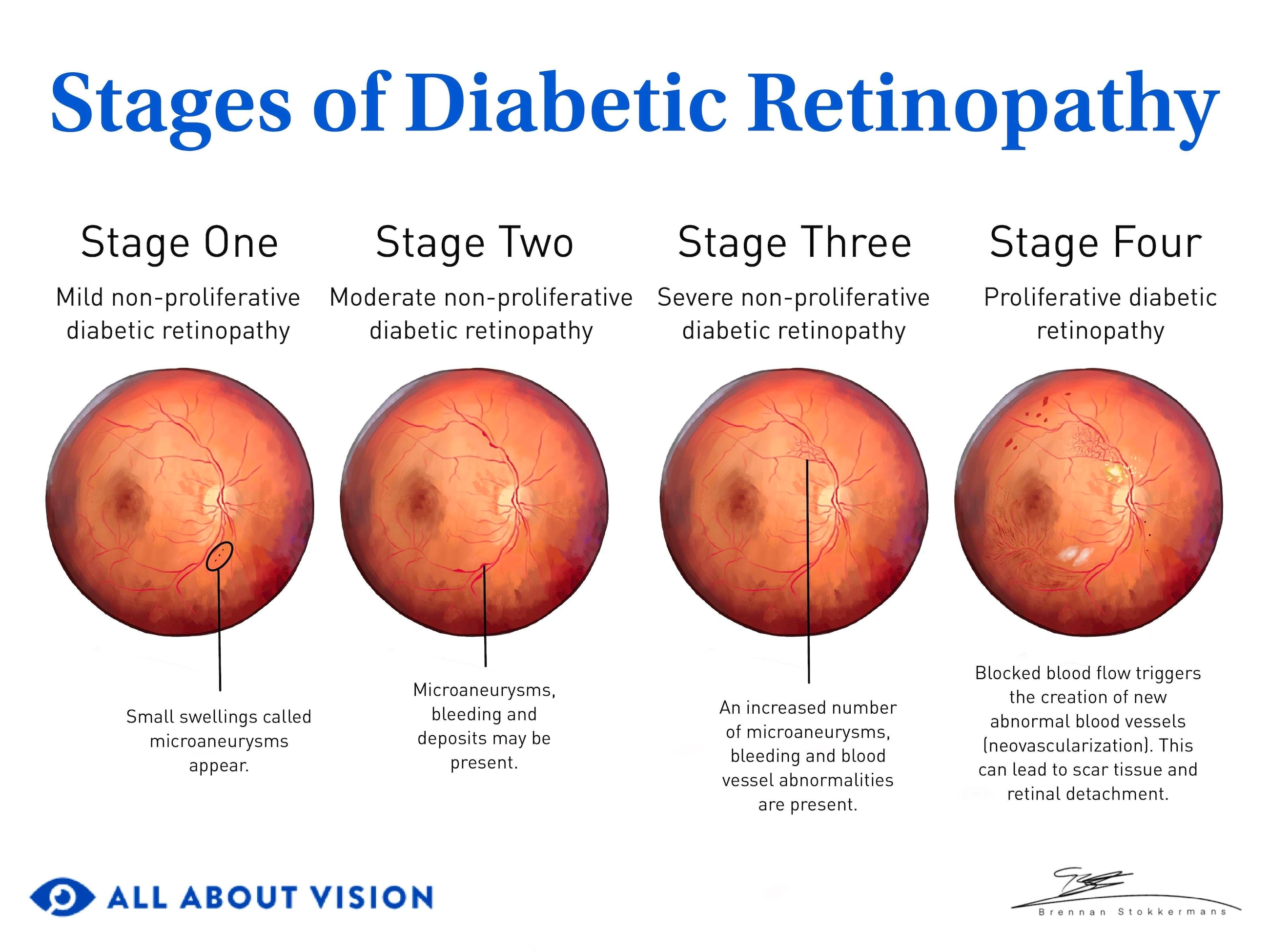
The condition is further grouped into stages, according to its progression. The four stages of diabetic retinopathy are:
- Stage 1 – Mild non-proliferative diabetic retinopathy
- Stage 2 – Moderate non-proliferative diabetic retinopathy
- Stage 3 – Severe non-proliferative diabetic retinopathy
- Stage 4 – Proliferative diabetic retinopathy
Click image to enlarge.
The earlier diabetic retinopathy is detected, the sooner treatment can begin — with the goal being to help slow or prevent it from progressing. Recognizing early-stage diabetes eye symptoms can lead to timely treatment and management. Your doctor may be able to detect signs of diabetic retinopathy, even before you have symptoms. Treatment does more than slow the disease. In some cases, timely treatment can actually improve the severity of diabetic retinopathy.
Diagnosis
Diagnosing diabetic retinopathy typically begins with a comprehensive eye exam. Your eye doctor may perform the following tests to diagnose the disease:
- Visual acuity exam – This exam uses eye charts to test your ability to see objects clearly at different distances.
- Amsler grid test – This test uses a square grid made of straight lines, rather than a chart with letters. It allows your eye doctor to assess your central vision for distortions or blind spots.
- Dilated retinal exam – Your eye doctor may place drops in your eyes to temporarily dilate (widen) your pupils. This allows them to better see into the back part of your eye, where the retina sits.
- Tonometry – Tonometry measures eye pressure (intraocular pressure) and is used to screen for glaucoma. It is typically part of the exam since people with diabetes have a higher risk of developing glaucoma.
- Ophthalmoscopy – With this technique, your eye doctor uses a special instrument called an ophthalmoscope. This device has magnifying lenses that allow them to examine the retina.
- Fundus photography – Your eye doctor may take pictures of the fundus (the back of the eye) to check the retina and other structures.
- Fluorescein angiography (FA) – Fluorescein (a yellow dye) is administered into a vein. The dye moves through your bloodstream and into the vessels in your eyes. A special camera is used to take pictures of the retina as the dye moves through the vessels. With this exam, your eye doctor can determine if new blood vessels have developed. It can also show any areas of blood vessel blockage or leakage.
- Optical coherence tomography (OCT) – Using light wave technology, OCT produces cross-sectional images of the eye’s structures. These images allow your eye doctor to view the layers of the retina to check for changes or damage. It also measures the thickness of the retinal layers.
- Optical coherence tomography angiography (OCTA) – A newer, noninvasive form of imaging that maps the retinal blood vessels in detail without the injected dye used in fluorescein angiography. It can reveal early microvascular changes and areas of reduced blood flow.
In some instances, artificial intelligence (AI) along with teleophthalmology and teleoptometry may be used to screen for diabetic retinopathy. Both teleophthalmology and teleoptometry are forms of telemedicine that allow doctors to deliver eye care through digital methods. Medical providers can use these tools to detect signs of the condition remotely, which could lead to an earlier diagnosis.
Comprehensive eye exams are essential to detect diabetic retinopathy vision problems early, monitor its progression and prevent further vision loss. Your doctor may use the results of your eye exam to determine if or which diabetic retinopathy treatments are needed.
Treatment options
Patients with mild diabetic retinopathy may not need treatment immediately. At this stage, controlling blood sugar, blood pressure, and blood cholesterol and lipid levels may help with managing the condition. Even at the mild stage, a small proportion of people progress to more serious, treatment-requiring disease over the following year. Recent screening data suggest this short-term risk is low, but it is not zero, which is why regular follow-up exams are still recommended.
Diabetic retinopathy treatment is most commonly provided by a retinal specialist, who is an ophthalmologist specializing in the retina. They may recommend one or more of the following treatments for advanced cases of the condition:
Anti-VEGF eye injections
Eye injections of medications known as anti-VEGFs can help stop the overproduction of vascular endothelial growth factor (VEGF) in the eye. They may help inhibit the growth of abnormal blood vessels and shrink those that have already developed.
Anti-VEGF drugs are injected into the vitreous of the eye. Along with slowing abnormal blood vessel growth, these eye injections may help control macular swelling. They can also reduce the risk of vessel leakage and retinal scarring.
Anti-VEGF injections used for diabetic retinopathy are often the same ones utilized for age-related macular degeneration (AMD). Anti-VEGFs used to treat diabetic retinopathy may include:
- Aflibercept
- Bevacizumab
- Brolucizumab
- Faricimab
- Ranibizumab
Eye injections are typically provided in your ophthalmologist’s office. They may be administered as often as monthly and, depending on the medication used and your treatment response, sometimes extended to about every three months, depending on the type of medication used and other factors. Your eye doctor can tell you how often you should have diabetic retinopathy medication injections.
Laser
Laser surgery closes off blood vessel leaks and reduces retinal swelling (macular edema). These procedures may also help shrink abnormal blood vessels and reduce the risk of retinal detachment.
There are two main types of laser treatment for diabetic retinopathy:
- Focal or grid laser photocoagulation – This treats certain cases of macular edema. It involves focusing laser energy directly on the affected area or in a grid-like pattern on the retina. Focal laser treatments target specific microaneurysms. Grid laser treatments address larger areas of blood vessel leakage.
- Scatter (panretinal) laser photocoagulation – Advanced cases, or proliferative diabetic retinopathy, may be treated with panretinal photocoagulation (PRP). This is a more rigorous type of laser diabetic retinopathy surgery. Laser surgery with PRP is designed to reduce the amount of VEGF produced by the retina. In this way, it may lower the risk of new, abnormal vessel development.
Laser diabetic retinopathy treatments are generally performed in an ophthalmologist’s office or an outpatient treatment center.
SEE RELATED: Laser coagulation eye surgery
Vitrectomy
A vitrectomy involves surgically removing some or all of the vitreous fluid from the eye. It may be recommended in advanced cases of diabetic retinopathy, such as when blood has leaked into the vitreous gel and is impairing vision. The vitreous cavity is then filled with a clear fluid to help maintain the shape of the eye.
A vitrectomy may also be conducted if scar tissue has formed in the retina.
Vitrectomies are typically performed at a hospital or an outpatient surgery center. It is a more involved operating-room procedure than cataract surgery.
Corticosteroids
Corticosteroid medications may also be used as a diabetic retinopathy treatment. Steroids may reduce fluid leakage and swelling in the retina to help control diabetic macular edema.
These treatments may be given in the form of an eye injection. As an alternative, an intraocular implant may be surgically placed in the eye. The implant is filled with a steroid, such as dexamethasone or fluocinolone acetonide. Steroid implants may offer a longer-acting option over monthly eye injections.
Corticosteroid implants are generally used in cases of diabetic retinopathy that are resistant to anti-VEGF injection therapy. They are designed to provide a controlled release of medication over a certain period of time.
Intravitreal steroid eye injections are typically performed in an ophthalmologist’s office. Implants may also be placed in an eye doctor’s office, although more complex implant procedures may require treatment in a hospital or surgical center.
Emerging and investigational treatments
Researchers are testing ways to reduce how often people need injections. These include longer-acting drug-delivery approaches (such as refillable eye implants) and investigational gene therapy designed to prompt the eye to produce its own anti-VEGF protein. These options are still emerging, and some remain investigational; your retina specialist can explain what is currently approved and appropriate for you
Health risks and complications
Diabetic retinopathy can threaten your vision and cause visual impairment. It can also increase the risk of other eye conditions. However, the risks and complications of the disease affect health factors that may go beyond your eyesight.
Diabetes-related retinopathy is a marker of more widespread blood-vessel damage from diabetes. It does not cause these conditions, but people who have diabetic retinopathy are more likely to also have:
- Cardiovascular (heart) disease
- Stroke
- Diabetic peripheral neuropathy (nerve dysfunction in the extremities)
- Diabetic nephropathy (kidney dysfunction)
- Diabetic foot syndrome
You could experience a range of other complications due to diabetes. Eye problems related to the disease can involve one or more of the following conditions:
- Dry eye disease
- Tractional retinal detachment
- Neovascular glaucoma
- Macular edema
- Vision impairment or blindness
There may be other medical problems associated with diabetic retinopathy. It is recommended to consult with your doctor to learn more about your personal risks or health concerns.
Prevention and management
It might not always be possible to prevent the onset of diabetic retinopathy, but you may be able to slow its progression and reduce the risk of vision loss. Although there is no cure, taking steps to manage the disease can help preserve your vision.
Many of the same healthy habits that may lower the risk of diabetic retinopathy may also help manage the condition. These include:
- Getting yearly comprehensive eye exams – Having your eyes examined at least once every 12 months allows your eye doctor to detect and treat problems as early as possible. In fact, many people first learn they have diabetes through a routine eye exam. The earlier diabetic retinopathy is detected, the sooner treatment for both the retinopathy and the underlying diabetes can begin.
- Managing diabetes and controlling blood sugar – Controlling diabetes is key to lowering the risks associated with diabetic retinopathy. It is important to monitor and control your blood sugar and take diabetes medication or insulin as prescribed by your doctor. A healthy diet and proper nutrition are also important for controlling blood sugar.
- Managing high blood pressure and blood lipids – Controlling your blood pressure and blood lipid levels can help lower the risk of diabetic retinopathy.
- Getting eye exams during pregnancy – If you are pregnant and have diabetes, it is recommended to get your eyes examined during each trimester. Inform your eye doctor if you develop gestational diabetes, as this is also a risk factor. Your eye doctor may recommend more frequent eye exams within the first year after giving birth.
- Following a healthy diet – Eating a healthy, balanced diet (such as the Mediterranean diet) may lower your risk for diabetes-related eye conditions. Certain vitamins and minerals may also help support your eye health. Talk to your doctor before taking any new dietary supplements.
- Getting regular exercise – Exercising regularly may lower the risk of eye diseases, such as diabetic retinopathy. Physical activity also helps lower blood glucose levels in the body.
- Avoiding tobacco use – Some studies have found that smoking may increase the risk of diabetic retinopathy. Quitting or avoiding smoking is important for your eye health and general wellness.
It is recommended to speak with your eye doctor for further details on lowering your risk of diabetic eye diseases and managing their effects.
Living with diabetic retinopathy
Diabetic retinopathy can affect daily life, especially if vision loss occurs. However, with the right support and strategies, many people with the condition can maintain their independence and quality of life.
Vision changes from diabetic retinopathy can vary, ranging from difficulty seeing in low light to vision loss, depending on its severity. If your vision is impacted, your eye doctor may refer you to a low vision specialist to help you optimize your remaining vision.
A low vision specialist may suggest practical changes you can make in your home and work environments to meet your specific needs. They may also recommend assistive low vision devices or optical aids that can make it easier to read print and see objects more clearly.
Seeking support from health care providers, family and friends may also help make daily life with diabetic retinopathy more manageable. There are also a wide variety of community resources that offer tools, education and encouragement for individuals impacted by vision loss, such as:
- Vision rehabilitation services
- State agencies and organizations
- Local and online support groups
- Transportation and independent living services
- Low vision resource centers
When to call an eye doctor
Diabetic retinopathy is a serious eye condition that requires prompt attention from an eye care professional. If you experience symptoms of the condition or notice any vision changes, it is important to visit an eye doctor right away.
Contact your eye doctor to schedule a comprehensive eye exam or to locate diabetic eye care services near you. Early diagnosis and treatment can help prevent vision loss from diabetic retinopathy and preserve your eyesight.
Frequently asked questions
People who are at risk for or diagnosed with diabetic eye disease often have additional questions about the condition. Some of these may include:
Is there a way to reverse diabetic retinopathy without surgery?
In some cases, the very early stage of diabetic retinopathy may be reversed with tight blood sugar control. Beyond the earliest stage, anti-VEGF injections can also regress the severity of diabetic retinopathy in many people; however, this controls the disease rather than curing it and generally depends on continued long-term treatment. For some patients, treatment can improve vision. But in many instances, vision impairment is permanent, and the goal of treatment is to prevent additional vision loss.
How effective are diet and lifestyle modifications in managing diabetic retinopathy?
Diabetes is a chronic, lifelong condition that can cause significant health issues if it is not controlled. Managing this disease through diet, exercise and medical care (including vision care) can help preserve your health and protect your eyesight. Controlling diabetes can slow the development and progression of diabetic eye diseases, such as retinopathy.
Why can people with diabetic retinopathy experience double vision?
Double vision can occur when diabetes damages the eye’s tissues or the nerves that control eye movement. When these nerves are affected, the eyes can become misaligned, so a single object appears as two images.
How often should people with diabetes get their eyes checked?
Yearly diabetic eye exams may allow your eye doctor to detect the condition as early as possible. Having regular eye exams (as recommended by your eye doctor) allows them to evaluate for signs of retinal diseases or other problems caused by diabetes. The earlier diabetes-related eye conditions are detected and treated, the greater the chances of preserving your vision.