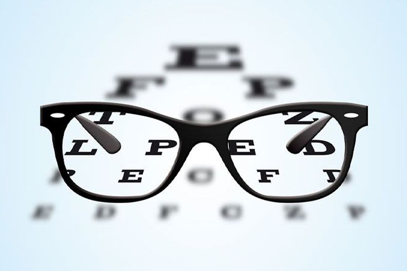
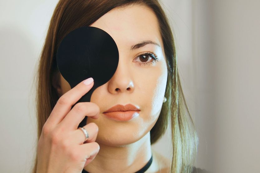
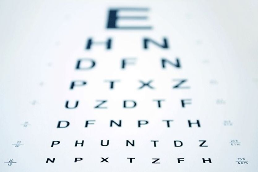
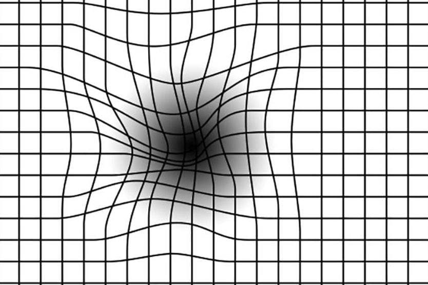
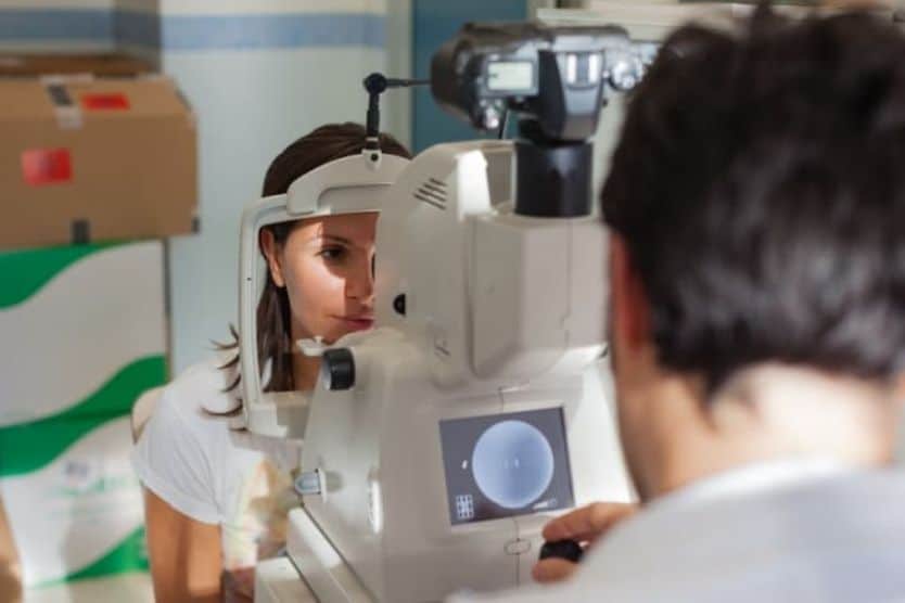
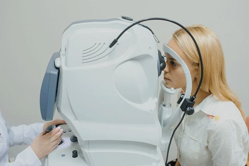
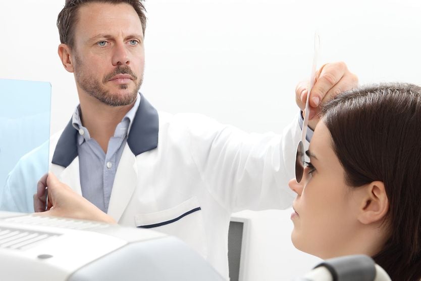
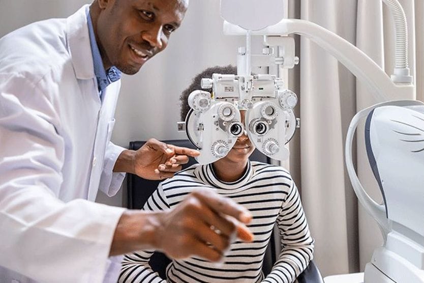
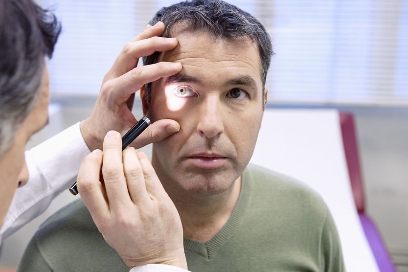
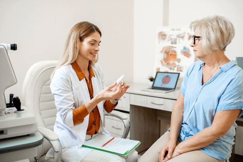
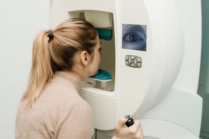
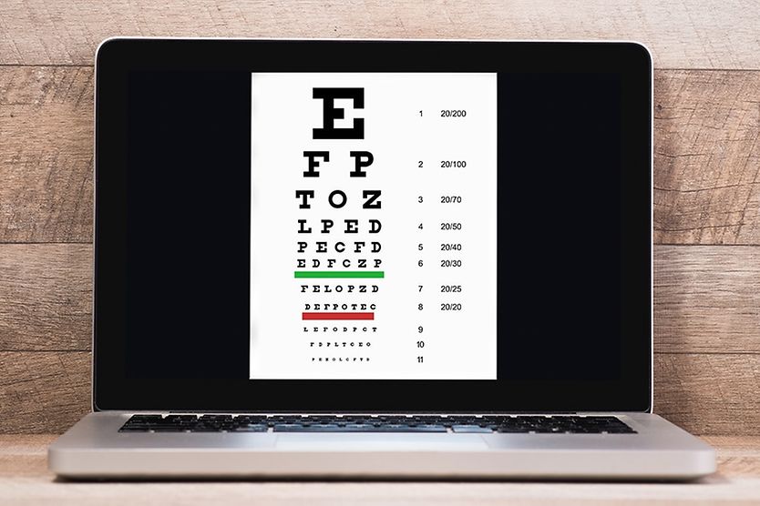
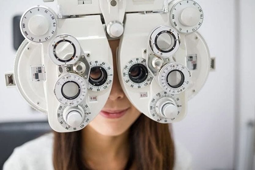
Home Conditions | Fluorescein angiography

Fluorescein angiography: Side effects, procedure details and what to expect during this type of eye test

Fluorescein angiography machine being used on a patient

What is fluorescein angiography (FA)?

Fluorescein angiography (FA) is a procedure that uses fluorescein dye to help view the tissue and blood vessels of the retina and choroid in the back of the eye. This test can help diagnose a variety of diseases and conditions that affect the eye.

Fluorescein angiography is not to be confused with a fluorescein eye stain test. This eye stain test uses a dye on the surface of the eye to detect issues on the cornea.

What can FA detect?

FA can detect many different eye diseases, including:

Diabetic retinopathy

Diabetic retinopathy is a microvascular disease that can occur from the long-term effects of diabetes. In the early stages of the disease, a patient may not exhibit any symptoms. An exam of the back of the eye may detect diabetic retinopathy. Symptoms may include blurry vision, floaters, and partial or total vision loss.

Retinal detachment

A retinal detachment can occur when the vitreous in your eye pulls on your retina. This happens more often in older individuals as the vitreous starts to thin and shrink. The fluid can then go behind the retina, tearing it. Symptoms include floaters, seeing flashes of light and seeing shadows in your peripheral vision. Vision loss is possible.

Retinitis pigmentosa

The term retinitis pigmentosa is used for a group of inherited retina diseases. Symptoms include trouble seeing at night or in low light, blind spots in your side vision, tunnel vision, photophobia, not being able to see color and low vision.

Retinal vein occlusion (RVO)

A blockage in the vein that carries blood from the retina is called a retinal vein occlusion (RVO). It usually happens to those in their 50s and 60s. Having diabetes, glaucoma or high blood pressure increases your risk. Symptoms include floaters, blurry vision, pain in the eye and vision loss.

Retinal artery occlusion (RAO)

Retinal artery occlusion is a serious condition that occurs when there is a blockage in an artery of the eye. It can be caused by a blood clot, inflammation of the blood vessels or spasm. Risk factors include smoking, diabetes, hypertension and being older. The main symptom is painless, sudden vision loss that happens in a few seconds. If this happens, you should go to the emergency room immediately.

Age-related macular degeneration (AMD)

Age-related macular degeneration (AMD) is a condition that may cause you to lose your central vision. It occurs when there is damage to your macula, which is located in the center of your retina. There are two types: dry and wet.

With dry AMD, vision loss tends to be gradual, and many people do not lose all of their central vision. Wet AMD is more severe and can lead to total central vision loss.

Risk factors include age, smoking, high blood pressure, being overweight and a family history of the condition. Symptoms include blurry vision, low vision, inability to see in low light and dark spots in your field of vision.

Macular edema

Macular edema is a condition that occurs when fluid that has leaked from damaged blood vessels builds up and causes swelling in the macula. Causes include AMD, diabetes, RVO, medication and trauma. Symptoms include blurry vision, seeing colors differently and wavy vision.

Ocular melanoma

Ocular melanoma is a rare type of cancer that forms on the uvea, or middle layer of the eye. It may not cause symptoms early on. FA can help detect it in its early stages. Risk factors include having light colored eyes, long-term exposure to sunlight and older age. Symptoms include seeing flashes of light, blurry vision, distorted vision, or a dark spot on the iris or conjunctiva.

Ocular ischemic syndrome (OIS)

OIS is a rare visual disorder that is caused by a hardening or narrowing of the carotid arteries. These arteries deliver blood and oxygen to the brain. FA is a critical test in helping diagnose OIS. The most common symptom is gradual vision loss over a few weeks to a few months. Other symptoms include loss of light perception, seeing afterimages and an achy feeling around the eye. OIS can cause vision loss.

Central serous chorioretinopathy (CSCR)

CSCR is a condition that occurs when fluid from the choroid leaks and builds up under the retina. This causes a small detachment of the retina. Risk factors include stress, insomnia, steroid use and hypertension. Those with autoimmune diseases and middle-aged males are also more at risk. Symptoms include distorted vision, seeing a dark area in your central vision, dimmed vision and blurry vision. Most people regain their vision without any treatment.

Unexplained vision loss

Unexplained vision loss can last for a few seconds to a few days. It can occur in one eye or both eyes. Vision loss can be partial or complete and can occur along with pain, eye redness or headache. Many things can cause temporary vision loss, such as dry eyes, ocular migraine, retinal artery occlusion, retinal detachment, glaucoma, trauma and stroke. If you experience unexplained vision loss, you should seek medical care immediately. 

Other conditions

FA can also be used to confirm a diagnosis or evaluate other eye diseases, which include but are not limited to:

  • Retinal vasculitis – A disorder of the retinal vessels. It is associated with a variety of autoimmune, inflammatory, infective and neoplastic disorders. Symptoms include seeing flashes, floaters, pain and blurry vision. It can threaten your sight.

  • Retinal dystrophiesA group of degenerative disorders that affect the retina. Symptoms include color blindness, night blindness and trouble with peripheral vision. It can lead to vision loss.

  • UveitisA group of diseases that usually affect the uvea. Other parts of the eye can also be affected. Symptoms include inflammation, red eyes and eye pain. It can lead to vision loss if not treated.

  • Posterior scleritisA condition that affects the back of the sclera, or white part of your eye. Symptoms include eye redness, a piercing pain in the eye and pain that gets worse with eye movement. It can cause vision loss if not treated.

  • Parafoveal telangiectasia – A rare disease in which the blood vessels of the fovea, inside the macula, are abnormal. It can cause vision loss in one or both eyes.

  • Ischemic optic neuropathy A condition in which blood flow to the optic nerve is reduced. This can cause vision loss in one or both eyes. People with high blood pressure, high cholesterol, diabetes, glaucoma, migraines, sleep apnea and heart disease are most at risk.

  • Neovascularization of the eye – A condition that occurs when blood vessels grow where they should not be growing. If it occurs in your macula or choroid, you may experience distorted vision or see a dark spot in your central vision. If it occurs in your cornea, you may experience eye pain, watery eyes, light sensitivity or red eyes.

  • Stargardt’s disease An inherited disease that affects children and young adults. Symptoms include blurry central vision and trouble seeing colors. Photos from FA will show a dark choroid, which should normally be visible.

What to expect during FA

Fluorescein angiography typically takes about 30 minutes. Your doctor will administer eye drops to dilate your pupils. This allows for a better view of the back of your eye. Then you will rest your forehead and chin in the appropriate places on a fundus camera. The doctor will then take photographs of your eyes.

After the first round of photos, a yellow colored dye will be injected into your arm. When the dye reaches the blood vessels in your eye, the fluorescein will cause the blood vessels to light up (fluorescence). A second round of photos will then be taken as this dye passes through your retina.

On a traditional FA, 30° to 50° of the retina can be viewed. On an ultra-wide-field FA, up to 200°, which is almost all of the retina, can be viewed.

What to expect after FA

Because your pupils will be dilated, you may experience blurry vision and light sensitivity for a few hours after FA. You will want to have someone drive you because it will be difficult to see. Wearing sunglasses afterward will help with light sensitivity.

Side effects and risks

There are very few side effects of FA. Some of these side effects could include:

  • Your skin could turn yellow from the dye traveling to the veins throughout your body. After a few hours, your skin should return to its normal color.

  • A burning sensation may be felt on your skin from the dye at the injection site. The dye is not harmful, and the sensation should go away after a few minutes.

  • Objects you look at may appear dark or tinted. This should also go away after a few minutes.

  • Your urine may turn yellow as your kidneys filter the dye from your blood. This could happen for up to 24 hours after the test.

There are also very few risks of FA. Some people may be sensitive to fluorescein. Sensitivity can lead to dry mouth, dizziness, an increased heart rate, having a metallic taste in the mouth or nausea. Other people may have an allergic reaction to the dye. This reaction can produce hives or itchy skin.

In very rare instances, a patient can experience breathing problems. If this happens, alert your doctor immediately or go to the emergency room.

Fluorescein angiography. Cleveland Clinic. December 2022.

Fluorescein angiography. StatPearls. February 2023.

Fluorescein eye stain. MedlinePlus. February 2023.

What is fluorescein angiography? American Academy of Ophthalmology. June 2021.

Diabetic retinopathy. StatPearls. May 2023.

Detached retina. American Academy of Ophthalmology. October 2022.

Retinitis pigmentosa. Cleveland Clinic. May 2022.

Retinal vein occlusion (RVO). Cleveland Clinic. June 2023.

Retinal artery occlusion. EyeWiki. American Academy of Ophthalmology. July 2023.

Macular degeneration. Cleveland Clinic. February 2023.

What is macular edema? American Academy of Ophthalmology. April 2023.

What is ocular melanoma? American Academy of Ophthalmology. November 2022.

Ocular ischemic syndrome. StatPearls. October 2022.

Carotid artery stenosis. Johns Hopkins Medicine. Accessed July 2023.

What is central serous chorioretinopathy? American Academy of Ophthalmology. September 2022.

Unexplained vision loss. Loyola Medicine. Accessed July 2023.

Retinal vasculitis. EyeWiki. American Academy of Ophthalmology. June 2023.

Retinal dystrophies. StatPearls. March 2023.

Uveitis. Cleveland Clinic. February 2021.

Scleritis. Cleveland Clinic. May 2023.

What is ischemic optic neuropathy? American Academy of Ophthalmology. April 2023.

Neovascularization of the eye. Cleveland Clinic. September 2022.

What is Stargardt disease? American Academy of Ophthalmology. December 2022.

Find Eye Doctor

Schedule an exam

Find Eye Doctor