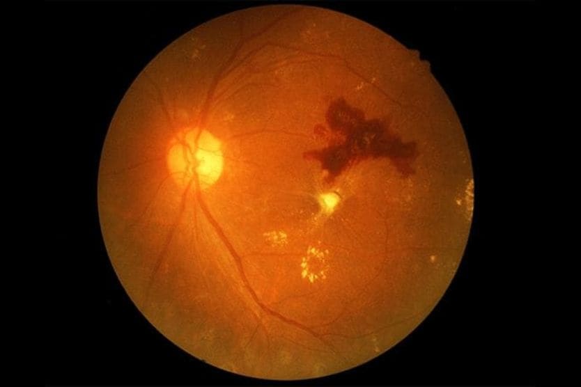
Retinitis pigmentosa


What is retinitis pigmentosa?
Retinitis pigmentosa (RP) is a group of rare hereditary eye conditions that cause the deterioration and dysfunction of cells in the retina (the light-sensing membrane at the back of the eye). As a progressive disease, RP can lead to vision loss and blindness over time.
The term retinitis pigmentosa is a bit of a misnomer. While the “-itis” suffix implies retinal inflammation, the condition is not inflammatory in nature. However, the disease typically causes pigmented (colored) clumps to form in the retina, which explains the “pigmentosa” part of its name.
The condition is also referred to as hereditary retinal dystrophy.
Researchers estimate that globally, more than 1.5 million people have RP. It’s also the most commonly inherited retinal disease worldwide.
RP can appear in two different forms:
- Non-syndromic – This is the most common form, affecting only the eyes.
- Syndromic – This form occurs alongside certain systemic conditions affecting other parts of the body. It’s commonly seen in Usher syndrome, which causes vision and hearing loss.
How it affects the eyes and vision
Retinitis pigmentosa develops when special cells in the retina called photoreceptors stop working properly. There are two types of photoreceptors:
- Rods – Cells that allow you to see in dim or low lighting.
- Cones – Cells that help you perceive color and see clearly in bright conditions.
Rods and cones are essential to your vision. They convert light that enters your eye into visual information, which your brain uses to interpret what you see.
The deterioration and loss of function in photoreceptors disrupts a layer of cells beneath the retina called the retinal pigment epithelium (RPE). This structure helps keep the retina healthy and supports rods and cones in detecting light. When the RPE is disrupted, it can migrate into the retina, contributing to the bone spicule-like pigment deposits that are characteristic of retinitis pigmentosa. Over time, the loss of photoreceptors and changes in the RPE can lead to vision loss.
What causes retinitis pigmentosa?
The primary cause of retinitis pigmentosa is genetic mutations (variants) that affect the rods and cones in the retina. Rods are usually impacted first, which is why night vision problems are often an initial symptom. Cones are usually affected later in the disease process, though this can vary from person to person.
Over 100 genes have been linked to RP, and there is no single mechanism for how this occurs. Parents can pass a mutated gene down to their child, or the mutation can happen spontaneously (on its own). In many cases, there is no known or obvious family history of the condition.
How RP is inherited
Three inheritance patterns are commonly associated with retinitis pigmentosa:
Autosomal dominant
With this pattern, only one parent carries a single copy of the altered gene. Each child has a one in two chance of inheriting the mutated gene and developing the condition.
RP associated with autosomal dominant inheritance is the least severe type. Symptoms of this form usually develop more slowly and appear in adulthood.
Autosomal recessive
Autosomal recessive inheritance means that both parents have a single copy of the variant gene and a single copy of the normal gene. While they are carriers, they are not affected by the condition. As a result, each child has a one in four chance of inheriting the gene mutation from each parent and developing the disease. A child who inherits a single copy of the gene variant will be a carrier but usually won’t develop the condition.
Symptoms of RP caused by autosomal recessive inheritance typically appear earlier in life. Night blindness and severe vision loss are also characteristic of this type.
X-linked
With X-linked inheritance, the variant gene is on the X chromosome. Since females have two X chromosomes (XX) and males have one X and one Y (XY), the inheritance pattern affects them differently. Men are more likely to develop the disease because they only have one X chromosome. But if a female carries the mutated gene, they are less likely to be affected by the disease since the other X chromosome will carry a healthy copy.
Males with an X-linked disease cannot pass the condition onto their sons because they give their sons a Y chromosome and not an X chromosome. However, their daughters will inherit their X chromosome and become carriers of the gene.
Females who carry the mutated gene have a one in two chance of passing it on to each child. If a daughter inherits it, she will be a carrier. If a son inherits it, he will be affected by the condition.
People with X-linked recessive retinitis pigmentosa typically experience the most significant vision loss.
What are the symptoms of retinitis pigmentosa?
RP symptoms typically involve vision-related changes and are often first noticed during childhood or adolescence. Symptoms develop slowly in some people and more rapidly in others, and commonly include:
Difficulty seeing at night and in low light
Nyctalopia, loss of night vision or night blindness, is often the first symptom of RP. It’s caused by the degeneration of rod cells, which help you see in dim light. Early changes in your night vision can make it harder for your eyes to adjust to darkness or low-light conditions. This can make it more difficult to perform everyday activities, such as navigating a dimly lit room or driving at night.
Peripheral vision loss
The loss of rod cells can also affect your peripheral (side) vision, or what you see out of the corners of your eyes. When this occurs, your field of vision gradually narrows, as though you’re looking through a tunnel. This type of vision loss is also referred to as tunnel vision.
Peripheral vision loss makes it difficult see objects around you and to navigate your surroundings. To get a clear view, you may need to turn your head to look directly at them.
Central vision changes
Gradual changes in your central vision can occur as RP progresses. Your central vision is your field of view when looking straight ahead and is what helps you see objects in fine detail.
Central vision loss can make everyday activities more challenging. It can make it harder to:
- Read text
- Perform close-up activities
- Recognize faces
- Carry out other daily tasks, such as driving and cooking
Color vision changes
Damage to your cone cells can impact your color vision, or your ability to perceive color. You might find it difficult to see or distinguish between certain colors, such as blue and green.
How is retinitis pigmentosa diagnosed?
Signs of retinitis pigmentosa may be detected during a comprehensive eye exam. To confirm a diagnosis, further retinal tests or imaging techniques may also be needed.
Eye exam and visual field testing
During the eye exam, your eye doctor may perform the following tests to check the health of your retina and other eye structures:
- Visual acuity testing – This measures how clearly you can see at different distances.
- Pupil dilation – Special eye drops are used to widen your pupils, giving your doctor a better view of your retina and other structures at the back of your eye.
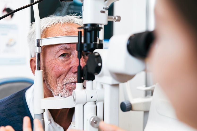
- Slit lamp exam – Your doctor uses a special microscope called a slit lamp to closely examine structures in your eye.
- Fundoscopy – This involves using an instrument called an ophthalmoscope to view the fundus, which is the back part of your eye.
- Color vision testing – Your doctor may perform various color vision tests to assess your ability to perceive color. This can help determine to what degree the cone cells are affected.
Visual field testing is also an important part of diagnosing retinitis pigmentosa. This involves tests that measure how well you can see in your peripheral and central vision.
Optical coherence tomography (OCT)
Optical coherence tomography is an advanced imaging technique that provides highly detailed pictures of the retina. With these images, your doctor can evaluate the individual layers of the retina and assess the health of the photoreceptor cells.
A similar technique, known as optical coherence tomography angiography (OCTA), detects changes in retinal blood vessels. This can help your doctor understand the severity and progression of the condition.
Fundus autofluorescence (FAF)
With this imaging technique, your eye doctor uses a special light to visualize the naturally fluorescing properties of lipofuscin (a byproduct of photoreceptor degeneration) in the retinal pigment epithelium. FAF can help your doctor determine the extent of photoreceptor degeneration and its progression.
Electroretinography (ERG)
ERG uses electrodes to measure the electrical signals produced by photoreceptor cells when exposed to flashes of light. The information produced by this test can help your doctor evaluate the health of those cells and whether they’re functioning properly.
Genetic testing
In addition to eye and vision tests, your doctor may recommend genetic or DNA testing to confirm an RP diagnosis. This information can also help you determine the potential risk of passing the condition on to your children.
Managing retinitis pigmentosa
Retinitis pigmentosa is typically managed by an ophthalmologist, who may also be a retinal specialist. But other healthcare professionals might also be involved in the overall care plan.
Scientists have not yet found a cure for retinitis pigmentosa. However, working with your eye doctor and healthcare team to manage its effects can help preserve your vision and quality of life. RP may increase the risk of developing other eye conditions, such as cataracts, refractive errors and cystoid macular edema, so treatment for those conditions may be needed.
Medical treatments and emerging therapies
Researchers are actively studying options for treating RP and restoring vision lost to the condition. These approaches include:
Gene therapy
Gene therapy works by introducing new genetic material into cells to help correct or compensate for faulty genes that cause disease. The exact approach varies, depending on the specific gene and mutation involved.
Many gene therapies are currently being studied as potential treatments for retinal diseases. So far, one therapy — voretigene neparvovec-ryzl — has received FDA approval for the treatment of RP caused by an RPE65 gene mutation. This gene therapy is administered during a surgery called a vitrectomy, which allows the treatment to be delivered directly to the retina.
Stem cell therapy
Scientists are also researching stem cell therapy as a way to treat and restore vision lost to RP. This therapy replaces damaged cells in the body with healthy cells to help regenerate new, healthy tissue. In the case of RP, the hope is for these new cells to transform into functioning photoreceptor cells, ultimately improving vision.
Retinal and cortical implants
Another area of RP treatment research involves visual prostheses, also referred to as “bionic eyes.” With this approach, an electronic device is surgically implanted into the retina. The device bypasses the damaged rods and cones, producing visual signals the brain can interpret. Studies have found that these retinal implants may help improve factors like visual acuity and color vision.
Other visual prostheses, called cortical implants, are surgically implanted in the brain’s visual cortex. These devices bypass the eyes altogether, receiving visual information from an external device and converting it into signals the brain can interpret.
Optogenetics
This is another potential therapeutic approach that helps the retina function, even when photoreceptor cells are damaged. It works by providing remaining healthy cells with light-sensitive proteins called opsins. These help the cells detect light and send visual signals to the brain to process what you see.
While optogenics likely won’t reverse the effects of RP, it could potentially help improve vision in some people with the condition.
Clinical trials and ongoing research
Research is ongoing to identify effective treatments for retinitis pigmentosa, and numerous therapies are being tested in clinical trials. Visit Clinicaltrials.gov to learn more about current studies.
Low vision aids and lifestyle support
Many people with retinitis pigmentosa use low vision devices and adaptive strategies to maximize their remaining vision and maintain an independent lifestyle.
Common approaches include:
- Low vision aids – These include tools such as magnifiers, telescopes, large print media and electronic devices. Digital technology, including accessibility tools, can make everyday tasks easier through features like text-to-speech and voice-activated controls.
- Home and work modifications – These may involve strategies like improving lighting and creating color contrast in indoor environments, and arranging furniture to minimize obstacles.
- Mobility training – This involves personalized instruction on navigating your surroundings safely and independently.
A low vision specialist can help you choose the options that best meet your needs.
Nutritional supplements
Certain supplements, including vitamins A and E, have been studied for their potential to slow the progression of vision loss in RP. In some studies, taking daily high doses of vitamin A appears to offer some benefit. Despite evidence that vitamin A is effective in slowing the disease, this approach is still being discussed within the eye care community.
Conversely, studies have shown that high-dose vitamin E supplementation can accelerate the disease and should be avoided.
When to see an eye doctor
Attending regular eye exams is essential for your eye health and vision. These exams help detect signs and symptoms of disease early, which could help inform management strategies.
If you experience sudden or rapid changes in your vision or notice increasing difficulty seeing in low-light conditions, contact your eye doctor right away. Early diagnosis and ongoing monitoring of retinitis pigmentosa allow your eye doctor to track vision changes, address potential complications, and recommend low vision strategies and lifestyle adjustments to support your daily needs.
Resources for living with retinitis pigmentosa
Retinitis pigmentosa can affect your life in many ways, and accessing the proper resources can ensure you get the support you need. Beyond medical care from eye doctors and other health professionals, there are numerous patient organizations, support networks and advocacy groups dedicated to helping people with vision loss due to conditions, such as RP.
Some of these resources include:
- Foundation Fighting Blindness
- National Institutes of Health
- Prevent Blindness
- American Foundation for the Blind
- American Printing House for the Blind
- Lighthouse Guild
- Hadley
- Retina International
- Global Genes
To further explore available resources for living with retinitis pigmentosa, talk with your eye doctor, low vision specialist or other members of your healthcare team.