Retinoschisis: Causes, symptoms and treatment options

On this page
Retinoschisis causes
Retinoschisis symptoms
How does retinoschisis affect vision?
Diagnosing retinoschisis
Retinoschisis treatment
Retinoschisis vs. retinal detachment
The importance of eye exams
On this page
Retinoschisis causes
Retinoschisis symptoms
How does retinoschisis affect vision?
Diagnosing retinoschisis
Retinoschisis treatment
Retinoschisis vs. retinal detachment
The importance of eye exams
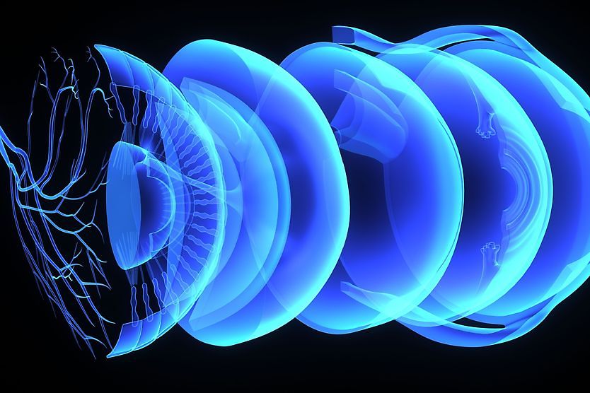
Retinoschisis is an eye condition that occurs when the retina (the tissue lining the back of the eye that converts light into images) is split into two layers. The condition can occur in any of the seven cellular layers of the retina. It may also occur for varying reasons.
The condition may be acquired due to the natural aging process (degenerative or senile retinoschisis) or genetically inherited (genetic retinoschisis). Cases of genetic retinoschisis generally occur more often in males than females, whereas degenerative retinoschisis seems to affect males and females equally.
Other names used for retinoschisis, which are given depending on the type and cause, include:
- Congenital X-linked retinoschisis or CXLRS
- X-linked retinoschisis or XLRS
- XJR
- Juvenile retinoschisis
- Degenerative retinoschisis
- Senile retinoschisis
Retinoschisis does not usually lead to blindness and is treatable with the aid of corrective eyewear. However, it can sometimes lead to poor vision or further complications that require additional monitoring and care.
Retinoschisis causes
Retinoschisis may be degenerative or genetic. The degenerative variety usually occurs in older patients, while genetic retinoschisis normally appears in younger people — particularly in young boys.
Degenerative retinoschisis
Degenerative retinoschisis, sometimes called senile retinoschisis, occurs in older adults and happens slowly over time due to aging rather than genetic reasons.
This type of the condition is naturally acquired in adults who are usually in their 50s, 60s and 70s, though it has been found in younger patients on some occasions.
Degenerative retinoschisis generally affects both men and women at the same rate.
Genetic retinoschisis (X-linked retinoschisis)
Genetic retinoschisis, also known as juvenile or x-linked retinoschisis, occurs due to a defect or mutation in the X chromosome. This type of retinoschisis is usually discovered in childhood, but it can affect children as young as infants. X-linked retinoschisis normally affects both eyes.
The reason X-linked retinoschisis is found more often in males is because males have one X chromosome, while females have two. When females have one altered X chromosome, the other one makes up for it and prevents retinoschisis from developing. Females can still carry the condition and pass it on to their children, however.
X-linked retinoschisis is considered a rare disease, affecting only about one in 5,000 to 25,000.
Retinoschisis symptoms
There are two primary symptoms of retinoschisis, including:
- Reduced central vision or visual acuity
- Decreased peripheral vision
These symptoms occur as the result of many eye conditions and may not indicate retinoschisis. However, you should let your eye doctor know if you (or your child) experience either or both at the same time.
Children with X-linked retinoschisis may also experience symptoms, such as:
- Crossed eyes (strabismus)
- Lazy eye (amblyopia)
- Involuntary eye movements (nystagmus)
SEE RELATED: Eye exams for children: Why they’re important
How does retinoschisis affect vision?
Because retinoschisis occurs in the retina, it can affect the clarity of how you see, in either your central or peripheral vision (or both).
Additional complications may occur in some patients, including:
- Retinal detachment — when the layers of the retina separate
- Vitreous hemorrhage — when blood vessels leak into the retina and bleeding in the eyeball occurs
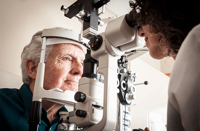
Diagnosing retinoschisis
How is retinoschisis diagnosed? Your eye doctor may notice signs of retinoschisis during a comprehensive eye examination. From there, an electroretinogram (ERG) test may be performed.
Your eye doctor may alternatively recommend optical coherence tomography (OCT) to help confirm a diagnosis.
Electroretinogram (ERG)
An electroretinogram (ERG) test may be performed to test the nerve tissue function of the retina.
During this process, special contact lenses that contain an electrode are placed on the eyes and worn as they are exposed to varying levels of light. The changes in light may cause your retinas to produce electrical activity, which is monitored and measured by the electrode in the lenses.
Levels of electrical activity in the retinas can determine their functionality and whether retinoschisis may be present.
ERG tests are often conducted on patients while they are awake without anesthesia. If your child is at risk for X-linked retinoschisis, vision researchers recommend having the test performed when they are two years old or younger, as it can be difficult for older toddlers and children.
Optical coherence tomography (OCT)
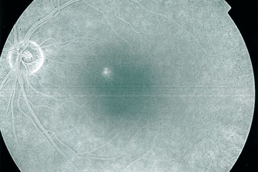
An imaging technique called optical coherence tomography (OCT), which uses light waves to capture 3-dimensional images of the retina, may be used to assess the structure and function of the retina.
Eye doctors may also use OCT to monitor the progression of retinoschisis in patients and help prevent further complications.
Retinoschisis treatment
Degenerative retinoschisis rarely requires treatment but should be monitored to ensure vision remains healthy.
Eyeglasses are often used to correct the vision impairment that comes with retinoschisis, although they do not cure the condition itself.
As previously mentioned, retinoschisis may cause additional problems for vision, including a detached retina or vitreous hemorrhage. These conditions may require immediate treatment to preserve vision. Retinal detachment can be treated with surgery, while vitreous hemorrhage is usually treated with cryotherapy or laser therapy.
Like other eye conditions, monitoring retinoschisis is an important component of treating it. See your eye doctor for annual exams, or as often as directed.
Retinoschisis vs. retinal detachment
Although they both affect the retina, retinal detachment is different from retinoschisis in a few ways:
- Retinoschisis: when the retina splits into two layers
- Retinal detachment: when the layers of the retina detach from the back of the eyeball
Retinal detachment is considered a medical emergency and should be treated promptly to keep vision healthy in both the short and long term. Retinoschisis, on the other hand, does not always require treatment and is typically not considered an emergency.
Both conditions are often rare, and vision should be diligently monitored under the care of an ophthalmologist after receiving a diagnosis and/or treatment. Retinoschisis may also lead to retinal detachment, making it especially important to monitor.
The importance of eye exams
Eye exams are an important first step in discovering conditions such as retinoschisis. Additionally, visiting your eye doctor on an annual basis can help detect any other vision problems and ensure the overall health of your eyes.
Retinoschisis doesn’t usually cause blindness, but both genetic and degenerative cases can lead to vision-threatening conditions, such as retinal detachment. This makes yearly eye exams more essential for adults, as well as children.
Contact your eye doctor if you have concerns before your next visit — your vision will thank you.
On this page:
Retinoschisis causes
Retinoschisis symptoms
How does retinoschisis affect vision?
Diagnosing retinoschisis
Retinoschisis treatment
Retinoschisis vs. retinal detachment
The importance of eye exams
On this page:
Retinoschisis causes
Retinoschisis symptoms
How does retinoschisis affect vision?
Diagnosing retinoschisis
Retinoschisis treatment
Retinoschisis vs. retinal detachment
The importance of eye exams