What is hyphema?
A hyphema is pooled blood inside the front part of the eye between the cornea and the iris. This space is called the anterior chamber. If the iris or nearby tissues are torn or damaged, they can bleed into this space. It is a painful medical emergency that can lead to vision loss if left untreated.
The anterior chamber is normally filled with a clear liquid called aqueous humor. It regulates eye pressure by steadily flowing into and out of the space. But pooled blood in the anterior chamber can block the flow, causing eye pressure to climb. This can lead to glaucoma and other serious complications.
A hyphema is not the same as a subconjunctival hemorrhage, which is blood between the thin, clear layer called the conjunctiva and the white part of your eye (the sclera). It’s usually due to something as simple as a sneeze or a cough. It can appear bright red and scary, but it is common, harmless and typically painless.
A hyphema is much more serious and requires immediate attention by your eye doctor.
Signs and symptoms of hyphema
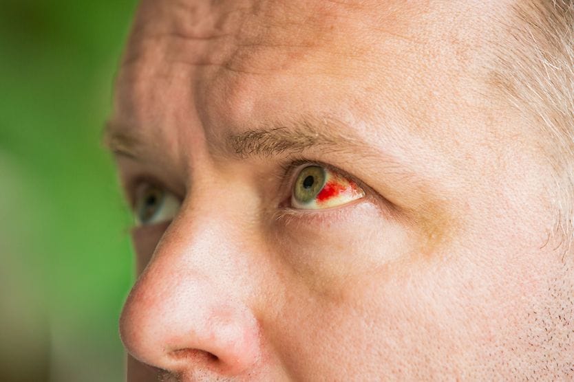
In some cases, there is enough blood in front of the iris that it’s visible to the naked eye.

It may be a small pool at the bottom of the iris, or it may even block part or all of the pupil. Visible hyphemas often appear to have layers. This is because the older blood at the bottom has a darker color.
In other cases, the red blood cells may only be visible to an eye doctor using special tools. This is called a microhyphema.
In addition to the bleeding, the following symptoms are usually associated with hyphema:
- Blurry or distorted vision (even if you can’t see the hyphema yourself)
- Eye pain
- Light sensitivity (photophobia)
- Headache
Eye pain, sensitivity to light and headache are especially likely if a hyphema is causing high eye pressure. The medical term for eye pressure is intraocular pressure (IOP). Extremely high IOP can also cause nausea and vomiting.
What causes hyphema?
The majority of hyphemas are due to blunt eye injury, but other types of eye injury can cause them, too. A hyphema can also be a complication of other health conditions, medication or eye surgery.
Eye injuries
The most common cause of hyphema is trauma to the eye. This is one of the reasons it is important to see your eye doctor right away if you have an injury that causes a black eye.
Blunt eye injuries cause the majority of hyphemas, and around 70% of these injuries occur in children’s sports. In older children and adults, the blunt eye injury is usually assault-related. Car accidents are also fairly common causes of blunt eye injuries.
Other types of eye injury that can cause hyphema include:
- Penetrating injury (something punctures the eye)
- Lacerating injury (something cuts or ruptures the eye)
Eye and health conditions
Certain eye and health conditions can be risk factors for hyphemas. They don’t cause them the way injuries do, but they can make it easier for a hyphema to develop spontaneously. Some of these conditions include:
- Blood clotting disorders, such as hemophilia
- Diabetic retinopathy
- Sickle cell disease
- Eye tumors, such as ocular melanoma
- Weakened or abnormal blood vessels
- Inflammation of the cornea and/or iris (anterior uveitis) from infection (such as ocular herpes)
Spontaneous hyphemas can also be possible in people who take anticoagulant medications (blood thinners).
Other causes
A hyphema — such as uveitis glaucoma hyphema syndrome (UGH syndrome) — can also occur during or after intraocular surgery. Certain procedures may carry a slightly higher risk of this complication, including:
- Laser peripheral iridotomy
- Other surgeries to treat conditions of the iris or ciliary body
- Cataract surgery (rarely; UGH syndrome can occur afterward if the implant rubs and chafes the iris)
How is hyphema diagnosed?
A hyphema diagnosis is fairly straightforward. If an eye doctor can see blood in the anterior chamber with their naked eye or with a slit-lamp exam, that is a hyphema. However, a microhyphema may be harder to identify and may be misdiagnosed as traumatic iritis.
However, that’s only a small part of the process. They need to perform a thorough eye exam to determine other very important factors, such as:
Whether your vision has been affected
They can usually check this with a standard visual acuity test.
Which tissue(s) in the eye are bleeding and damaged
The eye doctor will use eye drops to dilate the pupil. Then, they’ll use a slit-lamp to shine a bright light into the eye and look for any areas of damage.
Sometimes, a hyphema is deep enough to block part of the view through the pupil. In these cases, the eye doctor may also need to do an ultrasound to see everything.
If the hyphema was caused by an eye injury, they’ll also make sure to check the surface of the eye and eyelids for possible damage.
They may also need to use a gonioscope lens to look at the drainage angle around the edge of your iris. Depending on the severity of the hyphema and eye injury, they may wait a few days to do a gonioscopy.
Whether your eye pressure is in the normal range
Eye doctors measure eye pressure with a test called tonometry. One well-known method is the air puff test. Other methods involve a small device that touches the cornea very quickly to gauge its resistance.
Blood tests
In many cases, the eye doctor will need to check your blood for signs of any bleeding or clotting disorders. They may also need to check for sickle cell disease or sickle cell trait, as well as levels of blood thinner medications in your system.
The severity or “grade” of the hyphema
Eye doctors use a grading system to help determine your risk for complications related to the hyphema.
The severity is graded by how much blood accumulates in the eye:
- Grade 0 (microhyphema): No visible pooling of blood, but red blood cells can be seen in the anterior chamber with a slit-lamp.
- Grade 1: Pooling of blood in less than the lower third of the anterior chamber.
- Grade 2: Blood filling one third to one half of the anterior chamber.
- Grade 3: Blood filling one half to less than all of the anterior chamber.
- Grade 4: Total filling of the anterior chamber with blood. If the blood is bright red, this is called a total hyphema. If it is dark red-black blood, it’s sometimes called an “8-ball hyphema.”
In general, the higher the grade of hyphema, the greater the risk of vision loss and long-term damage to the eye.
The dark red or black color of an 8-ball hyphema (the most dangerous type) is associated with decreased circulation of aqueous humor and decreased oxygen in the anterior chamber of the eye. The iron in the blood is also toxic to the ocular tissues, especially the cornea and the trabecular meshwork.
Hyphema treatment
Treatment depends on the severity of the hyphema and the associated risk factors. In any case, treatment is intended to:
- Avoid a rebleed.
- Avoid glaucoma.
- Treat inflammation.
- Restore vision.
- Identify associated pathologies and disease conditions.
- Prevent corneal blood staining.
A low-grade, low-risk hyphema usually only needs at-home treatment and will heal on its own. More severe cases or those at risk of complications may need further treatment or surgery.
Medicine and other non-surgical options
At-home care usually includes a combination of the following precautions and treatments:
- Limited physical activity
- Head elevation (including when sleeping)
- Wearing an eye shield
- Frequent follow-up visits for a few weeks or months
- Pain medicine
- Anti-inflammatory medicine (topical or oral)
- Eye drops to help lower eye pressure
- Other medications
Never use over-the-counter pain medications that contain aspirin or nonsteroidal anti-inflammatory drugs (NSAIDs) if you have a hyphema. These medications can increase the risk of rebleeding in the eye.
Surgery for hyphema
Only about 5% of cases require surgery. These may include:
- Severe hyphemas
- Cases with uncontrolled high eye pressure
- Cases that don’t resolve with at-home care and medication
- Cases with continuing active bleeding
There are a few different types of hyphema surgery. The eye doctor may remove the blood via an incision in the cornea, or they may need to use a method similar to a vitrectomy or a trabeculectomy. The type of surgery will depend on the severity of the hyphema and the patient’s other risk factors.
Recovery and outlook
With proper treatment, your body will usually reabsorb the hyphema without permanent damage to the eye. Depending on the severity of the hyphema, it may take a few weeks to several months to reabsorb fully.
Possible complications
In some cases, clotting of the blood will clog or damage the drainage angle around the edge of the iris. This can cause increased eye pressure and lead to glaucoma and permanent vision loss.
Also, in some cases, rebleeding can occur inside the eye after the initial eye injury that caused the hyphema. Rebleeding usually occurs within a few days after the injury and can be more dangerous than the initial bleeding.
Rarely, severe or total hyphemas can cause corneal blood staining. If the blood remains in the anterior chamber too long, it can stain the back surface of the cornea. This can cause a long-term reddish tint to vision.
People with sickle cell disease and sickle cell trait have an increased risk of eye damage from a hyphema. Sickle cell is a hereditary disease in which red blood cells are crescent-shaped. This shape makes them more likely to cause clogging and high eye pressure.
How to prevent hyphema
The best way to avoid a hyphema is to wear safety glasses or other eye protection during any potentially hazardous activities.
Protective sports glasses should be worn when playing baseball, softball, racquetball, basketball, hockey or other sports that pose a risk of trauma to the eyes.
Also, be aware that sports, like boxing, significantly increase your risk of a traumatic hyphema. And if you participate in paintball games, wear protective headgear that includes a clear, impact-resistant shield that fully protects your face and eyes.
When to get help
Even if your eye feels fine and you don’t notice vision problems, see an eye doctor immediately if you have any eye trauma or see blood behind your cornea. Make sure to attend all follow-up visits your eye doctor or ophthalmologist recommends.
Also, routine eye exams are very important after having a hyphema. Your risk of elevated eye pressure and glaucoma may be higher, even years later.
")