How AI technology may help doctors detect and treat glaucoma sooner

Glaucoma is one of the leading causes of irreversible blindness globally – but it doesn’t have to be. Artificial intelligence (AI) is showing promise as a tool for earlier glaucoma detection and better long-term tracking — advances that could help fewer people lose their sight. It could also advance personalized treatments.
Glaucoma primarily affects adults and disproportionately impacts people from minority groups. An estimated 4.2 million Americans have glaucoma, with half unaware that they have it.
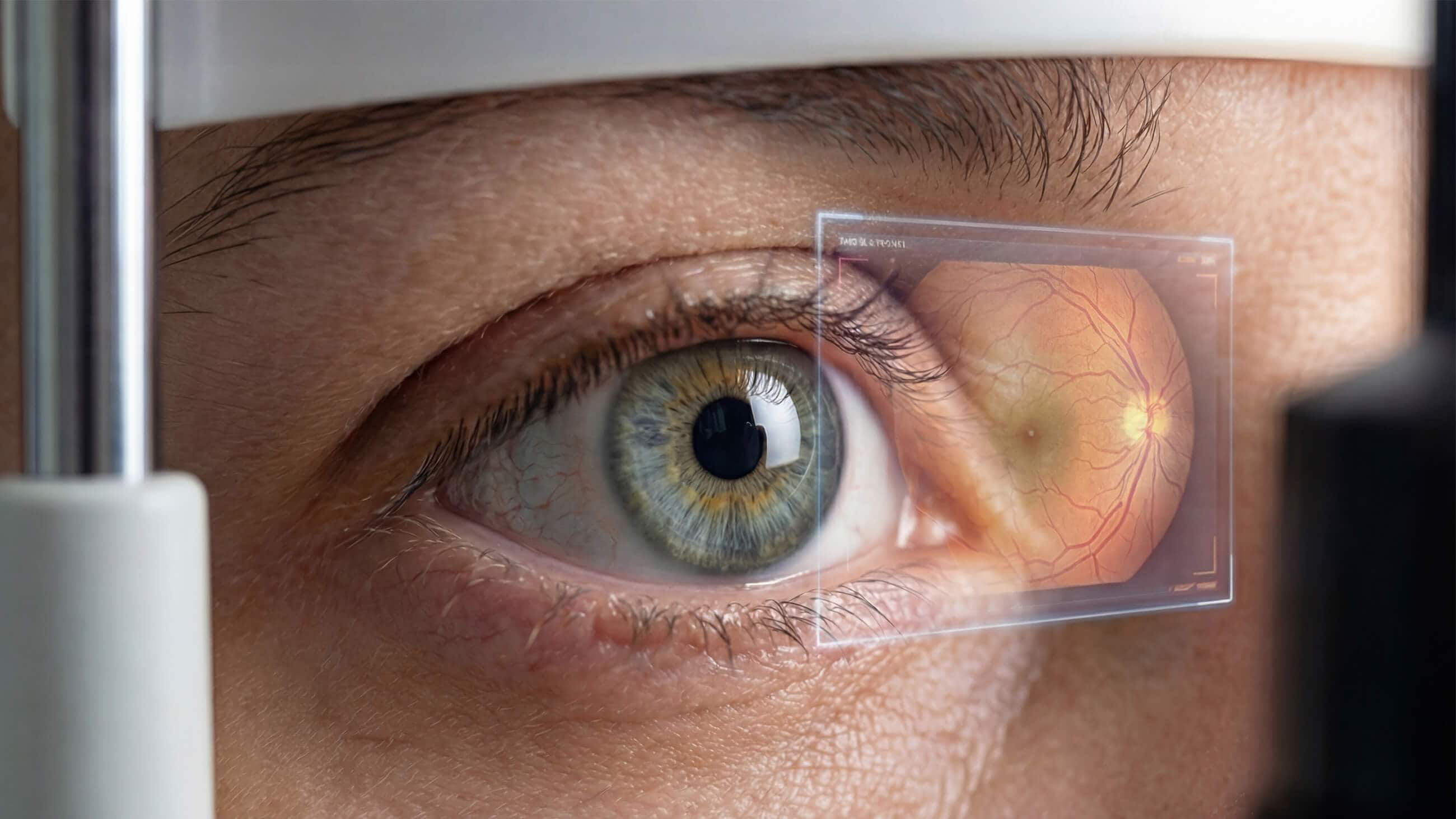
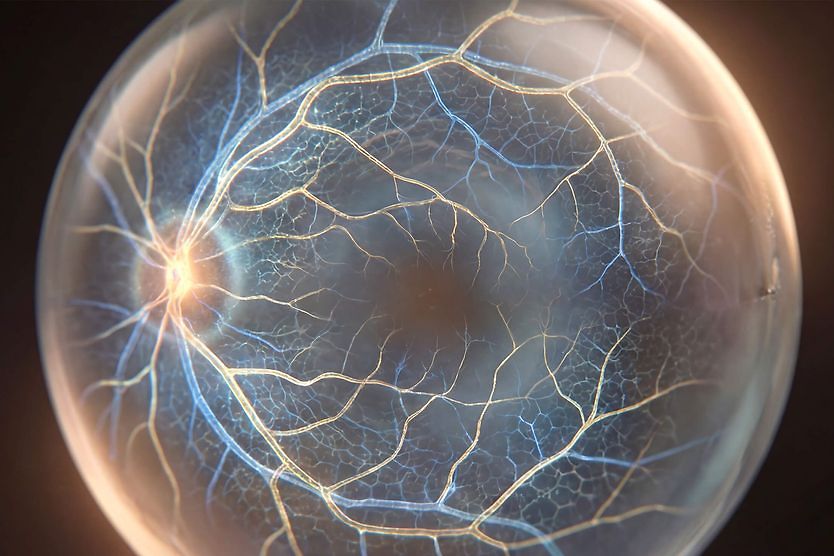
The disease damages the optic nerve, which transmits visual information from the eye to the brain. Because that damage cannot be reversed, early detection is vital to possibly prevent vision loss.
The research is promising, yet building AI systems to help doctors detect and treat glaucoma requires the right types of data from across siloed systems.
Challenges in detecting and treating glaucoma
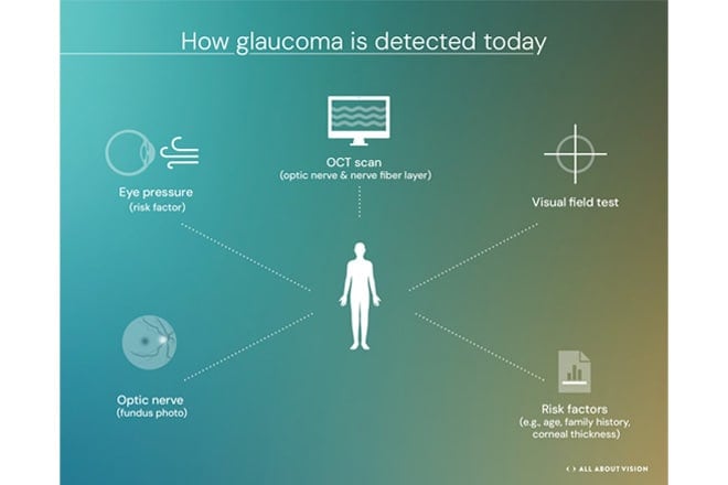
Eye care professionals consider eye pressure, signs of optic nerve damage, changes in peripheral vision, and other factors when determining if glaucoma is present and how quickly it’s progressing.
The two main types of glaucoma are open-angle glaucoma and angle-closure glaucoma. Open-angle glaucoma, the most common type worldwide, occurs when the drainage system gets clogged, preventing fluid from leaving the eye. Over time it can lead to increased intraocular pressure (IOP). Angle-closure glaucoma occurs when the iris blocks fluid from draining from the front of the eye. A form called acute angle-closure glaucoma is a medical emergency.
Because glaucoma often has no noticeable symptoms in its initial stages, early diagnosis is critical. Otherwise, a patient’s first warning may be irreversible blind spots in their peripheral vision.
The AI opportunity in screening and diagnosis
Daniel Liebman, MD, MBA, ophthalmologist at Massachusetts Eye and Ear and instructor in ophthalmology at Harvard Medical School, noted that AI innovation can help simplify the complexity of glaucoma diagnosis.
“AI helps us by synthesizing large amounts of data, finding patterns, and predicting in ways that can be difficult for us to do on our own. Glaucoma is a field full of large amounts of varied, and sometimes messy, data,” he said.
That “messy” data reflects the many ways clinicians assess whether a patient has glaucoma. Eye pressure is one key indicator, often rising when fluid doesn’t drain properly. Doctors also look for signs of optic nerve damage using optical coherence tomography (OCT) scans, which measure the thickness of retinal layers, tissues that may thin as glaucoma progresses.
Optometrists are typically the first to catch these signs, manage patients with therapeutic treatments, and refer them for further evaluation to comprehensive ophthalmologists or glaucoma specialists, when necessary.
This is why Michael Chaglasian, OD, FAAO, an associate professor of optometry and chief of innovative technology and data science at the Illinois College of Optometry, believes that AI tools could provide a clinical decision support function to optometrists by sorting through the mountains of available patient data. This would help optometrists make more informed decisions when diagnosing and treating patients.
“The need in optometry and ophthalmology is significant,” Dr. Chaglasian said. “We’re taking care of more patients, older patients, more complicated patients. And now we have access to overwhelming amounts of patient data [in the electronic health records]. Doctors need a clinical decision support system to read and review all that data and point them in the right direction; not to tell them what to do, but to suggest where they should look to make the right diagnosis.”
A single glaucoma visit can generate multiple streams of information that form part of a patient’s assessment, according to Dr. Chaglasian. Many factors are taken into account when determining the risk for glaucoma and glaucoma progression, sometimes making decision-making complex. Some of those variables include:
- Intraocular pressure (IOP)
- Visual field (including the type of testing done)
- Corneal thickness
- OCT/OCTA (OCT angiography) scans
- Optic nerve rim contour (using color retinal/fundus photographs and/or OCT measurements)
- Drainage angle (helps to determine open vs. closed angle glaucoma)
- Older age (especially over 60)
- Family history
- Being of African, Hispanic or Asian descent
- Having a history of diabetes, high blood pressure, severe nearsightedness (myopia), eye injury or long-term steroid use
Sophia Wang, MD, an assistant professor of ophthalmology at Stanford Medicine, studies AI in glaucoma care. She notes its potential use with basic screening tools that flag suspicious cases to models that pull multiple sources together to estimate a patient’s risk of progression.
“There’s screening diagnosis, where AI could read a fundus photograph or flag an OCT to say, ‘This person looks suspicious for glaucoma and therefore should be followed up with other testing,’” Dr. Wang said.
“Then there’s even greater potential: Could AI look at all the data and produce a risk score? Is this person a glaucoma suspect and non-progressive, or low risk? Or is this person at high risk, and we need to monitor them very closely?”
How AI could improve personalized patient care and monitoring
Doctors are optimistic that AI could provide more targeted and personalized care. Deciding when and how to use interventional treatments like surgery or less invasive measures is part of glaucoma care’s nuance. Predicting which patients are likely to worsen may help clinicians focus their efforts, according to Dr. Liebman.
“What we’re always debating in glaucoma care are questions like, who is stable? Who is not? Who is at risk and who we should be more proactive with?” Dr. Liebman said. “The hardest part is balancing the risk and benefit of intervention. If we could predict earlier who is likely to worsen on current treatment, we could determine who should be watched more frequently, who requires earlier intervention, and how to optimize resources and interventions.”
In health care, AI has taken on administrative and routine tasks for doctors, allowing them more time to focus on their patients. It’s no different for physicians specializing in glaucoma care. Rather than replace the doctor-patient relationship, AI aims to reduce the burden on doctors so they can do what AI can’t: provide a human touch. AI gives doctors more space to discuss diagnosis, treatment, medication, risks and benefits with their patients.
With more personalized care, patients could get the right intervention sooner — and that could be life-changing for many.
“The future of glaucoma care is going from a ‘one-size-fits-many’ approach to the optimal treatment for a given patient because we now have many different medicines and surgical options,” Dr. Liebman said. “That’s the big-picture promise. But there’s still a lot of work to get from here to there. AI isn’t magic. It’s math. There are limitations. ‘Data in, data out’ matters, and the quality of the data matters.”
Limits and challenges
Dr. Chaglasian noted that AI systems have already reached the market for another disease, diabetic retinopathy, in part because it’s diagnosed with standard fundus photographs of the back of the eye. Known as “single modality,” this information enables a more straightforward AI system focused on one element of diagnosis.
Glaucoma, by comparison, is typically assessed using multiple types of tests, often over many visits, which makes it harder to create the large, clean training sets that helped diabetic retinopathy tools get to market faster.
“These datasets are not widely available,” Dr. Chaglasian said. “With diabetic retinopathy, there were hundreds of thousands, millions, of retinal photographs, because that was a long-time standard of care, and it was easy to assemble datasets to train algorithms.”
Dr. Wang noted that one of the biggest obstacles is assembling the right pieces in a way that works across different health systems.
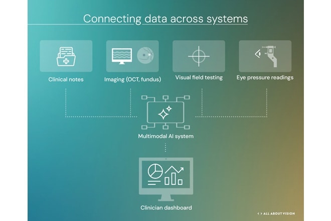
“If we develop AI models that can access multimodal information about a patient, there’s potential for AI to find patterns and support diagnosis. But there are challenges. In clinical systems, information is siloed. Electronic health record (EHR) data is in one system, photography in another. For AI to work across all of that, it’s quite challenging,” Dr. Wang said.
A glimpse into this future was offered by researchers at the University College London Institute of Ophthalmology and Moorfields Eye Hospital in the UK, who developed an algorithm that identified glaucoma 88% to 90% of the time using fundus images. They outperformed human graders who were correct 79% to 81% of the time.
Dr. Wang said strong early results can create pressure to deploy tools before they have been tested across different clinics and patient populations.
“Will [the data] work in my patient population and in my clinic? That comes down to representation in training data and careful evaluation of performance across demographic subgroups and disease subgroups. It also demands transparency from people releasing data models. It’s something the field needs to address,” she said.
Dr. Liebman said one of the trickiest issues is that glaucoma datasets don’t capture a disease’s natural progression because patients are usually undergoing treatment.
“One major hurdle is that much of the data used to train algorithms is retrospective. In real care, when glaucoma worsens, we intervene. We start medicines, do procedures and perform surgeries. So when you look back and try to predict who worsened, it’s not a purely natural progression because something was often done,” he said.
What AI may mean for patients today
Experts caution that it may be several years before AI is a feature of inpatient glaucoma exams.
Specialists anticipate both new innovations and disappointments as the field continues to develop.
“The hype cycle can lead to peaks and troughs, and that’s true with AI,” Dr. Liebman said. “We should recognize the real potential, while keeping a realistic understanding of current limitations,” he said.
Even with promising research, maintaining glaucoma follow-up visits and annual comprehensive eye exams remain essential.
“Optometry is in a good position to be a leader in safeguarding the vision of our aging population,” Dr. Chaglasian said. “Optometrists will use data analytics from a variety of sources. Not tomorrow, not next year — but within five years, who knows?”

