Custom-built vision: How 3D printing is personalizing eye care
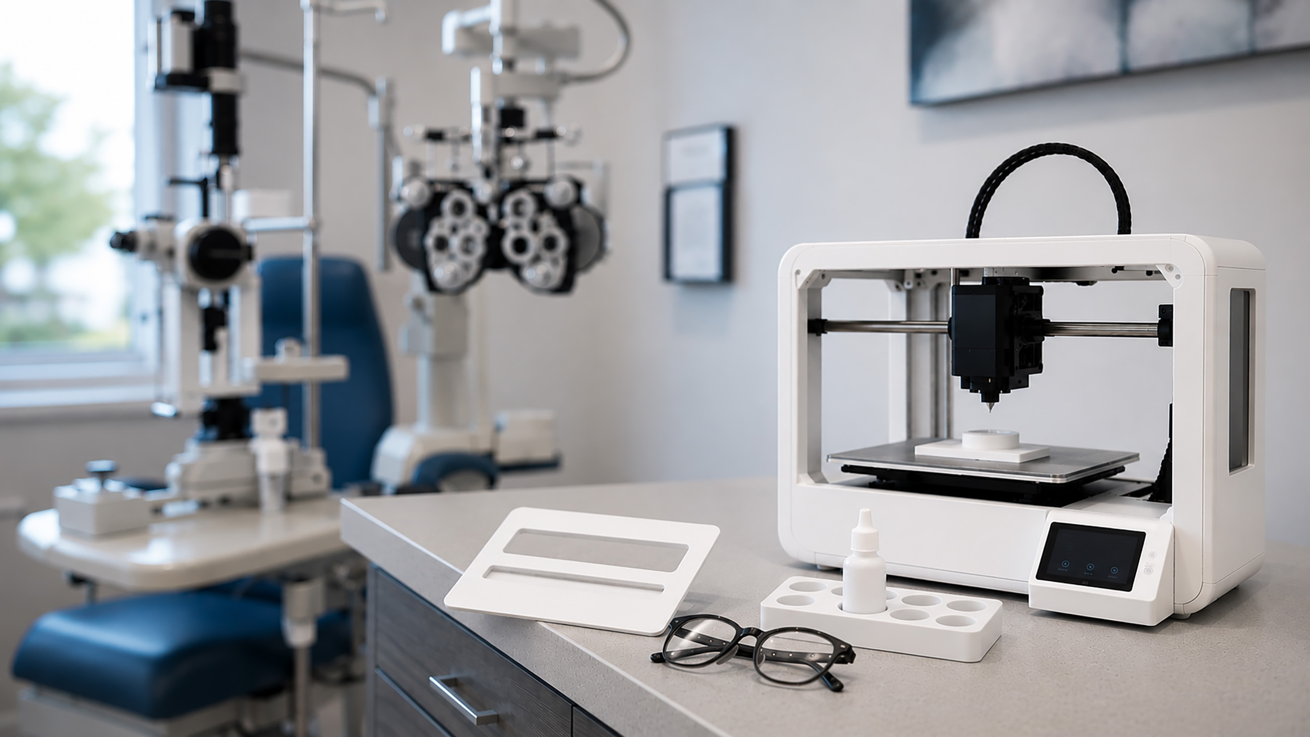

Gregory Hopkins, OD, MS, a clinical professor of practice and low vision rehabilitation specialist at the Ohio State University College of Optometry, is determined to personalize his patients’ care. The solution they need sometimes comes from his own 3D-printed solution and requires a little bit of creative thinking.
Dr. Hopkins has built a set of bespoke tools that patients appreciate for solving a specific problem. To help low vision patients find the elusive signature line on a form they need to sign, he designed and printed a credit-card-sized guide. The patient or someone helping them can mark where to sign the form. For added customization, it even doubles as a letter opener and is engraved with the eye clinic’s phone number.
As he learned more about 3D printing, Dr. Hopkins found his own creations had several advantages. They were often cheaper than retail products. They could be customized just the way he wanted them. Most importantly, they could be tailored to a patient’s exact needs.
In addition to his work at the university, Dr. Hopkins sees patients at the Department of Veterans Affairs. He wanted to accommodate those who arrived with a cane, military ball cap and multiple pairs of glasses but didn’t have an ideal place to put them during the eye exam. Often, a patient would set one or more items down by the sink, triggering the sink’s automatic sensor.
Working with biomedical engineers at the clinic, he created a small caddy that clips to the pole holding his exam instruments. No more running water.
As he has experimented further in creating practical and clinical tools, Dr. Hopkins regularly sees patient challenges as problems that can be solved with a 3D-printed tool.
“Low vision optometry involves a lot of problem solving,” he said. “Sometimes, there will be things where you kind of need to hack together, combining different optics, things that don’t exist.”
As his inventions have grown more complex to meet an array of challenges, many of his patients rely on the signature guide.
“It is the thing that impresses my patients almost the most,” he said. “I have all this expertise, all these devices and their favorite take-home is that little 3D-printed line guide.”
3D printing is used in multiple eye care settings. There are fixes like those in Dr. Hopkins’s practice, as well as advanced creations like prosthetic eyes. Some universities are testing 3D printing of anatomical models to help with surgical preparation.
The most experimental work is bioprinting, where researchers are developing cell-compatible materials, known as bioinks, that can form tissue-like material as a possible substitute for human-donor tissue. At each level, eye doctors and surgeons hope to design and implement treatment more tailored to each person’s needs.
Behind 3D printing techniques
3D printing is the common term for additive manufacturing. The process involves building three-dimensional objects one layer at a time from a digital file. Creating a 3D object requires designing it in CAD (computer-aided design) software and then, just like a document, assigning the printer to create it.
This design, however, doesn’t need to be built from scratch. There are many online communities where people share their designs with others, who can use them on their own printer. Designs for everything, from a hanger to a wall mount or smartphone stand, can be downloaded and then printed.
A 3D printer, like the one in Dr. Hopkins’s office, can handle a range of everyday tasks. More complex use cases involving prosthetics or surgical training require more sophisticated devices, materials and higher costs for a more complex printer.
The appeal to 3D printing is that it can make something personalized and specific. Dr. Hopkins solves many different practical needs: a magnifier that needs a better handle or a pair of telescope glasses that would hold better with a custom clip. A patient may find a 3D-printed stand makes it easier to use a tablet to magnify papers.
Building ocular prosthesis eyes with 3D printing
3D printing is transforming the prosthetics field. When someone loses an eye to injury or disease, they are often fitted with a prosthetic eye. While it doesn’t restore sight, it can be a critical treatment to help that person regain a sense of self and confidence as they recover and adapt.
Not everyone who needs a prosthetic eye has lost one to injury or illness, however. Some people are born with a missing or severely underdeveloped eye — congenital conditions such as anophthalmia or microphthalmia. Others can lose an eye later in life to trauma, cancer, or severe infection.
In 2021, Moorfields Eye Hospital in London fitted a patient with what was described as the world’s first fully 3D-printed prosthetic eye. It was built from a digital scan of the patient’s socket and a calibrated photo of their other eye to achieve the closest possible match.
Researchers at the same hospital later reported designing and printing custom prosthetics for 10 patients between 2022 and 2023, using the same approach.
The traditional method for a prosthetic eye requires a specialist, known as an ocularist, to hand-paint the prosthetic over a clinic visit, followed by hours of curing. The process can take as long as six weeks.
In contrast, 3D printing takes within two and a half hours, from scan to print. Once sent to the ocularist for finishing, the whole process takes half the time — two to three weeks. Though it remains a nascent technique that’s still being developed, 3D printing enables providers to speed up and scale prosthetic eye delivery to patients.
Looking forward: New surgical methods through bioprinting
Bioprinting is an advanced form of 3D printing that uses bioinks engineered to replicate human tissue. Many of the techniques that use this technology are in early stages. The research teams investigating bioprinting are tackling challenges that have previously lacked good surgical or treatment solutions.
David Myung, MD, PhD, is an associate professor of ophthalmology at the Byers Eye Institute at Stanford University and, by courtesy, of chemical engineering. He specializes in cataract and corneal surgery, external diseases of the eye and researches bioprinted solutions for difficult eye injuries.
A primary area of focus is corneal injuries. Dr. Myung said the area needs more attention because existing treatment options are limited. Currently, when someone has significant corneal scarring or damage, a corneal transplant is often the main technique to restore some level of vision — though the optimal approach depends on how much of the cornea is affected.
Superficial scarring may respond to laser surface treatment or specialty contact lenses, while eyes that repeatedly reject donor tissue may be candidates for an artificial cornea, called a keratoprosthesis.
Yet receiving a donor cornea can be difficult. It requires someone having agreed to donate their cornea after they die. The cornea must then be harvested, properly stored and sent for surgery within a strict time window.
Unlike other tissues in the body, the cornea has no blood vessels, making it an attractive target for 3D printing. Reconnecting a part of the body to the blood system is highly complex and, therefore, difficult to bioprint solutions for other organs.
Bioprinting may also help with another corneal transplant challenge. Post-transplant, many patients often struggle with vision challenges.
“Vision after a corneal transplant often needs further correcting, in particular due to astigmatism, and as a result, patients often require contact lenses.” Dr. Myung said. “With bioprinting, there’s the potential to be a lot more tailored and to give the patients a cornea that suits their refractive needs more precisely than we currently can do with cadaveric tissue. And it has the potential to enable the production of engineered donor corneas on a large scale to address the worldwide need.”
His team is working on a pair of techniques. One is a targeted repair for corneal blindness caused by a scar that doesn’t cover the entire cornea. Dr. Myung calls it a “spackle approach,” like spackling drywall. In this technique, the surgeon scrapes away the diseased tissue and backfills the defect with a bioprinted gel-like material that behaves like corneal tissue.
Another technique is what he calls a “fully baked” approach. His vision is an artificial cornea, bioprinted, then implanted and sutured or glued into place.
Dr. Myung said the challenge is designing a substance that can harden into shape without damaging the cells. His lab has created a bioink designed to keep cells healthy while the material sets.
“There's a lot to be excited about, and the technology is catching up and enabling things that previously weren’t really possible,” Dr. Myung said. “We’re still some ways away, but we have some amazing preclinical data showing that the materials we are developing mimic donor corneal tissue and can be printed on demand in the lab.”
For now, bioprinted corneas remain experimental. The most encouraging results so far come from laboratory and animal studies, and no bioprinted cornea is yet to be FDA-approved as a standard treatment or available to patients outside of research settings.
The role of 3D printing in surgical planning and training
Surgery’s delicate work often involves small, hard-to-reach structures. This can be especially challenging within eye care.
A 2023 study at Wills Eye Hospital tested a new approach using 3D printing to help doctors-in-training, noting that the bony anatomy around the eye is “complex and challenging for trainees to fully conceptualize.”
Twenty trainees reviewed a CT scan of an orbital fracture, which is a break in the bony socket that holds the eye, then reviewed it again alongside a 3D-printed model of the same fracture. Holding the physical model significantly improved their confidence in conceptualizing the fracture and planning the repair for most of the models tested. .
In a follow-up survey, about 95% rated the models useful for training. The models were most helpful in helping trainees grasp the anatomy, noted the study authors.
Researchers continue to study the value of 3D printed anatomy for surgical training. In a 2023 study at Wills Eye Hospital, the authors noted that the models are “not a stand-alone resource for trainees but a supplemental tool for enhancement.” In a different use case, researchers found that 3D-printed surgical guides can improve the accuracy of orbital reconstruction, but these efforts are early.
What to expect from the future
With its range of uses, 3D printing is on pace to grow in popularity. Bioprinting would create new solutions to challenging diagnoses. Everyday items, like those printed by Dr. Hopkins, could give a personalized touch to each eye care patient.
Dr. Hopkins sees the immediate benefits in his everyday clinical encounters. He recalled that 3D printing is now part of his mindset for helping patients. His clinical work extends beyond just diagnosing and treating conditions. He seeks to solve daily challenges and, when patients have challenges with functioning, finds original solutions.
“I would have someone say, ‘I can’t hold this magnifier. I love it, but it doesn’t come with a handle.’ As soon as the words leave my mouth that they don’t make a handle for that, I’m thinking to myself, ‘But I can,’” said Dr. Hopkins.