Ophthalmoscopy: What is it?
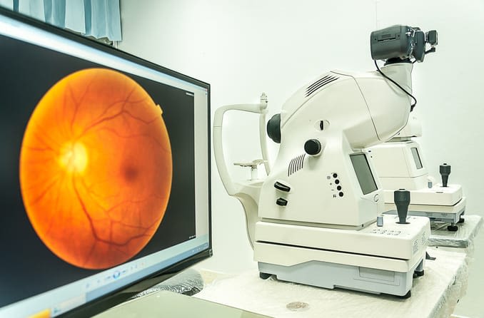

Ophthalmoscopy (also called fundoscopy or a fundoscopic exam) is a common procedure performed by an eye doctor. Pediatricians and general practitioners may also include ophthalmoscopy in routine physical exams.
The exam involves the use of special lenses and bright, direct light to provide a detailed view of the back of the eye — specifically, the retina, optic nerve and blood vessels.
What can ophthalmoscopy detect?
Fundoscopic exam findings can reveal or rule out many different diseases and conditions that manifest in the back of the eye, some of which may be more serious than others.
If ophthalmoscopy appears normal, your eye doctor will see a normally functioning retina and blood vessels that show no signs of concern.
If your doctor sees something abnormal, it may be a sign or result of one of the following eye conditions:
- Diabetic retinopathy
- Retinal inflammation due to a viral infection
- Retinal detachment
- Glaucoma
- Hypertension (high blood pressure)
- Macular degeneration
- Melanoma of the eye (a rare form of cancer)
- Optic nerve disease
SEE RELATED: Eye exams: 5 reasons why they are important
Types of ophthalmoscopy
There are three types of ophthalmoscopy or fundoscopic exam:
- Direct ophthalmoscopy, a common procedure, involves the use of a handheld tool called an ophthalmoscope to look inside the eye. This tool has special lenses and a bright light that enable your eye doctor to focus past your pupil and onto the back of your eye.
- Indirect ophthalmoscopy involves a brighter light that your doctor wears as a headlamp and a smaller handheld lens. In the case of a detached retina diagnosis, this test can be a critical first step. Your pupils will need to be dilated for this procedure.
- During slit lamp ophthalmoscopy, you typically place your chin on a chin rest while your eye doctor uses a table-mounted binocular microscope with a bright, narrow beam of light to examine your eyes. When performing a fundoscopic exam with the slit lamp, your doctor may hold a small lens close to your eye and direct the beam of light through the lens to illuminate the back of your eye.
Why is it called a fundoscopic exam?
Fundus is the medical term that eye doctors use to describe the inner portion of the back of the eye.
A fundoscopic exam (or fundoscopy) is the same thing as ophthalmoscopy; it’s simply a different name for the same procedure.
SEE RELATED: 8 things your eyes can reveal about your health
Does ophthalmoscopy hurt?
While there may be slight discomfort from the brightness of the ophthalmoscope’s light, these tests do not typically cause any pain. Indirect ophthalmoscopy may involve a slight pressure on the surface of your eye, but it generally is not painful.
If dilation is required, the accompanying temporary vision impairment may lead to slightly uncomfortable light sensitivity during that time. In the event that your eye doctor elects to dilate your eyes, you may be given disposable sunglasses before you leave the office to reduce any discomfort when you get outside.
Will my eyes need to be dilated?
Sometimes, but not always. Your optometrist or ophthalmologist can determine if pupil dilation is needed. Dilation is generally more common with indirect or slit lamp ophthalmoscopy.
Dilation can temporarily blur your vision, so it’s recommended to avoid driving until the effect has worn off. Since this effect can last several hours, it’s recommended to arrange for transportation from your doctor’s office.
READ MORE: What to avoid after eye dilation