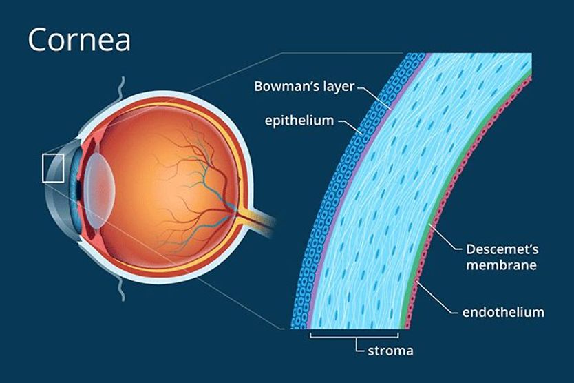
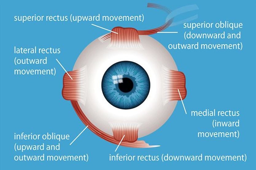
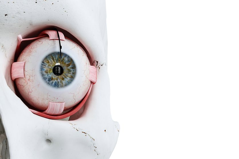
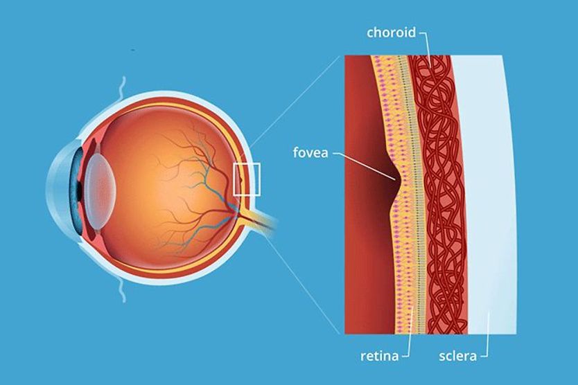
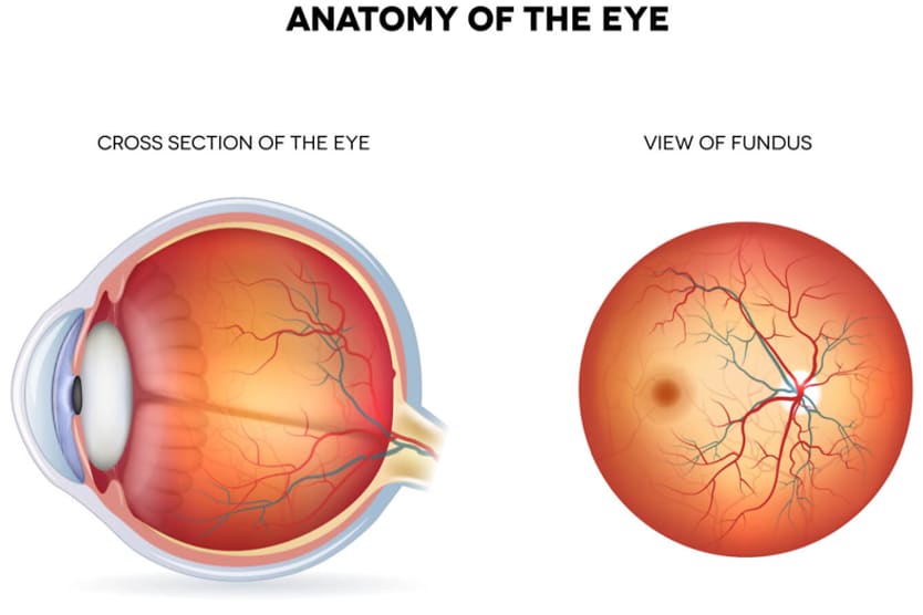
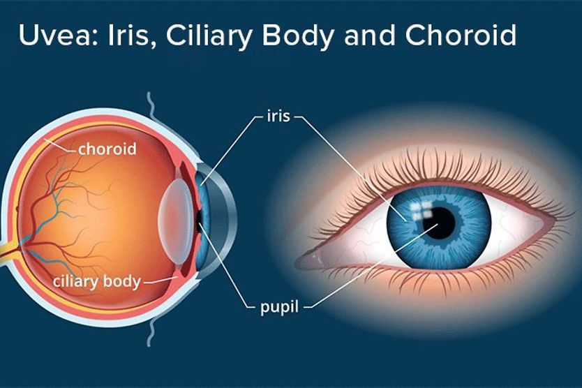
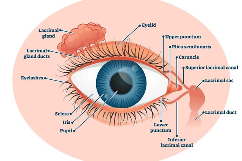
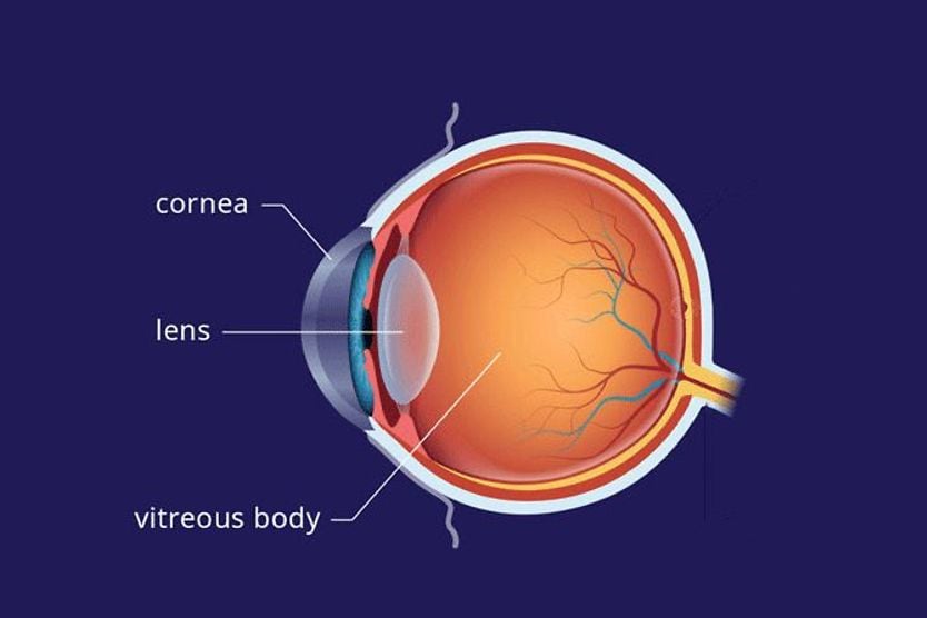
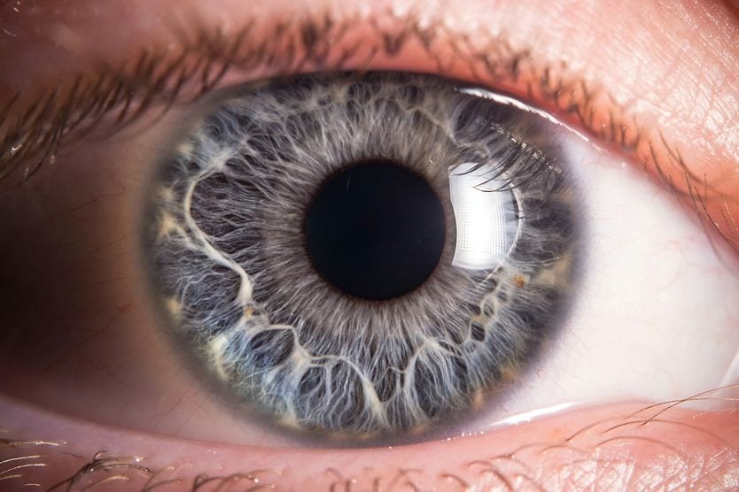
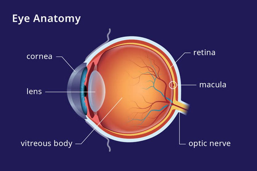
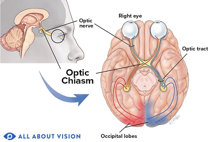
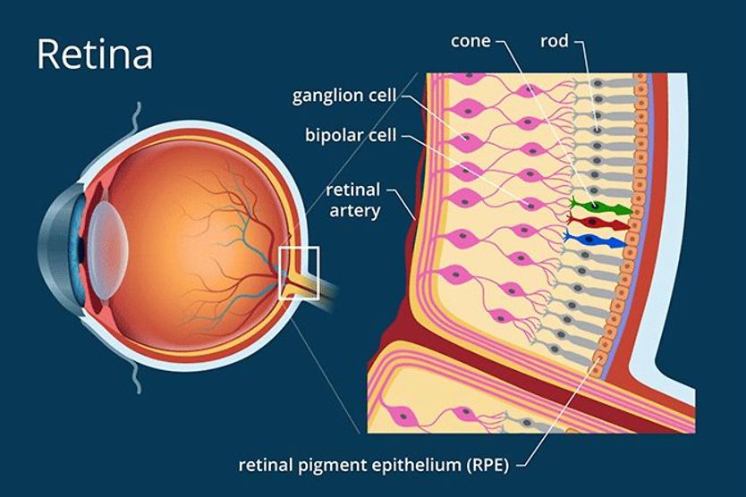
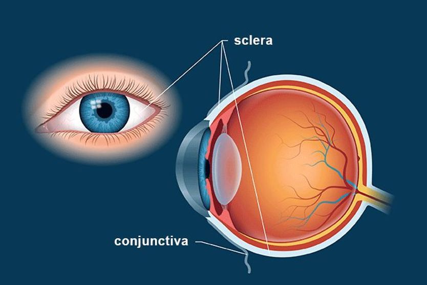
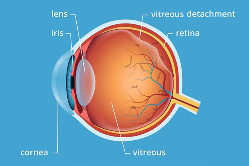
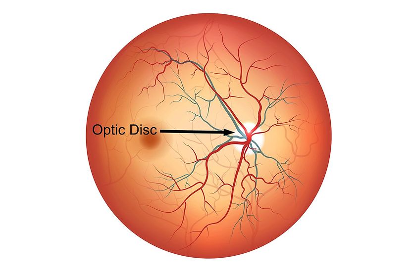
Home Conditions | Intraocular pressure

Intraocular pressure

woman getting an eye exam checking her intraocular pressure

What is intraocular pressure (IOP)?

Intraocular pressure (IOP) is the measurement of fluid pressure within the eyeball. Having normal eye pressure helps maintain the eye’s shape and supports the eye’s anatomy so it can function properly. If your IOP stays too high or too low, it can affect your eyes and put your vision at risk. 

IOP is measured in millimeters of Mercury (mmHg) and is considered normal in the 12-21 mmHg range. 

There are two different types of fluid that fill the eyeballs: aqueous humor (located in the front of the eye) and vitreous humor (located in the back of the eye). While both fluids have important roles within the eye, aqueous humor is responsible for intraocular pressure. 

Aqueous humor is a clear, watery fluid that fills the front part of the eye, between the cornea and the lens. Most of the aqueous humor is made up of water, with a small percentage consisting of proteins, sugars and other nutrients that the eye needs for optimal function.

The purpose of aqueous humor is to cycle through the front part of the eye, between the cornea and the front of the vitreous. During this cycle, aqueous humor provides nutrients to the lens and the cornea, since those structures don’t receive any kind of direct blood supply. 

SEE RELATED: Glaucoma and eye pressure Q&A

How IOP works

Healthy IOP is achieved when the level of aqueous humor production and drainage stay balanced. To fully grasp the balance, it’s important to understand the journey aqueous humor takes when completing its function. 

The eye is split into three parts, called chambers

  • The anterior chamber is between the cornea and the iris

  • The posterior chamber is between the iris and the lens. 

  • The vitreous chamber is between the lens and the retina

Aqueous humor is produced by a structure in the ciliary body called the ciliary epithelium. The ciliary epithelium is located in the posterior chamber. Aqueous humor collects in this area, then flows up through the pupil to the anterior chamber of the eye.

Once there, the aqueous humor drains from the anterior chamber, most commonly through the trabecular meshwork. The trabecular meshwork is a spongy tissue that is located at the angle of the cornea and iris. It is responsible for the majority of aqueous humor drainage. Improper function of the trabecular meshwork can work to elevate or decrease eye pressure.

The amount of aqueous humor produced and the amount drained must be even to keep intraocular pressure stable. If too little is being produced, IOP can be lower than normal — a condition called hypotony. Instances where aqueous humor is not draining effectively can cause abnormally high IOP, also called ocular hypertension

SEE RELATED: What is gonioscopy?

Risk factors and complications

There are certain characteristics that put someone at a higher risk for having an IOP that is too high or too low. Certain medical conditions can also raise the risk of IOP imbalance.

Risk factors for ocular hypertension

Ocular hypertension describes having intraocular pressure that is too high (higher than 21 mmHg). Some factors that put you at a higher risk of this include:

  • Being an older age

  • Having a family history of ocular hypertension or glaucoma

  • Being of African American or Latino descent

  • Having high myopia

  • Taking long-term use of a topical or systemic steroid medication

  • Having diabetes or high blood pressure

  • Experiencing eye surgery or an eye injury

  • Living with underlying conditions like pseudoexfoliation syndrome or pigment dispersion syndrome

  • Thinning of the central cornea

Complications of ocular hypertension 

Ocular hypertension can progress into the development of glaucoma. While ocular hypertension and glaucoma are related, they are not the same thing.

Glaucoma is a condition in which high eye pressure causes damage to the optic nerve, resulting in eventual vision loss. It is possible for someone to have ocular hypertension without having glaucoma. 

However, the diagnosis of ocular hypertension does put a person at a higher risk of developing glaucoma. 

READ NEXT: Hypotony: Low Eye Pressure

Risk factors for hypotony

Hypotony describes having an IOP that’s lower than normal. Characteristics that put someone at a higher risk for this include:

Complications of hypotony 

Hypotony complications may include:

SEE RELATED: Primary open-angle glaucoma: Causes, risks and treatment

How is my IOP checked?

Tonometry is the term used to describe methods of measuring eye pressure. While there are several ways to measure IOP, the two most common are called applanation tonometry and non-contact tonometry (NCT). NCT is sometimes referred to as “air puff” tonometry. 

Listed below are the different types of IOP measurements:

Goldmann/applanation tonometry

This method is the gold standard of IOP measurement. Anesthetic eye drops are used to numb the eyes and a non-toxic dye is instilled in the eye. 

In order to measure applanation tonometry, your doctor will use a device called a slit lamp to provide light and magnification. During this test, a tiny probe gently touches the eye’s surface and lightly flattens the center of the cornea. 

Your eye pressure depends on how much force is needed to flatten the cornea. Due to the use of a topical anesthetic (numbing agent), this procedure is done without pain or discomfort to the patient. 

Non-contact (air-puff) tonometry (NCT)

If you’ve had an eye exam, you’ve probably experienced the “air puff” test. This method of IOP measurement is convenient in that it doesn’t require any contact with the cornea, so no eye drops or dyes are required.

Using an NCT instrument, your eye doctor will have you rest your head on the testing device and focus on a tiny image of light. While focusing, a pulse of air is released into the eye to flatten the cornea. 

The cornea’s reaction to the puff of air is able to produce an IOP measurement. 

This method of IOP measurement is not uncomfortable but may cause a moment of surprise for some patients. 

Tono-Pen

The Tono-Pen is a portable, battery-powered, handheld device that allows an eye doctor to check a patient’s IOP. 

The Tono-Pen is another type of applanation tonometry that uses a topical anesthetic prior to the test. The tip of the Tono-Pen has a disposable cover for proper hygiene. This tip will gently press against the cornea and measure its resistance to determine IOP.

This method is repeated three or four times on each eye, allowing the Tono-Pen to calculate an average IOP based on those readings.

iCare tonometry

iCare is a portable battery-operated type of rebound tonometry that does not require the use of eye drops or numbing agents. Single-use probes allow for adequate hygiene measures. 

Rebound tonometry works by assessing the deceleration and rebound time of the probe, which makes brief contact with the cornea. iCare tonometry is suitable for all patients of all ages. 

When to see a doctor 

Most cases of abnormal IOP are not accompanied by symptoms. An elevation of IOP may be present for years, causing vision loss with no awareness from the patient. 

In order to monitor IOP and the issues surrounding it, a yearly comprehensive eye examination is suggested to establish a baseline IOP and evaluate your eye health. 

The best prevention for elevated IOP, glaucoma and the related loss of vision that can accompany these conditions is to get regular eye exams from your eye doctor

READ MORE: Glaucoma eye pressure vs. normal eye pressure

Eye pressure. American Academy of Ophthalmology. May 2022.

Ophthalmology terms. Emory Eye Center. Accessed August 2022.

Gross anatomy of the eye by Helga Kolb. Webvision. January 2012.

Intraocular pressure. StatPearls. July 2022.

What is ocular hypertension? American Academy of Ophthalmology. May 2022.

Ocular hypertension and glaucoma. BrightFocus Foundation. August 2021.

What is ocular hypertension? Medical News Today. November 2021.

Hypotony maculopathy. American Academy of Ophthalmology. July 2022.

Hypotony maculopathy: Clinical presentation and therapeutic methods. Ophthalmology and Therapy. August 2015.

Ocular hypotony. Medscape. December 2018.

How is eye pressure measured? BrightFocus Foundation. July 2021.

The do's and don'ts of measuring IOP. Review of Optometry. May 2015.

How to properly use a Tono-Pen. Automated Ophthalmics. November 2020.

What is considered normal eye pressure? Glaucoma Research Foundation. August 2022.

Find Eye Doctor

Schedule an exam

Find Eye Doctor