Eye muscles and their functions
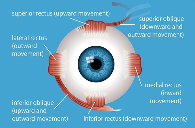

Intrinsic and extraocular muscles of the eye
There are two types of eye muscles: extrinsic muscles that control eye movement and position, and intrinsic muscles that control near focusing and how much light enters the eye.
Extrinsic eye muscles (also called extraocular muscles) are attached to the outside of the eyeball. They enable the eyes to move in all directions of sight.
There are six extraocular muscles around the eye and one muscle that controls movement in the upper eyelid. Though the extraocular muscles are found within the orbit of the eye, they are not located in the eyeball itself.
The main function of the extraocular muscles is to control eye movement and eye alignment. They are different from the intrinsic eye muscles, which enable the eye to focus on near objects and control how much light enters the eye.
Extraocular eye muscles and their functions
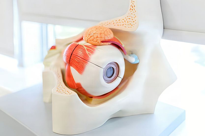
Among the extraocular muscles, there are four straight (rectus) muscles and two oblique muscles. They work together to move the eye from side to side, up and down, and to control its rotation.
Additionally, a muscle called the levator palpebrae superioris (LPS) raises the upper eyelid and keeps it in position.
All seven of these extraocular muscles enable eye movement and are controlled by three cranial nerves:
- Oculomotor nerve
- Trochlear nerve
- Abducens nerve
The ophthalmic artery helps supply blood to the extraocular muscles. The lacrimal and infraorbital arteries also contribute to their blood supply.
Recti muscles
The eye has four recti muscles, all of which attach to the front half of the eye (anterior to the equator of the eye). These muscles are:
- Superior rectus muscle
- Medial rectus muscle
- Lateral rectus muscle
- Inferior rectus muscle
Each of the eye’s recti muscles originates from the common tendinous ring (sometimes referred to as the annular tendon or annulus of Zinn). This is a fibrous ring of connective tissue that surrounds the optic nerve where it connects to the orbit.
Rectus is the Latin word for “straight,” which indicates that the recti muscles attach directly from the orbit to the sclera of the eye.
Superior rectus muscle
The superior rectus muscle is found at the top of the eye and controls upward movement of the eye. Movement of the superior rectus muscle is controlled by the oculomotor nerve.
Medial rectus muscle
The medial rectus eye muscle attaches to the side of the eye closest to the nose and moves the eye inward. Movement of the medial rectus muscle is controlled by the oculomotor nerve.
Lateral rectus muscle
The lateral rectus eye muscle attaches to the side of the eye closest to the temple and helps the eye move outward. It is controlled by the abducens nerve. Problems with this nerve can weaken the lateral rectus and cause double vision. Along with the medial rectus muscle, the lateral rectus muscle helps control horizontal saccades. These are quick eye movements that shift the eyes from side to side.
Inferior rectus muscle
The inferior rectus eye muscle is located at the bottom part of the eye and allows the eye to move downward. This muscle’s movement is controlled by the oculomotor nerve.
Oblique muscles
There are two oblique muscles of the eye. These muscles are:
- Superior oblique muscle
- Inferior oblique muscle
Unlike the recti muscles, the oblique muscles do not attach directly to the eye via the common tendinous ring. Instead, oblique muscles attach angularly to the eye and have separate origins.
Superior oblique muscle
The superior oblique eye muscle originates from the sphenoid bone, which is one of the seven bones that make up the eye socket. It is controlled by the fourth cranial nerve (trochlear nerve).
A small pulley structure in the eye called the trochlea connects the superior oblique muscle from the sphenoid bone to the top of the eye, near the nose.
When the eye is in the primary position (usually facing straight ahead), the main function of the superior oblique muscle is intorsion. (In other words, it rotates the 12 o’clock point of the vertical meridian of the cornea inward toward the nose.) It also moves the line of sight of the eye downward and outward.
The superior oblique also works with other extraocular muscles during vertical saccades to help the eyes move up or down.
Inferior oblique muscle
The inferior oblique eye muscle originates from the front of the orbital floor, close to the nose.
Its main function is to extort the eye when looking straight ahead (rotate the 12 o’clock position of the vertical meridian of the cornea toward the ear). It also elevates and abducts the eye (moves the direction of gaze upward and outward).
Action of the inferior oblique muscle is controlled by the third cranial nerve (oculomotor nerve).
Levator palpebrae superioris muscle
In addition to the six main extraocular muscles, the eye has another extrinsic muscle called the levator palpebrae superioris. This muscle is the single muscle responsible for elevating the upper eyelid and keeping it in position.
This muscle originates from the sphenoid bone in the eye’s orbital structure. Its movement is controlled by the oculomotor nerve.
Extrinsic eye muscles vs. intrinsic eye muscles
While extrinsic (extraocular) muscles control eye movement, the intrinsic eye muscles focus the eye and control the iris to allow a specific amount of light to enter it.
Of these muscle groups, the extrinsic muscles are the muscles around the eye. The intrinsic muscles are located in the eye. Extrinsic muscle movements are voluntary, while intrinsic muscle movements are involuntary.
The intrinsic eye muscles include:
Extraocular muscle function testing
The function of the extrinsic eye muscles can be evaluated during an extraocular muscle function test. This test is often called an extraocular motility exam.
During the test, a doctor or eye doctor holds a pen or other object in front of you and asks you to follow it with your eyes without moving your head. They observe your eye movements as the object is moved in different directions.
Different types of cover tests may also be performed to check eye movement and alignment. The doctor will cover each eye one at a time to look for gaze shifting when they uncover the eye.
The types of problems found during these tests can help determine where they originate:
- With the eye muscles themselves
- With the nerves that control the muscles
- In areas of the brain involved in eye movement
SEE RELATED: Ocular myasthenia gravis
Eye muscle surgery
If one of the extraocular muscles is too strong, too weak, or otherwise dysfunctional, an eye movement disorder can develop. Eye movement disorders can be mild or severe. If a case is severe enough, eye muscle surgery may be recommended.
Disorders that occur due to dysfunction of the extraocular muscles include:
- Nystagmus (involuntary eye movement)
- Strabismus (misaligned eyes)
- Esotropia (crossed eyes)
- Exotropia (wall-eyed)
- Amblyopia (lazy eye)
SEE RELATED: Strabismus surgery
Eye movement disorders are typically more common in childhood, but they can develop later in life as well. If you believe you may have an eye movement disorder, it is ideal to contact an eye doctor as soon as possible.
It is recommended to get regular eye exams to help maintain healthy vision, from visual acuity (how well you see) to ensuring the functions of your vision system (such as eye movement) are performing properly.
Advanced treatments for eye movement disorders
Treatment for eye movement disorders depends on the condition, its cause, and the muscles or nerves involved.
Advanced treatment options may include:
- Adjustable sutures – These sutures allow eye muscle positioning to be adjusted after strabismus surgery.
- Botulinum toxin – Injections of botulinum toxin may be an option during the early stages of strabismus.
- Muscle transposition – This treatment repositions working eye muscles to help improve eye alignment and movement.
- Latest treatment options – Experimental treatments using magnetic implants are being studied for nystagmus.
READ NEXT: What is ophthalmoplegia?