
What is ocular myasthenia gravis?
Ocular myasthenia gravis (OMG) is an autoimmune disorder. It attacks the muscles around the eye, leading to eye fatigue and weakness. It can be a precursor to the development of systemic or generalized myasthenia gravis (MG or gMG). The two main symptoms of OMG are ptosis (drooping eyelids) and diplopia (double vision).
What is systemic myasthenia gravis?
MG is an autoimmune disorder. It causes weakness in voluntary muscle groups throughout the body. A voluntary muscle is one that a person can consciously control. Autoimmune disorders cause an individual’s immune system to attack healthy body cells.
To move a voluntary muscle, the brain sends an impulse through a nerve to the muscle. The chemical acetylcholine is then released into the muscle where it attaches itself to receptors inside the muscle. When acetylcholine has attached to enough receptors, the muscle contracts.
In individuals with MG, the immune system produces antibodies that block or destroy these receptors. It can impact up to about 80% of the receptors in a particular muscle group. While the body is able to replace receptors, it cannot do so at a fast enough rate to maintain muscle strength. This causes the weakness associated with MG.
Myasthenia gravis is not genetically transmitted, nor is it contagious. It can occur at any age but tends to be more common in women under 40 and men over 60. Many individuals who develop MG initially experience ocular symptoms.
For around 15%, the condition will remain in the eyes; others will develop weakness in other areas of the body. However, if an individual has only experienced ocular symptoms for five years, they are unlikely to develop MG.
What causes ocular myasthenia gravis?

Illustration of ocular myasthenia gravis.
Eye muscles have certain features that can make them more susceptible to the effects of MG. They have fewer receptors compared to other muscles in the body, and may be one of the first tissues to be impacted when antibodies attack.
The muscles around the eye also contract at a much faster rate and are more easily fatigued than muscles that contract more slowly.
What are the symptoms of ocular myasthenia gravis?
There are two main symptoms of ocular myasthenia gravis:
- Ptosis (drooping eyelids)
- Diplopia (double vision)
The eyelid muscle weakness brought on by MG can cause the eyelids to droop. The eyelids can, in turn, obstruct vision by physically blocking all or part of the pupil.
Extraocular muscle weakness can interfere with the brain’s control over the movement of the eyes. When this happens, the eyes may no longer align They may also move at two different rates, leading to double vision.
Ocular myasthenia gravis symptoms may fluctuate throughout the day, sometimes worsening in the evening. Individuals can often find temporary relief by resting their eyes.
SEE RELATED: Apraxia eyelid opening
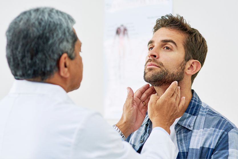
How is ocular myasthenia gravis diagnosed?
To diagnose OMG, an eye doctor will examine the eyes and ask about symptoms. They will compare performance of the eye muscles when they are rested and when they are tired. If the eye doctor believes that MG may be causing the symptoms, they will refer the patient to a neurologist or neuro-ophthalmologist.
The neurologist may use a diagnostic test called an electromyogram to measure the amount of electrical activity that appears when the muscle is at rest and contracted. This can help them identify how the muscle responds to stimulation.
There is also a blood test to check for MG. The test measures the number of anti-acetylcholine receptor antibodies. However, this test is less accurate in cases of ocular myasthenia gravis. A negative result does not necessarily mean the patient is negative for OMG.
What are the treatment options for ocular myasthenia gravis?
Treatment of OMG depends on the particular symptom and its impact on quality of life. For cosmetic concerns, the doctor may recommend treatments that are non-pharmaceutical.
Treatment options for common symptoms of ocular myasthenia gravis may include:
Ptosis
Each of these can physically help prevent the eyelid from obstructing vision.
- Eyelid tape
- Lundie loops (small wire hoops that can attach to glasses to hold the upper eyelid open)
- Eyelid crutches or ptosis crutches (bars that can be added to the top of eyeglasses to prop open the upper eyelid)
Double vision
An eye doctor may recommend an eye patch to help with double vision.The covered eye can grow weaker if it remains unused, so they may recommend alternating which eye is covered for younger patients.
Another option is the addition of prisms to the patient’s glasses, although this treatment is less common.
Other treatment options
Pyridostigmine is a medication that slows the breakdown of acetylcholine. It can be prescribed to temporarily help improve muscle strength.
The corticosteroid prednisone and the immunosuppressants azathioprine, mycophenolate and cyclosporine may be prescribed. They can help control the production of abnormal antibodies.
However, these drugs can cause significant side effects. Because of this, regular monitoring by your doctor is an important part of managing these medications safely.
In some cases, a thymectomy, or removal of the thymus gland, can be used to treat MG. The thymus gland is located in the upper chest and helps with the development of the body’s immune system during childhood.
The removal of this gland could help with reducing weakness in those suffering from MG. However, thymectomies are less commonly recommended for individuals suffering from purely ocular MG.
When should I see a doctor?
Comprehensive eye exams are a good place to bring up concerns regarding ptosis and/or double vision. If your eye doctor suspects myasthenia gravis, they will likely refer you to a neurologist for an official diagnosis. It is important to coordinate treatment with your eye doctor and neurologist for the best results.
SEE RELATED: Cocaine Effects on the Eyes and Vision


")