Neovascular glaucoma: Causes, symptoms and treatment options

What is neovascular glaucoma?
Neovascular glaucoma is a type of glaucoma, a group of eye diseases that damage the optic nerve, often because of high eye pressure. In neovascular glaucoma, damage to blood vessels in the retina causes new irregular vessels to form on the iris (the colored part of the eye) and in the eye's drainage angle. These vessels can bleed and block the eye from draining fluid, leading to higher pressure.
Increased eye pressure (called intraocular pressure, or IOP) can harm parts at the back of the eye, like the optic nerve and retina. The retina is tissue that senses light, and it sends information to the brain through the optic nerve. Too much damage to these structures can stop them from working. This could lead to permanent loss of vision.
Neovascular glaucoma is a type of secondary glaucoma. This means the disease is caused by another injury or condition. Neovascular glaucoma is typically caused by conditions that can lead to damage to blood vessels, including those in the eye.
Causes and risk factors of neovascular glaucoma
When blood vessels in the eye are damaged, new ones may be created. These new vessels are sometimes weaker than normal and may even bleed inside the eye. Growth of these vessels (neovascularization) can happen as a result of several different conditions.
One possible cause is ocular ischemic syndrome. Ischemia is what happens when a part of the body can’t get enough blood flow. This also means it isn’t getting enough oxygen. In ocular ischemic syndrome, this is often because the arteries that carry blood to your eyes are blocked or have become too narrow. Conditions that could lead to ocular ischemic syndrome include:
- High blood sugar (as in diabetes)
- High blood pressure (hypertension)
- Heart disease
- Elevated cholesterol levels and plaque formation
The same health problems can also cause other conditions that can lead to an ischemic retina and neovascular glaucoma. These include:
- Retinal vein occlusion – When a vein in the retina is blocked. A blockage of the main (central) retinal vein carries the highest risk.
- Diabetic retinopathy – When damage to retinal blood vessels happens as a result of diabetes.
Neovascular glaucoma is acquired, not something people are born with. It’s rare in children and is usually a complication of another serious eye or body-wide condition, such as certain retinal tumors.
SEE ALSO: Hypertension and eye health
Symptoms and diagnosis
Common symptoms of neovascular glaucoma include:
- Pain
- Redness
- Light sensitivity
- Blurry vision
- Complete loss of vision (especially if the disease has progressed)
However, there aren’t always symptoms in the early stages of neovascular glaucoma. Once eye pressure starts to rise, neovascular glaucoma can progress quickly and is considered an urgent eye condition. Patients may not notice until their symptoms are advanced or severe though. Early diagnosis is important for preserving vision and preventing further damage.
It is important to seek same-day care if you suddenly develop eye pain, redness or blurred vision, especially if you have diabetes or have had a retinal vein occlusion. Call your eye doctor right away, or go to an emergency facility if you can’t be seen quickly.
Diagnosis
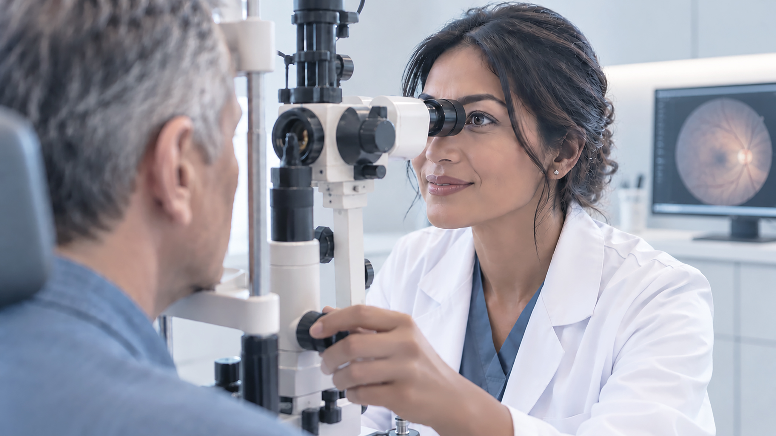
Neovascular glaucoma can be diagnosed clinically. Your eye doctor can use a tool called a slit lamp (a special microscope with a bright light) to look for signs of new blood vessels on the iris.
Another commonly used test is gonioscopy. This test lets your doctor see the drainage system in the eye in particular. It involves a special lens placed on the (numbed) surface of your eye, along with the use of a slit lamp.
Your doctor may also use imaging techniques, like fluorescein angiography, which uses a dye and special photographs to show areas of the retina that aren’t getting enough blood, and optical coherence tomography angiography (OCTA). OCTA can be used to examine blood vessels in the retina. A special type of ultrasound imaging called a B-scan may also be used to examine the back of the eye.
SEE ALSO: Retinal imaging and scans
Pathophysiology of neovascular glaucoma
Neovascular glaucoma usually happens when the retina becomes ischemic. If the retina isn’t getting enough blood, it tries to compensate by producing signaling proteins, like vascular endothelial growth factor (VEGF). VEGF helps create new blood vessels. The new vessels are supposed to help bring more oxygen to the retina. However, they are often fragile and bleed easily.
If the new vessels bleed enough, the blood can block a part of the eye called the vitreous. The VEGF then moves to the front of the eye, where it creates new blood vessels in the drainage angle.
When these irregular, sensitive, fragile vessels grow over the drainage angle, the angle can become narrow and scarred. If the drainage angle closes, fluid from inside the eye can’t drain, leading to higher eye pressure. These vessels may also bleed, resulting in layered blood in the front of the eye called a hyphema.
Treatment options for neovascular glaucoma
Treatment for neovascular glaucoma is focused on treating the cause of the neovascularization and lowering the intraocular pressure. While medications can help, surgery may be needed to improve drainage inside the eye. Treatment can also depend on what stage of closure is affecting the drainage angle.
READ MORE: Glaucoma treatment: Eye drops, oral medications, surgery and more
Medical management
Medications that lower eye pressure are often used for glaucoma. They can prevent the pressure from building up enough to cause damage or keep it from getting worse. They are often given as eye drops. Eye drops can also help with symptoms, like inflammation.
Another way to treat neovascular glaucoma is anti-VEGF therapy. Certain drugs can block VEGF, can quickly shrink the abnormal vessels and help lower eye pressure. These drugs are usually given via injection into the eye.
In the U.S., the anti-VEGF drugs used for neovascular glaucoma are U.S. Food and Drug Administration (FDA) approved for related retinal conditions, such as diabetic retinopathy and retinal vein occlusion, but not specifically for neovascular glaucoma. Using them for neovascular glaucoma is considered off-label, which is a common and accepted practice. Your doctor can determine whether this is a treatment option for you.
Laser and surgical interventions
The main laser treatment for neovascular glaucoma is called panretinal photocoagulation (PRP). It places many tiny laser spots on the outer, oxygen-starved parts of the retina. This lowers the retina’s production of VEGF, allowing the new, abnormal vessels to shrink over time. The goal of laser photocoagulation techniques like PRP is to treat the underlying cause and is considered the cornerstone of long-term control. It’s often combined with anti-VEGF injections, which work faster but wear off over time.
Medications that lower IOP are not always effective for neovascular glaucoma, and surgery may be needed. Glaucoma surgery typically aims to improve how your eye drains fluid or how much fluid your eyes produce.
One way to manage this is with drainage implants. Drainage implants are placed in the front of your eye to provide fluid with an alternate route to drain. A trabeculectomy may also be considered. This creates a small pocket in the conjunctiva called a bleb. The bleb is a new drain pathway that is surgically created. However in neovascular glaucoma, trabeculectomy is more likely to fail than in other glaucomas, especially when diabetes is the cause, so drainage implants are often preferred.
The new blood vessels can make treating neovascular glaucoma difficult. The blood vessels have a higher risk of bleeding during surgery. Additionally, surgery may lower intraocular pressure but not address the neovascularization.
For eyes with limited vision, or when other surgery isn’t a good option, doctors may use cyclophotocoagulation. This laser treats the part of the eye that makes fluid, so less fluid is produced.
A newer version, micropulse cyclophotocoagulation, delivers the laser in short bursts. In one study of eyes with neovascular glaucoma, it caused fewer serious complications than the traditional continuous laser, though the traditional method controlled pressure somewhat better over a year.
Living with neovascular glaucoma
Neovascular glaucoma may be treated with the help of many different specialists, and it will be important to follow the recommendations of your care team. Managing conditions, like high blood pressure or diabetes, is also important. In general, a healthy lifestyle will help keep your eyes healthy. If needed, support groups or counseling can be a valuable resource.
If neovascular glaucoma has caused lasting vision loss, consider asking about how to access vision rehabilitation (low vision) services. They can help with reading, daily tasks and safety, and they’re useful even when vision impairment is moderate.
People with diabetes should have a comprehensive eye exam with dilation at least once a year, starting at diagnosis for type 2 diabetes and within five years of diagnosis for type 1 diabetes. Your eye doctor may recommend more frequent checkups if you already have eye changes.
Prognosis and future directions
The prognosis of neovascular glaucoma depends on many factors. Generally, treatment outcomes depend on how well the disease is managed in the long term. Treatment can control eye pressure and stop new vessel growth, but it doesn’t cure neovascular glaucoma, and it can’t bring back vision that has already been lost. Most people need ongoing follow-up.
Advancements in treatment are helping with this, especially anti-VEGF treatments. Newer anti-VEGF drugs may require a lower frequency of injections than those that are currently used. Where in the eye the drug is injected might also make a difference. Injecting different drugs in different locations as part of the same treatment may result in better outcomes.
Newer types of surgeries that don’t require as invasive an approach can sometimes be used as an alternative. Specialized laser procedures, like cyclophotocoagulation, are sometimes used for eyes with poor visual potential or as an alternative to the prior described surgical procedures.
Advances in imaging are also helping identify neovascular glaucoma sooner. Advances in telemedicine and artificial intelligence technologies can help, especially in underserved areas.
READ MORE: How technology is transforming glaucoma diagnosis, management and treatment
Early detection is key
There are often no symptoms in early neovascular glaucoma. Sometimes, patients don’t see an eye doctor until the disease is at an advanced stage. But chances of preserving vision are better with early detection and treatment.
In general, controlling risk factors, like blood pressure and diabetes, is the most effective way to prevent or slow neovascular glaucoma. Having regular eye exams is important, especially if you have a condition like diabetes. In general, a healthy lifestyle and regular checkups will help you protect your eyes.
READ NEXT: Glaucoma causes: What we know about intraocular pressure (IOP) and optic nerve damage