What is glaucoma surgery?
Glaucoma surgery is a procedure your eye doctor may use to lower the pressure inside your eye. There are many different types of surgeries for glaucoma. A doctor can perform some procedures in their office or a surgery clinic, but some operations have to be done at the hospital.
Doctors usually try other kinds of treatment first, but sometimes, surgery is a highly suitable option.
How does surgery help glaucoma?
The front of each eye is filled with a gel-like fluid called aqueous humor, or aqueous for short. It keeps your eye nourished and helps give it its shape.
Aqueous drains out of your eye through a microscopic web called the trabecular meshwork. It drains out of the eye around the edges of your iris (the colored part of your eye).
The point where fluid drains is called the drainage angle. Once it's filtered, the fluid goes back into your bloodstream.
Glaucoma usually happens when too much pressure builds up inside your eye and damages your optic nerve.
Light signals from the outside world have to travel along your optic nerve to get to your brain. This is how you "see" something. When the optic nerve is damaged, you can permanently lose part or all of your vision.
High eye pressure usually happens for one or both of these reasons:
- Your trabecular meshwork doesn't let aqueous fluid drain out of your eye as well as it should.
- Your iris is blocking some of your drainage angle.
Glaucoma surgery helps your eye drain more of the backed-up aqueous fluid, so the pressure can go down in the rest of your eye. This protects your optic nerve from future damage.
Eye doctors can use many different types of glaucoma surgery to make this happen.
Do I need surgery for my glaucoma?
You may need surgery for glaucoma if other treatments aren't controlling your eye pressure very well. And if you have open-angle glaucoma, laser surgery may be effective compared to starting with other treatments. Your eye doctor will tell you if you need to consider surgery.
Your eye pressure number isn't always a sign that you do or do not need surgery. Normal eye pressure measures between 10 mmHg and 20 mmHg. But a number above 21 mmHg doesn't automatically mean that you need to have glaucoma surgery or even use treatment.
Some people have high eye pressure but no optic nerve damage. Damage can happen in the normal range, too. This is called normal-tension glaucoma.
Doctors take a lot of things into consideration before they decide which surgery to recommend. These factors can include:
- Which type of glaucoma you have
- How severe your glaucoma is
- Your success with other treatments
- Your medical history and health risk factors
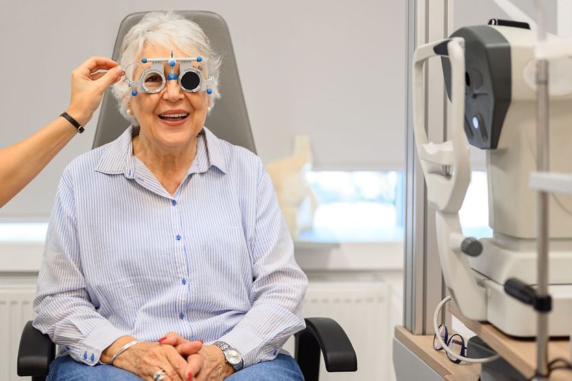
Different glaucoma surgery types use different techniques to lower the pressure inside your eye. Many types of glaucoma surgery are performed by glaucoma specialists or comprehensive ophthalmologists. But some states in the US allow optometrists to do certain laser surgeries.
If glaucoma is in both eyes, eye doctors usually operate on one eye first, then wait until a later date to finish the second eye.
Doctors can perform some procedures in their office or at an outpatient clinic. Some surgeries are done at the hospital.
The procedure itself can take anywhere from five minutes to one hour or longer, depending on the type of surgery.
SEE RELATED: 5 things that can cause high eye pressure
Incisional glaucoma surgery
Incisional surgery gets its name from the incision (cut) a doctor uses to open the surface of your eye. It can be called filtering surgery, too.
The main type of incisional surgery is a trabeculectomy.
Trabeculectomy
- Used often for: Open-angle glaucoma
- Type: Outpatient (you'll go home the same day)
- Eye incision: Yes
- Location: Hospital
- State during surgery: May be awake with eye numbed, relaxed (sedated) or asleep (general anesthesia)
- Typical operating time: 1 hour or less
Trabeculectomy is a common type of glaucoma surgery. It's usually very effective at lowering eye pressure. Your doctor may recommend it if they can't control your eye pressure with less invasive surgeries or eye drops alone.

Click on illustration to enlarge.
During a trabeculectomy, a doctor will cut a small flap in part of your eye that's usually hidden by your top eyelid. They access the trabecular meshwork through this opening.
They'll remove a piece of the meshwork so the aqueous fluid can flow out of your eye more easily.
After trabeculectomy surgery, they'll close the flap and it will heal. The flap collects and filters aqueous fluid as it leaves the eye, creating a small fluid blister called a bleb. It will be covered by your eyelid, so you won't be able to see it.
A healed bleb shouldn't be uncomfortable, but it can still get infected or even break and leak. If it becomes flat, this may be a sign that the surgery isn't working anymore. It's important to have regular checkups with your eye doctor so they can monitor the bleb.
Glaucoma laser surgery
Some surgeries use lasers designed to change the way fluid drains inside your eye.
Doctors can perform many of these procedures without making an incision in your eye. They do this by using a special laser that only changes a certain part of your eye without damaging the front of your eye as it passes through.
Types of glaucoma laser surgery include:
- Selective laser trabeculoplasty (SLT)
- Micropulse laser trabeculoplasty (MLT)
- Argon laser trabeculoplasty (ALT)
- Laser peripheral iridotomy (LPI)
- Cyclophotocoagulation (CPC)
Selective laser trabeculoplasty (SLT)
- Used often for: Open-angle glaucoma
- Type: Outpatient
- Eye incision: No
- Location: Eye doctor's office or outpatient surgery center
- State during surgery: Awake with eye numbed
- Typical operating time: 5-10 minutes

Click on illustration to enlarge.
SLT is one of three common types of laser trabeculoplasty. During SLT, an eye doctor sends short laser pulses through a lens to burn tiny holes in your trabecular meshwork. That lets the fluid in your eye drain effectively.
More doctors now choose SLT as their first treatment option for open-angle glaucoma.
The effects of SLT can last two to three years or longer. If you respond well to the first procedure, it may be safe to do the same procedure again if your eye doctor thinks you need it.
Micropulse laser trabeculoplasty (MLT)
- Used often for: Open-angle glaucoma
- Type: Outpatient
- Eye incision: No
- Location: Eye doctor's office or outpatient surgery center
- State during surgery: Awake with eye numbed
- Typical operating time: 5-10 minutes
MLT is a newer type of laser trabeculoplasty. It uses a laser in quick bursts instead of a constant laser beam. This lets the trabecular meshwork in your eye cool down between laser pulses.
MLT has a high success rate. Many patients handle it well, since the short laser pulses are gentler on nearby parts of the eye.
Argon laser trabeculoplasty (ALT)
- Used often for: Open-angle glaucoma
- Type: Outpatient
- Eye incision: No
- Location: Eye doctor's office or outpatient surgery center
- State during surgery: Awake with eye numbed
- Typical operating time: 5-10 minutes
ALT is the oldest kind of laser trabeculoplasty. It uses a laser made of a chemical called argon to burn little holes in your trabecular meshwork.
The argon laser used during ALT can cause more damage to the surrounding areas than the other two laser procedures like it. SLT laser surgery is more common as a first treatment, but many doctors still recommend argon laser trabeculoplasty to their patients. ALT can only be done once.
Laser peripheral iridotomy (LPI)
- Used often for: Angle-closure glaucoma
- Type: Outpatient
- Eye incision: No
- Location: Eye doctor's office or outpatient surgery center
- State during surgery: Awake with eye numbed
- Typical operating time: 5-10 minutes
LPI is used for another type of glaucoma called angle-closure glaucoma. It happens when your iris bulges and blocks your eye's drainage angle.

Click on illustration to enlarge.
This glaucoma can progress for a long time (chronic) or happen very suddenly (acute). Acute angle-closure glaucoma is usually very painful and can quickly cause blindness. It's a medical emergency.
During LPI, a doctor uses a laser to make a hole in the outer edge of your iris. This exposes the trabecular meshwork and lets more aqueous fluid drain in and out.
A laser iridotomy is different from an iridectomy, which is when an eye doctor surgically cuts out a piece of the iris. Both may be used to prevent an acute attack of angle-closure glaucoma.
Cyclophotocoagulation (CPC)
- Used often for: Refractory glaucoma (doesn’t respond to other treatment or surgery)
- Type: Outpatient
- Eye incision: Sometimes
- Location: Varies
- State during surgery: May be awake with eye numbed or relaxed (sedated)
- Typical operating time: 10-15 minutes
CPC targets the tiny ridges in your eye that make aqueous fluid. They're called the ciliary processes. You can find them behind the iris and above the drainage angle.
Doctors mainly use CPC to treat refractory glaucoma, but it could be a good option for other types of glaucoma, too. Someone who has refractory glaucoma has tried as many treatments as they could safely try, but they still have high eye pressure.
There are three types of CPC laser surgery:
- Transcleral diode cyclophotocoagulation (TSCPC) – A laser is used to shrink the ciliary processes. Doctors usually pick TSCPC when the patient's vision has been damaged a lot.
- Micropulse transscleral cyclophotocoagulation (MP-TSCPC) – A similar, non-invasive procedure that uses short bursts of a laser instead of a constant beam. MP-TSCPC may be preferred for patients without vision loss who have a good chance of keeping their vision in the future.
- Endoscopic cyclophotocoagulation (ECP) – A doctor guides a tiny camera into the eye to help them find the exact location of the ciliary processes, then shrink them. An incision is needed during ECP. It can also be called endocyclophotocoagulation.
SEE RELATED: How many glaucoma suspects eventually develop glaucoma?
Minimally invasive glaucoma surgery (MIGS)
- Used often for: Open-angle glaucoma
- Type: Outpatient
- Eye incision: Yes, minimal
- Location: Varies
- State during surgery: Varies
- Typical operating time: Varies, depending on the type of surgery
Some newer procedures are known as minimally invasive glaucoma surgery, or MIGS for short. There are many different types of MIGS. Doctors usually only recommend them for people with less severe glaucoma.
MIGS usually comes with less risk and a quicker recovery time than more invasive types of glaucoma eye surgery. A doctor will still need to make a cut in the surface of your eye, but the incision is smaller than in older glaucoma surgeries. That's why MIGS is known as "minimally invasive" or "micro-invasive."
Many MIGS procedures use a tiny implanted device called a shunt or stent. This lets aqueous fluid drain more easily and helps your eye pressure go down.
Sometimes, doctors perform MIGS at the same time as cataract surgery.
Some current MIGS procedures are:
- Micro trabeculectomy
- Trabecular micro-bypass stent
- Suprachoroidal shunts
You won't always hear your eye doctor use these terms. Instead, they might call them by their surgery or device names.
Trabecular MIGS
This group of surgeries focuses on the web of trabecular meshwork near the edge of your iris.
Some trabecular MIGS remove part of the meshwork. Others use a device called a stent to open up parts of the meshwork and let more fluid flow out. The medical name for this is trabecular micro-bypass stent.
Suprachoroidal shunts
There's a thin layer between the outside-facing wall of your eye and the inner wall. It's called the suprachoroidal space, and it helps aqueous fluid flow out of your eye.
Small tubes called suprachoroidal shunts can be used to connect this space to the front of your eye, which helps your eyes drain aqueous fluid.
SEE RELATED: Can glaucoma cause headaches?
Other types of glaucoma surgery
Some surgeries aren't an incisional surgery or a MIGS.
These other glaucoma surgery types include:
- Glaucoma implant surgery
- Deep sclerectomy
- Procedures that are less common or still being developed
Glaucoma implant surgery
- Used often for: Less common types of glaucoma
- Type: Outpatient
- Eye incision: Yes, minimal
- Location: Outpatient surgery center or hospital
- State during surgery: Awake or relaxed (sedated) with eye numbed
- Typical operating time: 1 hour or less
During glaucoma implant surgery, a doctor inserts a tiny tube called a shunt near the edge of your iris. The tube is connected to a small plate that's implanted nearby on the surface of your eye.
You won't be able to feel the tube or plate after surgery. You can't see the plate unless you open your eyes very wide.
These glaucoma implants are also known as aqueous shunts or tube shunts. There are many different types, but they may help lower eye pressure by filtering more aqueous fluid out of the eye.
Many people who have implant surgery have a less common kind of glaucoma. These can include glaucoma that happens after an injury, is caused by diabetes or is present at birth.
Deep sclerectomy
- Used most often for: Open-angle glaucoma
- Type: Outpatient
- Eye incision: Yes, minimal
- Location: Outpatient surgery center
- State during surgery: Awake with eye numbed
- Typical operating time: 30-45 minutes
A deep sclerectomy involves making a small opening in the white part of your eye to help the aqueous fluid drain. Sometimes, the doctor will place a tiny drain or implant in your eye. Some doctors in the United States use deep sclerectomies, but it's more common in other parts of the world.
Its medical name is non-penetrating deep sclerectomy, or NPDS. It's called a non-penetrating surgery because the surgeon doesn't need to cut the entire way through the lower layers of your eye wall.
This is why people are less likely to have complications with a deep sclerectomy than they are with a trabeculectomy. But a trabeculectomy can be better at lowering eye pressure when people need it.
Your eye doctor will help you decide which procedure is suitable for your condition.
Others
There are many more glaucoma surgery types than the ones listed here.
Many newer procedures aren't used by a lot of doctors yet but could be in the future. Some aren't very common because they're only used for rarer cases of glaucoma. Other surgeries are still being researched or developed.
Your eye doctor will use their medical knowledge and experience to help you decide which specific type of procedure has a high chance of treating your glaucoma.
Recovery
Glaucoma surgery recovery is different for each type of procedure. It usually takes a few days to a few weeks for your vision to look normal again. It can take longer than this, but it's rare.
In general, recovery time is usually faster with less invasive procedures and more mild glaucoma.
Your doctor will give you detailed instructions to follow during your recovery. These instructions can help speed up the healing process and lower your risk of complications.
Risks and complications
Having surgery for glaucoma is safer today than it's ever been. But all surgeries come with a risk of complications. Your risk may depend on your type of surgery, past treatments, medical history and other factors.
Some side effects should go away on their own after a little while. These might include:
- Eye redness
- Eye tearing
- Eye pain or irritation
- Swollen eyelid
- Feeling like something is stuck in your eye
Other complications can include:
- High eye pressure, even after surgery
- Low eye pressure
- Problems on the front of your eye (cornea)
- Infection
- Cataracts
- Vision loss
Your eye doctor will give you information about the risks and complications involved with your surgery. They'll also tell you how to reduce any uncomfortable side effects.
SEE RELATED: A utologous serum eye drops
Surgery costs
Your out-of-pocket surgery costs will depend on many things. It can be very different from person to person, even when they have the same kind of surgery.
Glaucoma surgery is usually covered by medical insurance, not vision insurance. Many plans will cover surgery if your eye doctor says you need it, but you could still have out-of-pocket costs.
These factors may affect your total cost:
- Type of surgery
- Who helps perform your procedure
- Where you have your surgery
- Your medical insurance coverage
- Your insurance plan's copays, deductible, coinsurance and out-of-pocket maximum
If you have insurance, talk to your insurance company and your doctor's billing office beforehand. Make sure the specific procedure, surgery facility, surgeon, anesthesiologist and any other factors involved in your glaucoma surgery are covered in your network.
Talk to an eye doctor
Not everyone who has glaucoma needs to have surgery. Many people work with their eye doctors to safely manage their condition with medicated eye drops and regular follow-up visits alone.
If your doctor thinks you need to have surgery, they may recommend a procedure to protect your eyesight in the future.
SEE RELATED: How to find a glaucoma specialist