What is Best disease?
Best disease, also called Best vitelliform macular dystrophy, is an eye condition that damages a small part of the retina called the macula. The macula is responsible for your central vision, which lets you see fine details and things directly in front of you.
The condition is hereditary, which means it's passed down from parents to children. It can start to affect someone's eyesight during childhood or adolescence, but the most noticeable vision changes typically don't happen until middle age.
Best disease is rare. Some researchers estimate it affects about 1 in every 10,000 to 20,000 people.
The condition is named for Dr. Friedrich Best, a German eye doctor who described it in detail in 1905.
Causes of Best disease
Best disease is caused by a change (mutation) in one copy of a gene called BEST1.
It follows an autosomal dominant inheritance pattern. That means if one parent carries the mutation, there's a 50% chance their child will inherit it and develop Best disease.
In Best disease, a yellow pigment called lipofuscin builds up beneath the macula. Over time, this buildup forms vitelliform lesions — yellow deposits on the macula that look like an egg yolk.
The word vitelliform means "resembles an egg yolk" in Latin.
Best disease is one type of macular dystrophy. All macular dystrophies cause the macula to break down over time, which worsens central vision.
Signs and symptoms of Best disease
Best disease signs and symptoms usually affect both eyes but no other part of the body.
Eye changes may start between ages 5 and 20, but the most noticeable eyesight symptoms typically don't start until age 40 or older.
You can have Best disease without any noticeable symptoms. In this case, an eye doctor may detect signs during a routine comprehensive eye exam.
Once you start to notice symptoms, they might include:
- Blurry vision
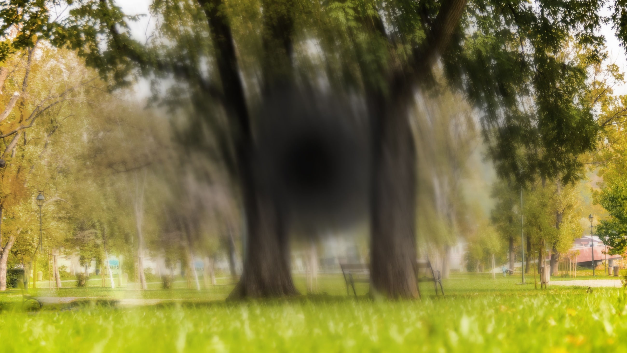
- Straight lines look wavy or distorted (metamorphopsia)
- Colors look dim or less vibrant
- Vision loss in the center of your view
While Best disease can cause low vision, it typically doesn’t cause complete blindness. People with advanced Best disease usually retain their peripheral (side) vision and can still see at night.
There's another type of vitelliform macular dystrophy that doesn't cause vision problems until mid-adulthood, but this is not Best disease. It's known as adult-onset vitelliform macular dystrophy. This condition is called a pattern dystrophy because the pigment buildup shows up in a pattern, unlike Best disease.
Stages of Best disease
Best disease symptoms vary depending on how far the condition has progressed.
It usually happens in five or six stages:
- Stage 1 (previtelliform) – The macula looks normal. Vision isn't affected.
- Stage 2 (vitelliform) – A yellow lesion shaped like an egg yolk appears on the macula. Some people may have more than one lesion. Vision is still normal or mostly normal.
- Stage 3 (pseudohypopyon) – The round lesion appears to be partially filled with yellow fluid that has settled toward the bottom. Vision may become a little blurry or distorted but is still good overall.
- Stage 4 (vitelliruptive) – The lesion starts to break apart, which can make it look like a scrambled egg. Vision may appear blurrier or more distorted.
- Stage 5 (atrophic) – The lesion fades away but leaves scarring on the macula. Central vision looks very blurry or dark.
- Stage 6 (choroidal neovascularization/CNV) – In about 1 in 5 cases, abnormal blood vessels grow under the retina, which can leak and cause swelling. Central vision loss can quickly worsen, often causing blind spots or dark areas.
In many cases, the stages of Best disease that affect vision the most (stages 5 and 6) don't start until someone is at least 40 years old.
The disease progresses at different speeds in each person. Some people notice their vision changing slowly, while others may see changes more quickly.
Diagnosis
Since changes in your eyes can start long before you notice vision problems, diagnosing Best disease often begins with a routine eye exam.
An eye doctor may want to run certain tests during or after your exam. These tests can give them a clearer view of any eye changes caused by Best disease and may include:
- Electro-oculography (EOG) – Measures electrical activity in the deepest layer of the retina as it reacts to different light levels. It can help detect Best disease before changes are visible inside the eye.
- Optical coherence tomography (OCT) – Takes detailed, cross-sectional pictures of the retina to help your doctor identify changes.
- Fluorescein angiography – Involves injecting a special dye into your arm while your pupils are dilated. The dye travels to the blood vessels in your eyes and helps your eye doctor detect leaks or other signs of late Best disease.
- Genetic testing – Uses a blood test to check for changes in the BEST1 gene, which can confirm whether Best disease runs in your family. Genetic testing is also used to help diagnose other genetic eye conditions, like retinitis pigmentosa or Stargardt disease.
Prevention
When at least one parent carries a mutated BEST1 gene, there is no way to lower a child's risk or prevent them from inheriting Best disease.
A genetic counselor can help you learn about this and other genetic risks before or during pregnancy.
Management and treatment
There is no cure for Best disease yet, but it's still important to track its changes with regular eye exams. This helps your eye doctor diagnose complications early on.
People with larger macular lesions may need to avoid high-intensity exercises or contact sports. An eye injury could make the lesion break open and worsen vision problems.
An eye doctor may recommend different procedures to slow vision loss in people who have CNV (stage 6). These might include:
- Anti-vascular endothelial growth factor (VEGF) injections – Medicine is injected into the eye to prevent the growth of abnormal blood vessels.
- Laser photocoagulation – A special laser is used to seal leaky blood vessels.
- Photodynamic therapy – A laser and medicine are used together to destroy faulty cells and close off leaky blood vessels.
Anti-VEGF injections are usually the preferred treatment. The other two treatments have been used successfully in the past, and may still be used in some cases, but they're less common now.
Researchers are exploring options that use stem cell and gene therapy to treat Best disease. However, this research is still early and experimental.
Living with Best disease
It's normal to feel frustrated or worried about changes to your vision. Finding support from family, friends and low vision specialists can help you with the transition.
Many people use lifestyle adjustments and low vision aids to adapt to changes in their central vision. Your provider may recommend techniques such as:
- Increasing the font size and contrast level on digital screens
- Using electronic device features that read text out loud, like screen readers or text-to-speech software
- Using magnifying glasses or large-print books
- Exploring new ways to do daily tasks like cooking and cleaning
- Finding ways to use your peripheral vision more during everyday activities
- Joining a low vision support group
- Meeting with a counselor for help in coping with vision loss
While low vision comes with challenges, you can still live a happy and fulfilling life. If you've been diagnosed with Best disease, talk to your eye doctor or a low vision specialist about ways they can help you adapt to your specific symptoms.
When should you see a doctor?
Both types of vitelliform macular dystrophy — including Best disease — tend not to impact vision until later in life. Routine eye exams can help you keep track of any eye changes and manage new symptoms as soon as they appear.
Eye exams are also used to diagnose other macular problems, such as age-related macular degeneration.
If your vision becomes blurry, distorted or dark, it's very important to schedule an eye exam with an eye doctor as soon as possible so you can get the care you need.

")