What is pseudopapilledema?
Pseudopapilledema is a condition where the front part of the optic nerve, the optic disc, appears elevated, even though it isn’t swollen. The most common cause is the buildup of deposits called optic disc drusen. It is typically harmless and does not produce noticeable symptoms for most people.
The optic disc, or optic nerve head, is where the optic nerve connects to the back of the eye. This nerve carries visual information from the eye to the brain, allowing us to process what we see. Eye doctors closely examine the optic disc during exams because changes here can signal health issues. While pseudopapilledema means the optic disc appears swollen, it does not involve true swelling or pose any urgent health risks, which is why it’s called “pseudo,” meaning “false.”
This is different from true papilledema, which means the optic disc is actually swollen. Papilledema happens because of a dangerous rise in pressure within the skull called increased intracranial pressure (ICP). True papilledema is often accompanied by headaches and vision problems. It is a serious condition that can indicate an urgent underlying health issue. It can also result in vision loss. Pseudopapilledema, however, typically doesn’t cause noticeable problems or serious health risks.
Causes of pseudopapilledema
Pseudopapilledema most commonly results from optic disc drusen. However, several other structural variations can also create the appearance of an elevated optic disc.
Optic disc drusen
The most common cause of pseudopapilledema is optic disc drusen (ODD). These deposits are found in about 0.2% to 3.1% of the world’s population. They are made of fatty proteins and calcium. With age, ODD can enlarge and move from being buried deep to becoming more superficial and visible. This can cause the optic disc to appear elevated.
Congenital variations
Some people are born with structural differences in the optic disc that can cause parts of the disc to appear raised. These include:
- Small, crowded optic discs – Where nerve fibers squeeze through smaller openings, commonly seen in farsighted people
- Tilted optic discs – Where one part sits higher than another, creating an elevated appearance
- Myelinated nerve fibers – When incomplete nerve development can cause obscured disc margins
Cell growth near the optic disc
Growth of cells near the optic nerve — from inflammation or a mass — can make the optic nerve look raised during an eye exam.
Traction
As people get older, the gel inside the eye called the vitreous begins to pull away from the back of the eye (the retina). This traction can give the optic disc a raised appearance. In other cases, a thin layer of scar tissue can form on the retina and pull on the disc. This happens more often in people with diabetes or problems with blood vessels in the retina.
Genetic conditions
People with Down syndrome may sometimes have an elevated optic disc because of optic disc drusen. However, true swelling can also occur from health problems, so additional tests may be necessary. Rare conditions, like Leber’s hereditary optic neuropathy, can also cause the optic nerve to look raised without true swelling.
Risk factors
Optic disc drusen are the most common reason for pseudopapilledema. They affect about 1 out of every 40 to 300 people and occur equally in males and females. Drusen might be hidden deep initially but can become more visible with age. Some risk factors for ODD include:
- Being Caucasian
- Family history
- Certain vision or health conditions (retinitis pigmentosa, angioid streaks or Usher syndrome)
Symptoms
This condition is usually discovered during an eye exam because people are often unaware of it. Proper diagnosis requires specialized tests to differentiate it from serious conditions.
Recognizing symptoms of pseudopapilledema
Most individuals with pseudopapilledema don’t notice any symptoms. However, those with optic disc drusen might experience small areas of vision loss, like an enlarged blind spot. Nearly 90% of people with optic disc drusen have some visual field defect, though these changes are usually mild and progress slowly.
Possible symptoms, if any, include:
- Short episodes of dim, blurry or flickering vision
- Gradual side vision reduction, affecting about 70% of people (often goes unnoticed)
Recognizing symptoms of true papilledema
True papilledema is a serious medical emergency. It causes symptoms that can affect both vision and overall health, such as:
- Headaches
- Nausea
- Vision disturbances
- Tinnitus (a rhythmic “whooshing” sound in the ear)
- Confusion
- Difficulty with movement and speech
- Drowsiness or tiredness
Diagnosis
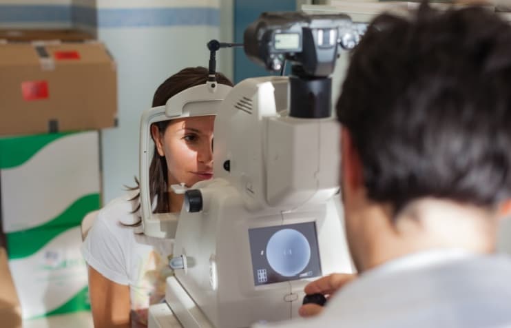
To confirm a diagnosis of pseudopapilledema, you may be referred to a neuro-ophthalmologist, a doctor with specialized training in optic nerve conditions. They will conduct a thorough eye examination that includes several important steps.
History
Your doctor will start with a detailed medical history to assess your risk factors for papilledema. They will ask about:
- Medical conditions
- Headaches and vision changes
- Medications and supplements
- Family history of eye conditions
- Symptoms
Physical exam
Your doctor will use specialized equipment to examine your optic nerve head. They will also measure the pressure inside your eyes. Managing eye pressure helps prevent additional vision loss when you have optic disc drusen. They will also look for any rare complications that might need special attention.
Optic disc assessment
An optic disc with pseudopapilledema may look similar to papilledema. However, there are key features that help doctors tell them apart:
- Blood vessels that cross the optic disc remain visible and are not hidden by swelling.
- There are no small spots of blood (hemorrhages) or leaking fluid (exudates) around blood vessels.
- Some people may have superficial optic disc drusen that can be seen.
- There may be a gentle visible pulse in the veins.
In papilledema, blood vessels can appear blurry and show signs of leaking.
Eye pressure measurement
Your eye doctor will measure the pressure inside your eyes during the exam. If you have optic disc drusen and high eye pressure, you may need eye drops to lower it. High pressure combined with optic disc drusen can lead to significant side vision loss.
Checking for rare complications
Although uncommon, abnormal blood vessels may grow around the optic disc drusen. This can interfere with vision. Early detection and treatment can prevent serious vision loss.
Rarely, optic disc drusen can block a blood vessel supplying the optic nerve. This can lead to sudden vision loss in a condition called “nonarteritic anterior ischemic optic neuropathy” (NAION).
Specialized testing
Your eye doctor may use advanced retinal imaging and scans to confirm the diagnosis. This includes:
- Visual field testing – Maps your side vision to identify any subtle blind spots that might result from drusen pressing on nerve fibers
- Fluorescein angiography – Uses a special injected dye to photograph blood flow in the retina (true optic disc swelling will show leakage)
- Optical coherence tomography (OCT) – Creates detailed cross-sectional images of your optic nerve and surrounding tissues
- B-scan ophthalmic ultrasound – Shows drusen as highly reflective bright spots that create characteristic shadows on the images
This technology allows doctors to see structures deep within the optic nerve that may not otherwise be visible.
Treatment and management
After optic disc drusen is diagnosed, eye doctors usually recommend regular checkups. This helps them manage any related issues and monitor vision changes.
Treatment options
Currently, optic disc drusen are not treated, and there is no way to prevent them from getting larger over time. Routine exams are important to ensure any problems are found early and managed appropriately.
Even though optic disc drusen contain calcium, having them does not mean there is a problem with calcium levels in your body. It does not increase the risk for kidney stones or gallstones.
Managing your condition
Vision changes due to pseudopapilledema are usually subtle. They don’t typically interfere with daily activities. It’s important to let doctors know if you have already been diagnosed with this condition. Sharing this information helps your health care team interpret your eye findings correctly.
Understand your condition and learn warning signs, so you know when to seek immediate medical attention. Contact your eye doctor immediately if you notice:
- An onset of vision loss or disturbances.
- New or severe headaches, especially when lying down.
- Nausea and vomiting.
Understanding a diagnosis of pseudopapilledema
The most frequent cause of pseudopapilledema is optic disc drusen. Most people with ODD do not notice major changes in their vision. Occasionally, some may experience brief episodes where their vision dims or flickers for just a few seconds. Or, they may notice small areas where their vision is missing.
About 7 out of 10 people with this condition may experience some mild loss of side vision over time. Comprehensive eye exams and visual field tests allow your eye doctor to monitor these areas and detect any changes early.
It’s important to share this information with all your health care providers. This helps them correctly interpret your eye exam results and avoid unnecessary concern or testing.