Macular Dystrophy: How This Rare Eye Disease Causes Blindness
On this page
What causes macular dystrophies?
Types of macular dystrophy
Treatments for macular dystrophy
How macular dystrophy is diagnosed
On this page
What causes macular dystrophies?
Types of macular dystrophy
Treatments for macular dystrophy
How macular dystrophy is diagnosed
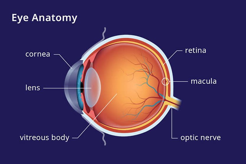
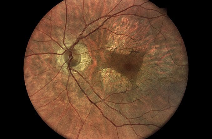
Macular dystrophies are a relatively rare set of eye conditions. Linked to inherited genetic mutations, macular dystrophies can cause deterioration of the most sensitive part of the central retina (macula), which has the highest concentration of light-sensitive cells (photoreceptors).
As the name implies, the macula of the retina is affected in a macular dystrophy. The macula is the central area of the retina that contains photoreceptors responsible for central vision and color perception. When the macula is damaged or scarred due to macular dystrophy, your central vision is affected — and this can lead to blindness in some cases.
What causes macular dystrophies?
Macular dystrophies differs from a far more common eye disease known as age-related macular degeneration (AMD), often caused by age-related deterioration of the retina and macula.
While aging, family genetics and risk factors such as smoking can cause common forms of AMD, macular dystrophy is linked to genetic mutations that — for no apparent reason — trigger degradation of retinal cells. Some forms of macular dystrophy appear in childhood, and other forms appear in adulthood.
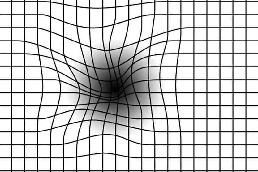
However, it is sometimes difficult to distinguish AMD from inherited macular dystrophy because of the similarity of symptoms, including decreased visual acuity and loss of central vision.
One of the most common forms of macular dystrophy is Stargardt's disease, which accounts for about 7 percent of all macular dystrophy cases and commonly occurs in childhood.
Types of macular dystrophy
Many forms of macular dystrophy have been identified, including:
- Stargardt's disease, the most common type of macular dystrophy, which usually occurs in childhood. A different form of Stargardt's disease, called fundus flavimaculatus, is typically found in adults. Stargardt's disease is characterized by formation of pigmented waste cells in the retina.
- Vitelliform macular dystrophy (VTM), which generally is discovered first with the presence of a large, yellow oval lesion (vitelliform) in an egg yolk shape that shows up in the center of the macula. Many genetic mutations of this form of macular dystrophy have been identified, including Best's disease, which affects children and young people. A different version of the disease can also appear in adults, with macular lesions that vary in size and shape.
- North Carolina macular dystrophy, which is an extremely rare form of macular dystrophy identified by a very specific genetic marker. While named for North Carolina family members who have this inherited form, the disease has been found in other locations worldwide.
Other types of macular dystrophy can cause specific degeneration of light-sensitive cells known as cones. The cones are responsible for color vision and are most concentrated in the macular area of the retina.
While not technically macular dystrophy, retinitis pigmentosa (RP) is an inherited photoreceptor dystrophy that destroys light-sensitive cells in the eye.
Treatments for macular dystrophy
If you have macular dystrophy, an eye doctor will refer you to a retina specialist. They can help you determine the exact nature of the disease. For example, some types are progressive and some aren't.
Genetic analysis and counseling may be needed to help you determine the type of macular dystrophy you have and whether the eye condition is likely to be passed on to your children and descendants. Also, you can make better decisions about family planning if you have an idea of the degree of vision loss associated with your type of macular dystrophy.
At this time, there is no proven treatment for macular dystrophy. However, gene therapiesy and stem cell research are being studied in clinical trials and have shown promise. Your retina specialist can help you decide if you're eligible for a clinical trial. Many trials target specific types of macular dystrophy, offering a way to access experimental therapies before they are widely available.
For example, several prospective trials have evaluated the use of retinal pigment epithelial (RPE) cells derived from human embryonic stem cells for the treatment of Stargardt's disease. So far, 31 patients have been treated, all of whom experienced improved or stable, best-corrected visual acuity (BCVA).
How macular dystrophy is diagnosed
Symptoms of macular dystrophy can include decreased visual acuity with no obvious cause, such as refractive errors or cataracts.
If your eye doctor suspects you have macular dystrophy, he or she may order special eye tests that are not part of a routine eye examination for a definitive diagnosis. For example, a test called fluorescein angiography can detect retinal damage from macular dystrophy.
A test using optical coherence tomography (OCT) can also be performed to analyze eye tissue for the possible presence of a yellow-brown pigment (lipofuscin) found in the retinal pigment epithelium (RPE). Lipofuscin is waste material sloughed off from deteriorating eye tissue.
Still another option is an electroretinogram (ERG) test that involves placing an electrode on your eye's outer, clear surface (cornea) to measure how well photoreceptors in your retina respond to light.
On this page:
What causes macular dystrophies?
Types of macular dystrophy
Treatments for macular dystrophy
How macular dystrophy is diagnosed
On this page:
What causes macular dystrophies?
Types of macular dystrophy
Treatments for macular dystrophy
How macular dystrophy is diagnosed