How OCT and OCTA provide different information about the eye’s structure and blood flow

The names don’t exactly help
Optical coherence tomography (OCT) and optical coherence tomography angiography (OCTA) are closely related and built on the same imaging technology, but designed to provide different views.
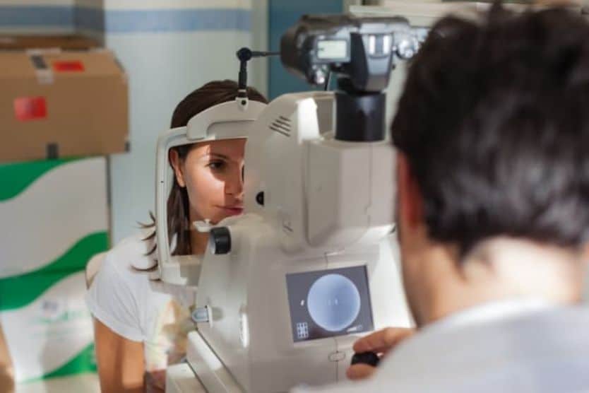
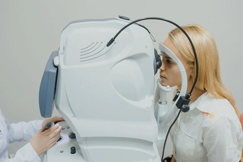
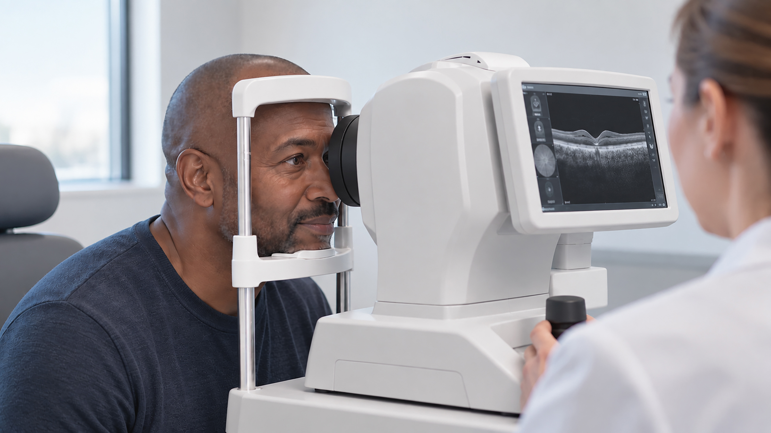
These painless and noninvasive imaging scans can be performed at your eye doctor’s office. For either test, you sit in front of a scanner, rest your chin and forehead on supports and look at a target for two to four minutes while images are taken.
OCT shows the eye’s structure
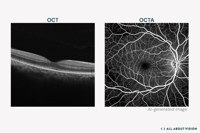
OCT uses light to create detailed cross-sectional images, almost like slices, of structures inside the eye.
Back of the eye
OCT can show the retina, the light-sensitive tissue at the back of the eye, and the optic nerve, which carries visual information from the eye to the brain. When OCT is used to image these structures, it is called posterior segment OCT.
It can reveal swelling, fluid, thinning and other changes that help diagnose and monitor conditions such as:
- Age-related macular degeneration
- Diabetic retinopathy
- Glaucoma
- Geographic atrophy
- Inherited retinal diseases (IRDs) like retinitis pigmentosa
Front of the eye
OCT can also image the cornea, the eye's clear outer surface, as well as the iris, the colored part of the eye, and the area where fluid drains from the eye. When OCT is used to image these structures, it is called anterior segment OCT.
It can also measure corneal thickness. This is called optical pachymetry. These images and measurements can help evaluate risk for corneal conditions (such as keratoconus) and glaucoma.
For cataract surgery
During surgery, OCT can give the surgeon real-time images of the structures inside the eye.
OCTA shows blood vessels and blood flow
OCTA repeatedly scans the same area and detects changes caused by moving blood cells. Software uses that information to map tiny blood vessels in the retina and the choroid, the blood vessel layer beneath it.
It can also show vessels at different depths. Eye doctors may use OCTA to detect abnormal blood vessels or areas of reduced blood flow.
OCT vs. OCTA at a glance
What to know | OCT | OCTA |
Main view | Eye structure | Blood vessels and blood flow |
Can show | Structures in the front and back of the eye, thickness, fluid and thinning | Tiny vessels, reduced blood flow and abnormal vessel growth |
How it works | Uses light to create images of eye tissue | Repeats OCT scans to detect moving blood cells |
Why might your eye doctor use both?
OCT can show changes in the eye’s tissues. OCTA can add information about the blood vessels in the same area.
A quick note
OCTA does have limits. It cannot show blood vessel leakage. Another test, called fluorescein angiography (FA), uses dye injected into a vein to show where vessels may be leaking.