Fovea centralis

Fovea definition
The fovea is a tiny part of the eye’s anatomy that makes a significant difference in our eyesight. Resting inside the macula, the fovea (also called fovea centralis) provides our sharpest vision.
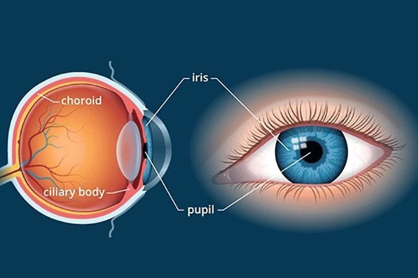
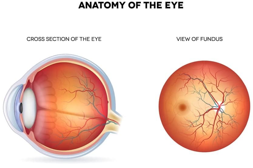
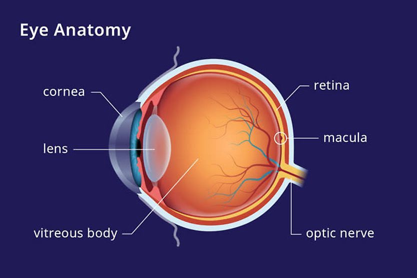
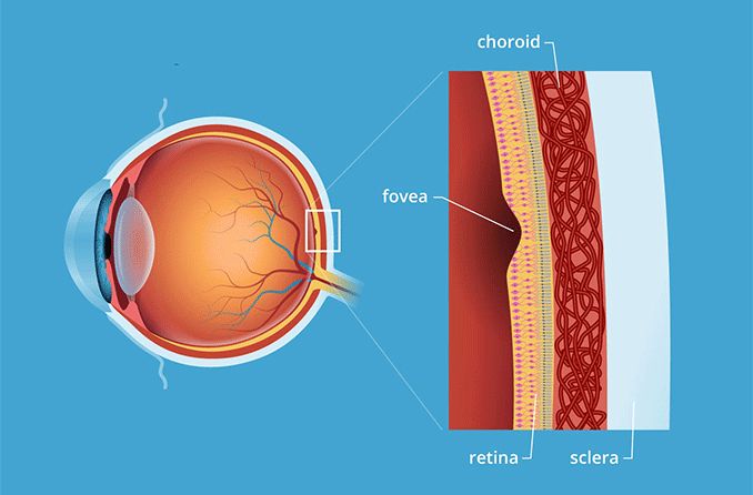
The name fovea comes from the Greek word meaning “small pit.” This is an appropriate name, as the fovea is a tiny depression (or pit) in the macula, a small structure located in the center of the retina, the light-sensitive tissue that lines the back of the eye.
The retina, macula and fovea work together to provide optimal central and peripheral vision.
Fovea function
To fully grasp the fovea’s function, it’s important to understand its anatomy related to the macula.
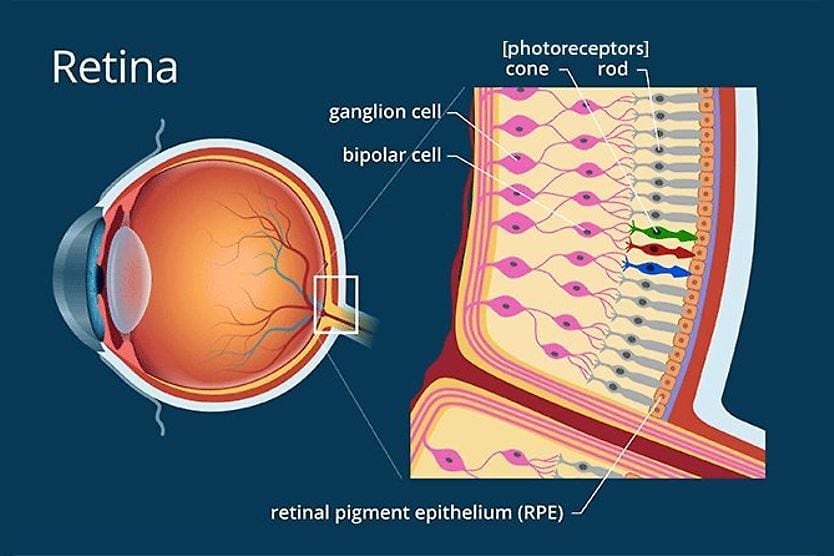
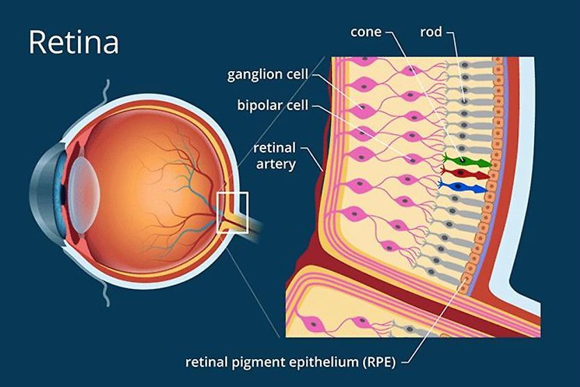
The macula is a very light-sensitive part of the retina that contains two types of photoreceptor cells: cones and rods. This dynamic duo of cells takes light rays and converts them into signals that are then sent to the brain.
Cone cells are responsible for producing color and fine details, while rods provide peripheral vision, movement and shades of grey. Rods are mostly located outside the macula, and the cones are located inside.
The fovea does not have any rods or other neurons, only millions of tightly packed cones. By grouping en masse, cones get optimal exposure to soak up light as it comes into the eye, allowing them to create the sharpest possible image.
Fovea centralis function also includes the discernment of other image details, such as distinguishing between different colors and sensing three-dimensional depth.
Fovea vs. macula
Fovea anatomy can be tricky because the retina and macula are also light-sensitive parts of the eye that create sharp vision. So, where does the fovea come into play, and how is it different from the macula and retina?
Think of it as “sharp, sharper, sharpest.” The retina is the tissue that lines the back of the eye and produces sharp vision whenever light hits it correctly. The macula is the center portion of the retina that produces even sharper vision with its rods and cones. The fovea is the pit inside the macula with only cones, so vision can be at its sharpest.
While the fovea and the macula have the same objective of providing clear vision, they achieve that goal in different ways.
Conditions that may affect the fovea
Because the fovea is such an essential part of a person’s vision, it’s important to prevent and/or monitor the conditions that may jeopardize its function. Conditions that may affect the fovea include:
- Macular degeneration (both wet and dry forms) - Age-related thinning and abnormal protein growth on the macula (dry) or macular scarring due to abnormal blood vessel growth and leakage (wet).
- Stargardt's disease - Form of macular degeneration in which photoreceptors in the macula die off. Unlike age-related macular degeneration (AMD), it can affect young adults and children.
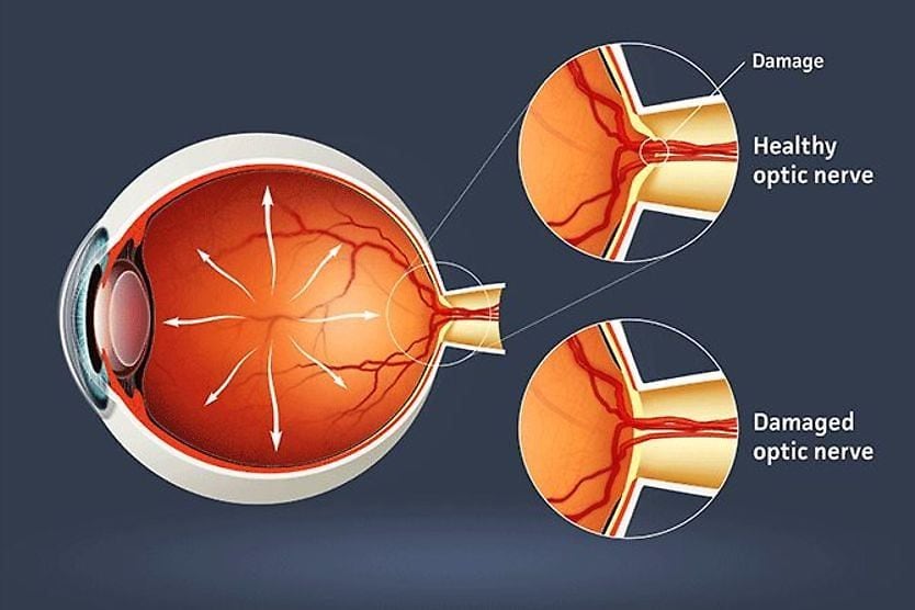
- Diabetic retinopathy - Occurs when consistently high blood sugar levels cause damage to the retina.
- Macular hole - A tear or hole that forms in the macula.
- Macular pucker - When a bulge, wrinkle or crease develops on the macula and affects central vision.
- Macular edema - Fluid buildup in the macula.
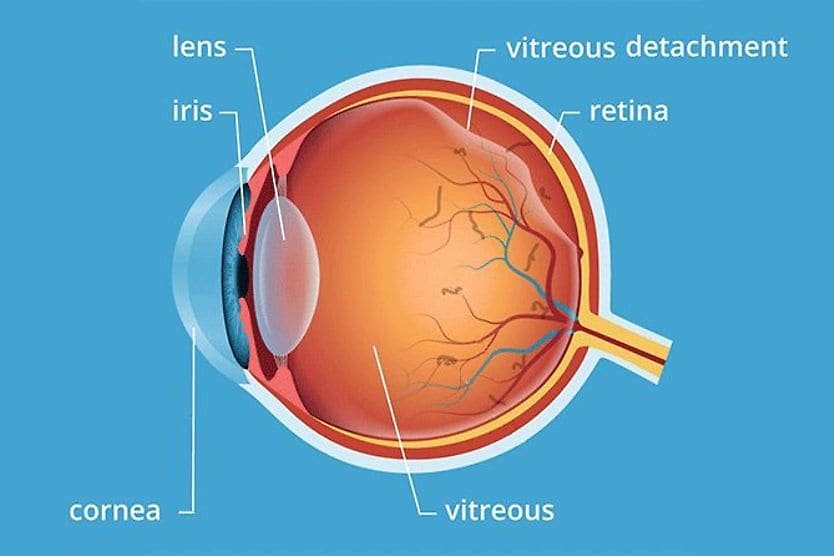
- Retinal detachment - When the retina lifts or tears away from the back of the eye.
- Retinoblastoma - Cancer of the eye that begins in the retina.
- Retinal vein occlusion (RVO) - When a vein in the retina becomes blocked.
- Retinal artery occlusion (RAO) – When an artery in the retina becomes blocked.
- Retinopathy of prematurity (ROP) - Condition in which abnormal blood vessels grow on the retina; it can affect babies born prematurely.
- Choroidal neovascular membranes (CNVM) - Irregular, damaged blood vessels that begin to develop under the retina in an area called the choroid.
- Cytomegalovirus retinitis (CMV) - Viral infection that affects the retina.
- Retinitis pigmentosa (RP) - Genetic condition that affects how the retina responds to light; commonly seen in individuals with Usher syndrome.
- Macular telangiectasia types 1 and 2– A condition that causes blood vessels around the fovea to dilate and leak. Type 2 has a shortened name called “MacTel.”
Importance of routine eye exams
A key step in protecting your sight and keeping your eyes healthy is to have yearly comprehensive eye exams. An eye doctor can detect early signs of a fovea-related condition before you notice any symptoms.
READ MORE: Eye anatomy: A closer look at the parts of the eye