Retinal hole: Is it serious?


Is a retinal hole as serious as it sounds? Read on to learn about retinal holes — including types, symptoms, causes and treatment — and if you may be at risk of developing a hole in your retina.
What is a retinal hole?
A retinal hole is a small break or defect in the light-sensitive retina that lines the inside of the back of the eye.
Retinal holes can occur anywhere in the retina. When a hole develops in the macula lutea (the more sensitive part of the central retina), it’s called a macular hole.
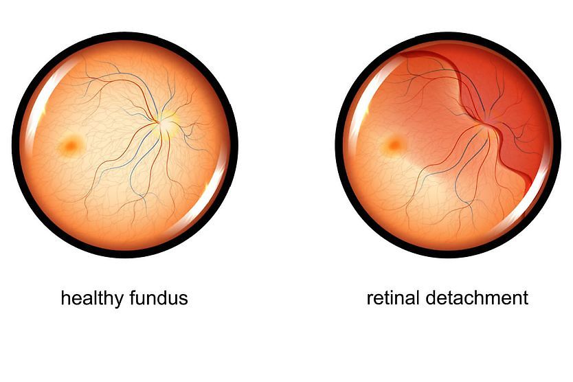
Many retinal holes are harmless, but some are of concern because they may accompany or cause a retinal tear or retinal detachment.
Types of retinal holes
There are two basic types of holes in the retina:
Atrophic retinal hole
Atrophic retinal holes are small round or oval holes that typically occur in the peripheral retina. This type of retinal hole is associated with degeneration (atrophy) of retinal tissue. Atrophic retinal holes are usually harmless and don’t require treatment. An estimated 5% of the general population has atrophic retinal holes.
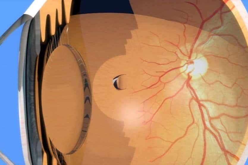
Operculated retinal hole
Operculated retinal holes are round, oval or out-of-round holes where a plug or “cap” (operculum) of retinal tissue is pulled forward into the vitreous body of the eye where it floats above the hole. Like atrophic holes, operculated retinal holes occur more often in the peripheral retina.
Retinal hole symptoms
Symptoms of a hole in the retina depend largely on the type of hole:
- Atrophic retinal holes typically have no symptoms. They are typically discovered during a routine comprehensive eye exam.
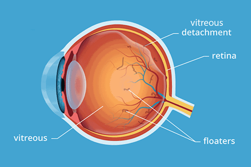
- Operculated retinal holes usually have the symptom of a noticeable vitreous floater. This is due to the cap of retinal tissue that has detached from the hole and is floating in the vitreous. Symptoms of operculated retinal holes can also sometimes include flashes of light.
Hole in retina causes
Risk factors for retinal holes include high myopia and trauma to the head or eyes. Causes of retinal holes vary by type:
- Atrophic retinal holes are caused by a localized degeneration of a small spot in the retina. The underlying cause for this atrophy is usually unknown but is generally associated with aging.
- Operculated retinal holes are caused by traction on the retina by the vitreous humor (the gel-like fluid inside the eye) as it liquifies with age. This type of retinal hole is often associated with posterior vitreous detachment. Trauma to the head or eye also may cause an operculated retinal hole.
Retinal hole surgery
In many cases, no treatment for retinal holes is required. Your eye doctor may just note the finding of a retinal hole and monitor it with routine comprehensive eye exams.
However, in some cases — for example, if vitreous fluid is seeping under the border of a retinal hole, which could increase the risk of a retinal detachment — retinal hole surgery may be performed.
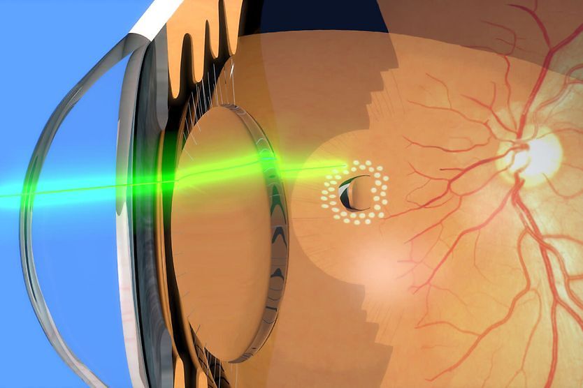
Typically, retinal hole surgery is performed with a laser, which is used to seal the retinal tissue around the hole to the back of the eye.
A retina specialist may also recommend surgery to treat a hole in the retina if the patient is experiencing flashes of light or has already had a retinal detachment in the other eye.