Lattice corneal dystrophy: Symptoms, causes and treatment options


What is lattice corneal dystrophy?
Lattice corneal dystrophy is a genetic condition that affects the cornea of both eyes (the clear protective dome at the front of the eye). There are many different corneal dystrophies. In this type, material builds up in the cornea in a pattern that looks like a lattice. This can cause irritation and vision problems over time. Both eyes are affected, although one can progress faster than the other.
Lattice corneal dystrophy is caused by a specific gene and mutation. Mutations can create abnormal versions of a protein. Sometimes these abnormal proteins don’t fold correctly which means they don’t form the right structure to do their job. They can then clump together, forming amyloid fibers, causing the corneal deposits seen in lattice corneal dystrophy.
Causes and risk factors of lattice corneal dystrophy
Lattice corneal dystrophy (type 1 or LCD1) is caused by mutations in the TGFBI gene. This gene produces a special protein called kerato-epithelin. It normally helps keep the cornea clear. But abnormal forms of it can build up, leading to lattice corneal dystrophy.
Another form of lattice corneal dystrophy (type 2 or LCD2) is caused when abnormal forms of a protein called gelsolin build up. This is caused by a mutation of the gelsolin gene (GSN) on chromosome 9. However, this type has been re-classified as a systemic disorder with effects in the cornea in addition to other parts of the body, rather than a condition that is isolated to the cornea itself. The systemic condition goes by a few names:
- Hereditary gelsolin amyloidosis (AGel)
- Familial amyloidosis of the Finnish type (FAF)
- Meretoja syndrome
Besides the cornea, it can affect the facial nerves causing drooping or weakness along with hearing loss, the nerves of the arms and legs, the heart’s electrical system and the kidneys.
Having a family history of lattice corneal dystrophy is a primary risk factor. It’s typically inherited in an autosomal dominant pattern. This means you only have to inherit one copy of the gene from a parent to get the condition.
Symptoms and diagnosis of lattice corneal dystrophy
Lattice corneal dystrophy may cause eye discomfort or problems with sight. It can be diagnosed during an eye examination or using genetic testing.
Symptoms to watch for
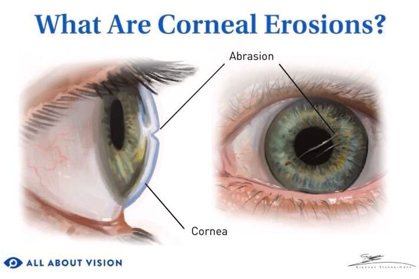
Lattice corneal dystrophy typically causes eye irritation. One source of this irritation is corneal erosion, or wearing away of the corneal surface. Corneal erosions can lead to symptoms like:
- The sensation of something in the eye
- Eye pain (especially upon waking)
- Light sensitivity
- Watery eyes
Your sight may also become blurry or hazy as material builds up in the cornea over time.
Diagnostic tools and tests
Lattice corneal dystrophy can be diagnosed during an eye examination. Your eye doctor can use a special microscope called a slit lamp to examine your eyes. Special imaging techniques like optical coherence tomography (OCT) may also be used to see different layers of the cornea.
In rare cases, your doctor may also perform a corneal biopsy for a sample of your corneal tissue. Laboratory testing can recognize certain characteristics of the disease. DNA testing may also be used to confirm the diagnosis. It’s helpful because lattice corneal dystrophy can resemble many other forms of dystrophy.
SEE RELATED: Tests in an eye exam
Treatment options for lattice corneal dystrophy
There are several treatment options for lattice corneal dystrophy, including non-surgical and surgical options. It is important to note that any non-surgical options do not treat the condition itself, but rather the effects of the condition, such as recurrent corneal erosions. Surgical intervention is the only way to remove the lattice buildup of amyloid fibers in the cornea.
However, the underlying genetic mutation remains, so it is possible for the condition to return even after surgery. Recurrence is not unusual — it is expected. After laser treatment for lattice corneal dystrophy, roughly half of eyes show a recurrence within five years and the large majority within 10. No medical or surgical treatment available today corrects the gene change itself.
Pitfall of laser vision correction
Laser vision correction, including laser-assisted in-situ keratomileusis (LASIK), photorefractive keratectomy (PRK) and small incision lenticule extraction (SMILE), are not appropriate if you have lattice corneal dystrophy or any other corneal dystrophy caused by a change in the TGFBI gene. Both the flap the surgeon creates and the laser reshaping itself can sharply accelerate deposit buildup resulting in cloudiness that can be severe and permanent.
If you have lattice corneal dystrophy or a family history of any corneal dystrophy, it is important to share this information before you are scheduled for laser vision correction. Some surgeons now recommend genetic screening before refractive surgery.
Non-surgical treatments
Non-surgical treatment of lattice corneal dystrophy typically aims to delay surgery and provide comfort. These treatments could include topical lubricants, topical antibiotics to prevent infection, topical steroids or bandage contact lenses. These are special lenses that protect the cornea, which allow healing to take place while providing pain relief. Oral antibiotics, such as doxycycline, are sometimes used; however, this is to reduce inflammation rather than treat an infection.
Symptoms of dry eye disease are especially common with the systemic kind of lattice corneal dystrophy (type 2). These can be managed with lubricating eye drops or methods that keep the eye from losing tears (like scleral contact lenses or special plugs), alongside other prescription therapies or in-office treatments.
Surgical interventions
Lattice corneal dystrophy is progressive (meaning it slowly gets worse over time). For this reason, surgical intervention may eventually be needed. In many cases, it isn’t needed until a person is in their 40s or older.
Laser treatment, called phototherapeutic keratectomy (PTK), may be used. Phototherapeutic keratectomy uses a laser to remove damaged areas of the cornea and replaces them with healthy corneal tissue from a donor. This can improve comfort by decreasing erosions and vision by removing deposits and corneal irregularities.
Lattice corneal dystrophy is typically treated with PTK, but a keratoplasty, also known as a corneal transplant, may be required if the condition becomes more severe. The procedure typically has favorable outcomes. Deposits may build up again in the years following treatment, but in many cases, they can be removed and managed with additional treatment like phototherapeutic keratectomy.
Living with lattice corneal dystrophy
Lattice corneal dystrophy is a progressive, lifelong condition. It could come back even after surgical treatment. Regular treatments to address issues such as dryness and corneal erosions are often necessary, and additional laser or surgical interventions may be needed over time.
Lifestyle adjustments
With lattice corneal dystrophy, it’s important to have regular checkups with your doctor so your treatment can be changed as needed. You should take any medications exactly as prescribed (especially if you have received a corneal transplant). This will help you manage symptoms.
You may also benefit from genetic counseling. Genetic counseling can help you understand how your disease is inherited and passed on.
Support and resources
If your sight is affected, especially after treatment, low vision clinics can be a helpful resource. Other groups, including educational bodies and charitable organizations, can provide support.
SEE ALSO: Resources for the visually impaired
Advances in research and emerging treatments
Researchers are making progress to find ways to correct the gene change itself. In research published in 2026, a technique called base editing corrected the two most common TGFBI gene changes in human corneal cells grown in a dish, fixing the altered copy of the gene while leaving the healthy copy untouched.
In some studies, gene editing in animal models and human cells affected by dystrophy was shown to be possible. Gene editing involves targeted changes in a gene sequence. This is accomplished through delivery of a special molecular system.
For treatment, stem cells, which are able to become specialized cells, may provide a useful material for future grafts. The availability of corneal graft tissue can be scarce, since it must come from a healthy donor, and there is a shortage of donor tissue in general.
READ MORE: Stem cell therapy for vision: Breakthroughs in restoring eyesight
Managing lattice corneal dystrophy for better vision
Lattice corneal dystrophy is genetic, so it cannot be prevented. It’s also progressive, so it can cause greater vision loss or worsening symptoms over time. However, many patients are able to manage this condition with minimal loss of vision. It’s important to see an eye doctor regularly to stay on top of the condition.