Corneal dystrophy: Symptoms, causes and treatments


What is corneal dystrophy?
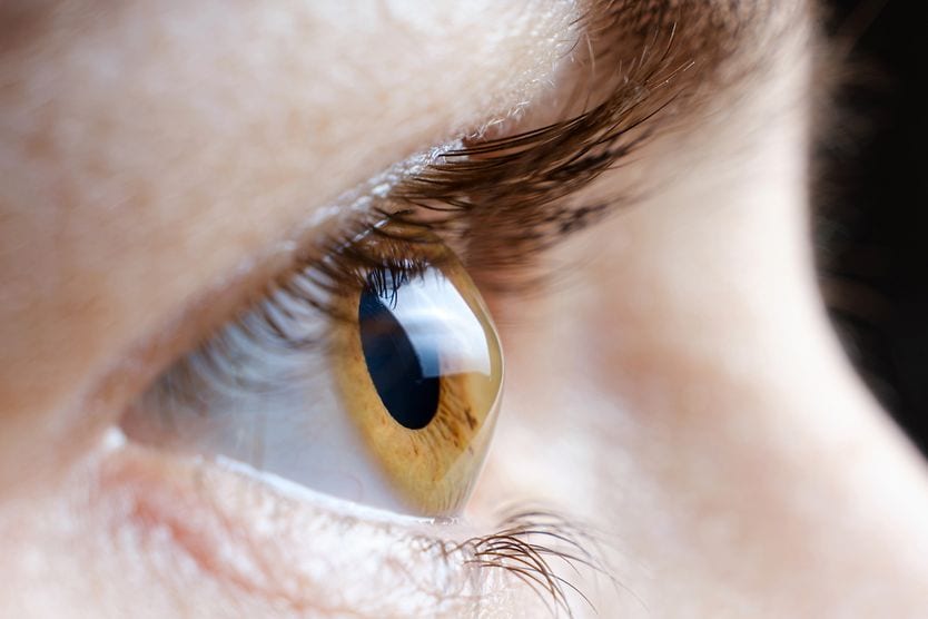
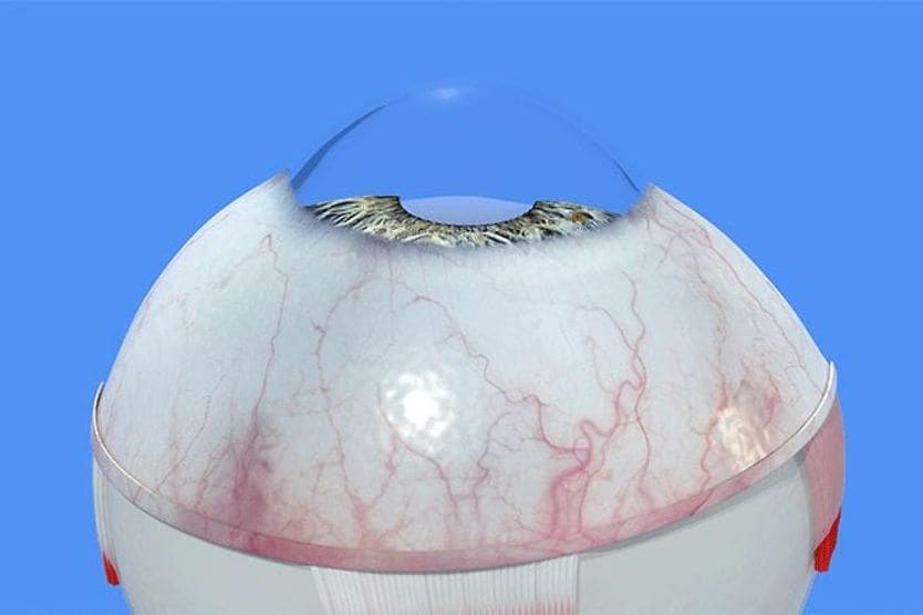
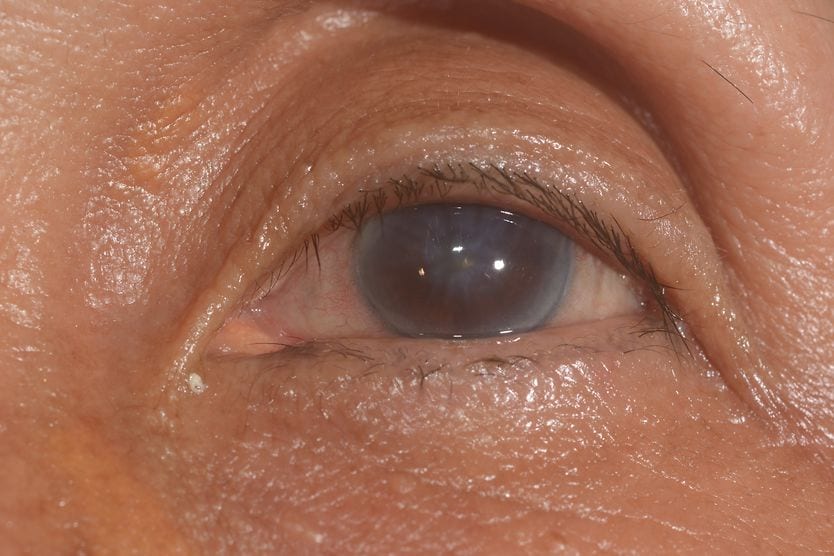
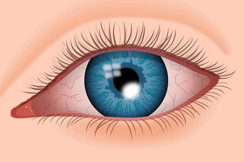
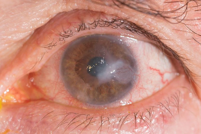
Corneal dystrophy is a group of mostly inherited conditions in which material builds up in the cornea, the clear, protective dome on the front of the eye. There are several different types based on genetic mutation and which layer of the cornea is affected. The types are grouped and named under an international classification that is updated as the genetics become clearer.
A genetic mutation is a change in a DNA sequence, which can cause your genes to function differently. This leads to a genetic condition. These can happen without any family history. There are several genetic mutations that can lead to corneal dystrophy.
Corneal dystrophies are different from corneal degenerations, which are a more common cause of deposits and clouding in the cornea. Dystrophies are usually inherited, affect both eyes in a roughly symmetric pattern, are not caused by inflammation or injury, and often begin long before symptoms appear. Degenerations, such as arcus, band keratopathy or Salzmann nodular degeneration (SND), are generally acquired, may affect one eye more than the other, and are linked to aging, previous inflammation or injury.
Corneal dystrophies can cause symptoms, like blurry vision. Although corneal dystrophy can cause some vision loss or visual impairment, it’s rare for it to cause complete blindness. With the right treatment, good vision is possible.
SEE RELATED: What are corneal deposits?
Types of corneal dystrophy
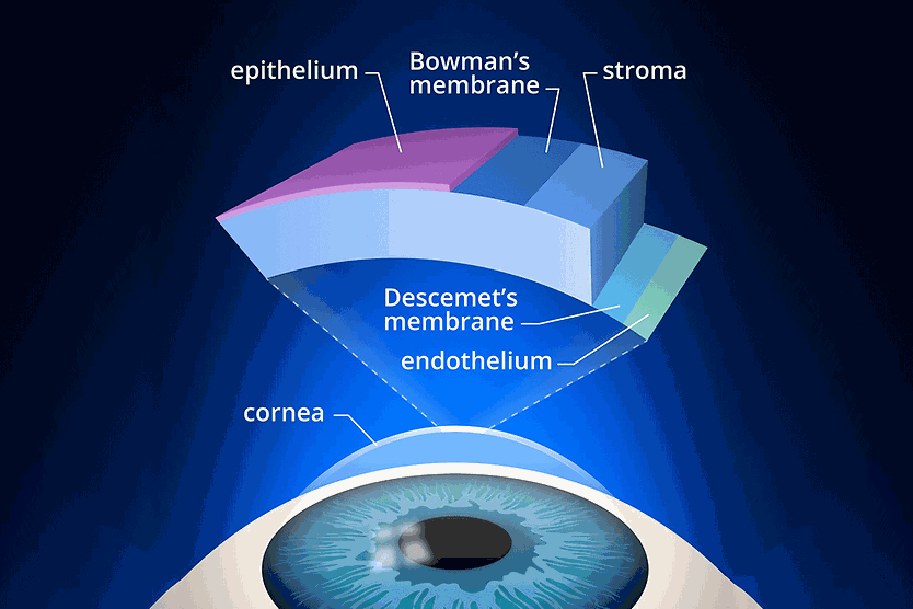
There are several different layers of the cornea that a corneal dystrophy may affect.
Epithelial and subepithelial dystrophies
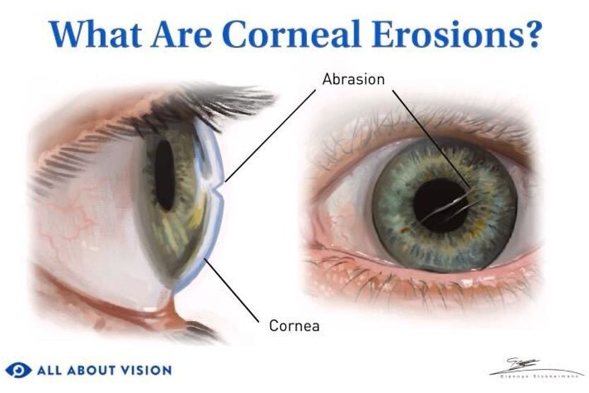
The epithelium is the outermost layer. It protects the rest of the cornea from outside irritants. An epithelial dystrophy can cause corneal erosion or wearing away of the surface. This can also expose nerve endings in the cornea, leading to pain or foreign body sensation.
Some kinds of epithelial dystrophy include:
- Map-dot-fingerprint dystrophy – When the epithelium develops folds, which may look like groups of dots or fingerprints. It is also called anterior or epithelial basement membrane dystrophy (ABMD or EBMD). It is the most common of these conditions. It is worth noting that most cases of map-dot-fingerprint dystrophy do not run in families. Current classification treats it as usually acquired rather than inherited, even though the name still describes it as a dystrophy.
- Meesmann dystrophy – When small collections of fluid called microcysts form in the epithelium. These microcysts may eventually break open, causing a flare-up in symptoms, like watery eyes or foreign body sensation.
Stromal dystrophies
The stroma is the middle layer of the cornea. It is also the thickest and the strongest. When the stromal layer is affected, it is called stromal dystrophy. Changes in the stroma may also affect other layers of the cornea:
- Lattice dystrophy – When proteins that have been “misfolded” clump together as deposits called amyloid. The buildup forms a lattice or grid. This can cause vision problems.
- Granular dystrophy – When there are granular deposits (that might look like sugar granules or crumbs) on the stroma. They grow in number and clump together over time.
- Macular corneal dystrophy – When cloudy, gray-white deposits spread across the stroma and, unlike other stromal types, extend all the way to the edge of the cornea. It usually causes more severe vision loss than lattice or granular dystrophy, and it is inherited.
Endothelial dystrophies
The endothelium is the innermost layer of the cornea. This layer is responsible for controlling the flow of nutrients and water in and out of the cornea, which is important for keeping it clear. Conditions that affect the endothelium can cause swelling, which could lead to vision loss.
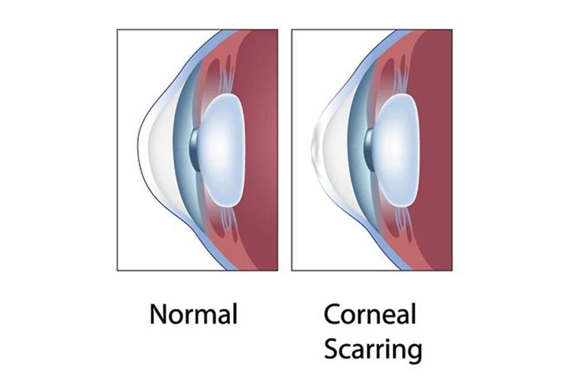
The most common type is Fuchs endothelial corneal dystrophy (FECD). In this type of dystrophy, the process of losing endothelial cells is sped up. This is a normal part of aging, but in Fuchs endothelial dystrophy, cells are lost faster and in greater numbers than normal. This can lead to swelling, which can then lead to scarring and possible vision loss.
One symptomatic pattern is classic for people with FECD. Vision is often at its worst on waking and improves over the first few hours of the day because the closed eyelid stops the cornea from drying overnight. If that sounds familiar to you, it might be helpful to mention at your next eye exam.
Congenital hereditary endothelial dystrophy is a rarer type in which the cornea is cloudy from birth or early infancy.
Symptoms of corneal dystrophy
The symptoms will depend on the specific type of corneal dystrophy. Some cases may have no symptoms. Common symptoms could include:
- Haziness or decreased clarity of vision
- Blurry vision
- Epiphora (watery eyes)
- Dry eyes
- Eye pain or the sensation of something in the eye
- Photophobia (sensitivity to light)
- Nystagmus (uncontrolled side-to-side eye movements)
- Corneal edema (swelling)
- Corneal erosion
Causes and risk factors
Corneal dystrophy is a genetic condition caused by a change in your genes. The changed gene could be inherited or due to a spontaneous change (meaning you’re the first in your family to have it). The type of corneal dystrophy will depend on the gene involved.
Some genes that could be involved include:
- TGFBI – This gene helps create a protein that connects to collagen and helps strengthen the cornea. Alterations in this gene can lead to epithelial or stromal dystrophies.
- KRT3 and KRT12 – These genes, both in the keratin gene family, help form and strengthen the corneal epithelium. Alterations in these genes can lead to Meesmann dystrophy.
- SLCA411 – This gene usually creates a protein that helps transport materials between the stroma and epithelial cells. A mutation in this gene and COL8A2 can lead to congenital hereditary endothelial dystrophy (CHED).
The majority of corneal dystrophies are inherited in an autosomal dominant pattern (you only have to inherit it from one parent to get it). Other rarer types are inherited in an X-linked dominant pattern. This type can only be inherited through genes located on the X chromosome, although inheriting just one copy is still enough to get it.
Corneal dystrophy is a genetic disease, so a family history will increase your risk. In general, it doesn’t affect people differently based on factors, such as sex or race (although Fuchs endothelial dystrophy is more likely to affect people who are white, female or over the age of 30).
Diagnosis of corneal dystrophy
Corneal dystrophy can be diagnosed during a regular comprehensive eye exam. During a comprehensive eye examination, your eye doctor uses special tools to look inside your eye. They may further investigate your cornea using tests, such as:
- Optical coherence tomography (OCT) – A special imaging test that lets doctors see parts of the eye, like the retina or cornea, in detail.
- Corneal topography – An imaging technique that provides a “map” of the cornea’s surface, showing irregularities in the curvature.
- Pachymetry – A technique that measures corneal thickness. Pachymetry can be performed with imaging, like OCT, or with a handheld ultrasonic pachymeter tool.
- Specular microscopy – A test that photographs the endothelial cells on the back of the cornea and counts them. The endothelial cell count is the key measurement in FECD and other endothelial types, and it helps your doctor judge how the condition is progressing and when treatment may be needed.
Genetic testing may also be performed using a blood or saliva sample to confirm the specific type of corneal dystrophy. Most corneal dystrophies are diagnosed during the eye exam itself, so genetic testing is not usually needed to make the diagnosis.
It is used when the picture is unclear, when family planning questions come up, and importantly before laser vision correction, where the result can change the recommendation.
SEE ALSO: How DNA health testing can detect eye diseases
Treatment options for corneal dystrophy
Treatment for corneal dystrophy will depend on its type and severity.
Noninvasive treatments
For epithelial dystrophies, you may be given surface treatments, like special contact lenses meant to reduce pain and help your corneas heal. Eye drops or artificial tears may help eye dryness, pain or foreign body sensation. Hypertonic solution may be used to treat Fuchs dystrophy. Treatments like these can help with corneal erosion, which is a common symptom of corneal dystrophy.
In some cases, damaged areas of the cornea can be removed using laser treatment, like phototherapeutic keratectomy.
Surgical treatments
If the cornea is damaged enough, a corneal transplant (keratoplasty) may be needed. This procedure replaces damaged areas of the cornea with healthy tissue from a donor.
If the endothelium is affected, partial corneal transplantation may be used:
- Descemet’s membrane endothelial keratoplasty (DMEK) – This type removes the diseased corneal endothelium along with the Descemet membrane, the thin layer it sits on. They are replaced with donor tissue made up of endothelium and Descemet's membrane, with no other layers attached.
- Descemet’s stripping endothelial keratoplasty (DSEK) – This type removes the same tissue as DMEK, but the donor graft includes a thin layer of corneal stroma along with endothelium and Descemet’s membrane. While the extra layer makes the DSEK graft easier to handle during surgery, DMEK’s thinner graft tends to give sharper vision long-term.
Rejection is still possible at any point, and the majority of people typically use corticosteroid eye drops long-term to lower that risk. Grafts can also fail years later and be repeated.
Living with corneal dystrophy
Many people with corneal dystrophy have good vision with the right treatment. But corneal dystrophy is lifelong, and you should see your eye doctor regularly to monitor the condition and update treatment as needed. Even successful treatments may need to be maintained.
It’s important to follow any treatment directions from your care provider and to seek help from a specialist when needed. Numerous organizations can help you find treatment or cope with any vision loss.
Advances in research and treatment
Many other ways of treating corneal dystrophy are being researched.
For one, it might be possible to correct the genetic mutations that cause corneal dystrophy. Methods like gene therapy and engineering special molecules for correcting mutations may help with this. For another, special medications called neuroprotective agents may help cells survive and function properly, which could help in degenerative conditions, like corneal dystrophy.
Stem cells, which are special cells that can turn into a variety of different specialized cells, are another avenue. Using stem cells as graft material may prove to be more beneficial than a typical corneal graft.
For nonsurgical methods, injectable corneal endothelial cells may be a future alternative to surgery. Trials with injectable cells that have been engineered to go to a specific area are underway.
Note: None of the approaches are currently approved by the U.S. Food and Drug Administration (FDA) as a treatment for corneal dystrophy.
READ MORE: Stem cell therapy for vision: Breakthroughs in restoring eyesight
Protecting your vision
There are several types of corneal dystrophy, and symptoms, causes and treatments will vary for each. Even if there are no symptoms or they are being managed, regular follow-up with an eye care professional is important to monitor progression. An eye care specialist will be able to help determine the best way to monitor and treat. If you are considering laser vision correction, it is recommended to inform your surgeon about any family history of corneal dystrophy before you are screened.