An overview of corneal diseases

What is corneal disease?
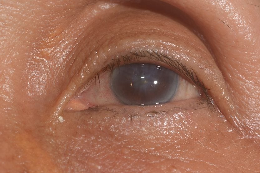
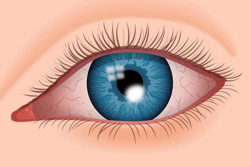
Corneal diseases refer to conditions that affect and damage the cornea. This damage can be caused by factors, such as inflammation, infection, dryness, dystrophy or degeneration. These diseases can lead to pain, discomfort and potential vision loss due to permanent clouding or scarring.
Another name for corneal disease is keratopathy. About 3% to 5% of vision loss and blindness worldwide is due to corneal opacities resulting from corneal disease. Nearly 80% of these cases are preventable or treatable.
RELATED: 15 keratopathies: Causes, symptoms and treatment
What is the cornea?
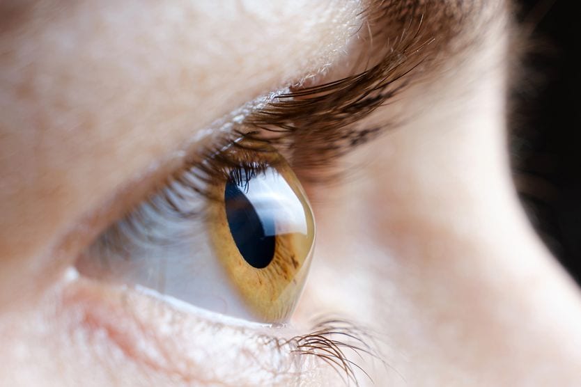
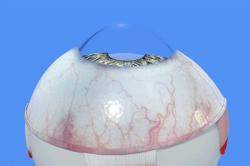
The cornea is the transparent, dome-shaped tissue at the front of the eye. It serves as a protective barrier against bacteria, viruses and particles. It also filters some ultraviolet (UV) radiation.
It is highly sensitive to pain due to its large number of nerve endings, which help protect the eyes from injury. These nerve endings respond to temperature, touch and chemicals, often triggering an involuntary blink when stimulated. The cornea can heal superficial injuries very quickly, typically regenerating within 24 to 48 hours.
The cornea also plays an important role in focusing light that enters the eye. Because of its dome shape, it bends incoming light. This helps focus light rays on the light-sensitive tissue, the retina, at the back of the eye.
Why is the cornea clear?
The cornea is clear because it is mainly composed of well-organized collagen fibers and water. The orderly arrangement of these fibers allows light rays to pass through. A transparent cornea is essential for clear vision.
To ensure nothing obstructs the path of the light rays, the healthy cornea is avascular, meaning it lacks blood vessels. Instead, the cornea receives oxygen from the tear film and atmosphere along with nutrients from the fluid inside the eye called aqueous humor.
With every blink, tears spread across the cornea, keeping it moist and nourished while clearing away debris. The cornea receives additional support from a nourishing fluid called aqueous humor, which is located inside the eye.
How does corneal disease affect vision and eye health?
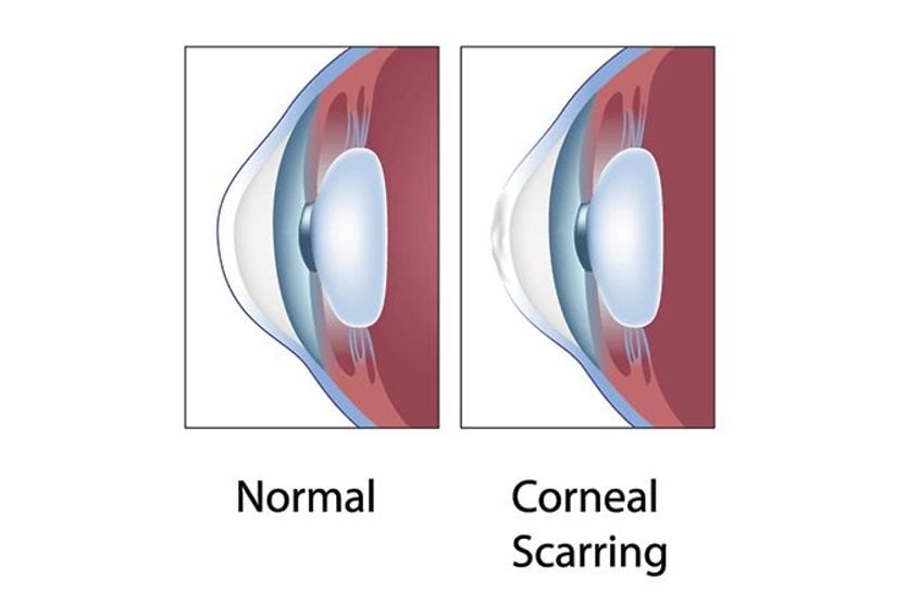
Conditions that affect the cornea can decrease its clarity. Severe, untreated corneal disease can lead to vision loss.
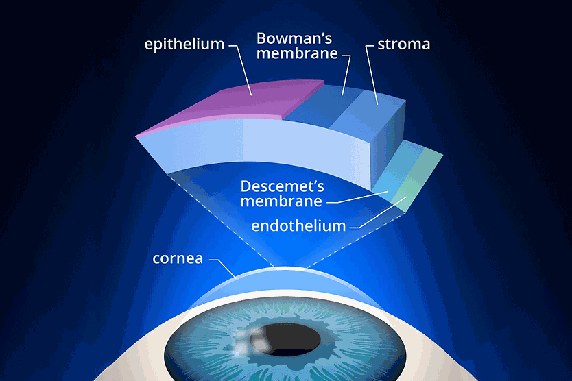
Corneal diseases often cause corneal inflammation, a condition known as keratitis. This inflammation can occur for various reasons, including infections, allergies, injuries or other health issues. Each of these conditions affects different parts of the cornea, which is composed of distinct layers. These layers provide structure and help protect against particles and germs. Damage to each layer of the cornea affects vision and overall eye health in unique ways.
The layers of the cornea are:
- Epithelium – This is the smooth outer layer. It serves as a barrier, absorbing nutrients and oxygen from tears. It can start to heal in 24 to 48 hours from superficial injuries with complete regeneration around seven to 14 days. It is rich in nerve endings, making it highly sensitive to pain.
- Bowman’s layer – A transparent protective layer made of collagen. Unlike the epithelium, Bowman's layer does not regenerate; injuries to this layer heal by scarring.
- Stroma – The middle layer made of water and collagen. It accounts for approximately 90% of the cornea’s thickness, providing it with structure and elasticity. It is important in keeping the cornea transparent.
- Pre-Descemet’s layer – Also known as Dua’s layer, experts debate whether this is a separate sixth layer or part of the stroma. However, research indicates it has distinct features.
- Descemet’s membrane – This is the protective layer of collagen fibers. This membrane recovers well from injury.
- Endothelium – A single layer of cells that pumps excess water out of the stroma. It keeps the stroma from absorbing too much fluid (which would cause swelling and hazy vision). The corneal endothelium does not have the ability to regenerate.
What are the common symptoms of corneal diseases?
Symptoms of corneal disease can vary based on the specific condition. Some conditions may not cause symptoms initially but still require medical care. However, due to the large number of nerve endings, many corneal problems can cause significant pain and discomfort. Symptoms typically include:
- Eye pain (mild to severe)
- Blurry vision or vision that gets progressively worse
- Sensitivity to light (photophobia)
- A foreign body sensation, described as feeling grit or something stuck in the eye
- Red or bloodshot eyes
- Watery eyes or excessive tearing
- Pus or discharge from the eye
- Trouble keeping the eye open
What causes corneal diseases?
Corneal diseases develop from a variety of conditions that contribute to corneal damage. For example, injury to the cornea from trauma can lead to complications, such as infection and an ulcer. Common causes of corneal disease may include:
- Infectious/microbial keratitis (bacterial, viral, fungal or parasitic)
- Noninfectious keratitis
- Corneal dystrophy (which runs in families)
- Corneal degeneration due to aging or disease
- Dry eye
- Systemic conditions, such as autoimmune diseases (including rheumatoid arthritis)
Risk factors
Several factors can increase the likelihood of developing corneal disease, including:
- Improper contact lens hygiene – Increases the risk of developing corneal infections and other complications. This includes not cleaning lenses properly, wearing them too long or swimming with contacts.
- Dry eye – Occurs when the eyes don’t produce enough tears. This can lead to dryness and increase the chance of corneal issues.
- Environmental exposures – Prolonged exposure to dust, chemicals or ultraviolet light can damage the corneal surface over time. People who work outdoors or in industrial settings face higher risks from these environmental factors.
- Medical conditions – These include a weakened immune system, autoimmune diseases, connective tissue diseases and previous eye injuries.
- Repeated eye rubbing – This is associated with certain corneal diseases, including keratoconus. The risk is higher for those with severe allergies or atopic keratoconjunctivitis, a condition associated with eczema.
- Use of corticosteroid eye drops – Long-term use may cause thinning of the cornea. Steroids may also increase the risk of steroid-induced ocular hypertension/glaucoma, posterior subcapsular cataract and susceptibility to infection (especially herpetic and fungal keratitis).
Examples of corneal diseases
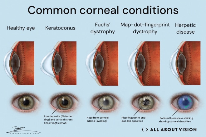
Although there is a great deal of overlap, corneal conditions can be categorized into broad categories:
Corneal ulcer
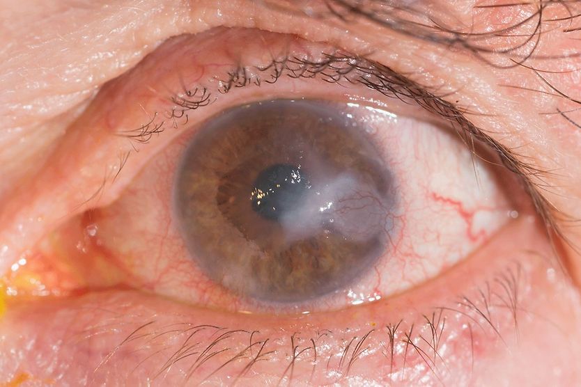
A corneal ulcer is an open sore on the surface of the cornea. It is often the result of infectious keratitis, but it can also develop from other disorders. A corneal ulcer can quickly threaten your vision, so you need to see a doctor right away. Treatment generally requires antibiotics or antifungals.
Infectious (microbial) keratitis
Infections caused by bacteria, viruses, fungi or parasites result in infectious keratitis. These infections can rapidly develop into a corneal ulcer.
Bacterial keratitis
Bacterial infections can cause severe corneal damage. These infections can lead to vision loss if left untreated.
Risk factors include:
- Poor contact lens hygiene
- Weak immune system
- Underlying eye disease
- Recent eye injury
Viral keratitis
Herpes simplex virus type 1 (HSV-1) — the same virus that causes cold sores — is the most common cause of viral keratitis, though HSV-2 can occasionally be responsible. The cornea can become infected when the virus travels from another part of the body, such as the mouth. Most cases heal without lasting effects. However, severe infections can cause corneal scarring and lead to permanent vision problems.
Varicella-zoster virus (VZV), which causes chickenpox, can reactivate later in life as herpes zoster, known commonly as shingles. It may affect the eye as a condition called herpes zoster ophthalmicus (HZO).
Herpesviruses may lead to corneal ulcers or ongoing keratitis.
Fungal keratitis and Acanthamoeba keratitis
Fungal keratitis and Acanthamoeba keratitis are not as common as bacterial or viral keratitis:
- Fungal keratitis – An eye injury, especially from plants, can lead to fungal keratitis. It must be treated promptly to prevent potential vision loss.
- Acanthamoeba keratitis – Most cases occur in contact lens wearers who have not handled their lenses properly. It can also happen if the lenses are exposed to water, such as during showering or swimming. While it is rare, it is a serious condition that requires immediate medical attention.
Noninfectious keratitis
Noninfectious keratitis can be due to several underlying causes, including:
- Eye injury or foreign body in the eye
- Severe dry eye
- Photokeratitis (damage from intense ultraviolet radiation)
- Contact lens overwear
- Vitamin A deficiency
The leading cause of preventable vision loss in children globally is vitamin A deficiency.
Ectasia
Keratoconus
Keratoconus is the most common form of corneal ectasia worldwide. Ectasia means the cornea becomes thinner and bulges outward, which in the case of keratoconus is in a cone-like shape. This change can lead to blurry vision, often starting in the late teens to early 20s. Its progression can be linked to genetics, frequent eye rubbing or other environmental factors.
Signs that doctors look for during an eye exam include:
- Rizzuti sign – A bright, cone‑shaped light reflex near the inner edge of the cornea.
- Fleischer ring – A brownish ring of iron deposits around the base of the cone on the cornea.
- Vogt striae – Fine, vertical “stress lines” inside the cornea that can come and go with gentle pressure on the eye.
It can be managed with glasses, special contact lenses or outpatient procedures. There are several surgical options including corneal cross-linking (CXL). This procedure strengthens the corneal fibers and can slow or stop disease progression. It can help many people avoid a corneal transplant.
Corneal dystrophies
Corneal dystrophies are rare, slow-progressing disorders that run in families. They typically affect both eyes and are non-inflammatory. They cause the buildup of abnormal material within one or more corneal layers, leading to clouding and vision loss. There are over 20 corneal dystrophies that affect the anterior, middle and posterior parts of the cornea.
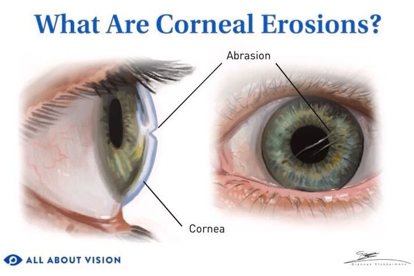
A common symptom of corneal dystrophies is recurrent corneal erosion (RCE). This occurs when the corneal epithelium doesn’t adhere properly to the eye. It separates from the layers underneath when the eyes are opened in the morning. This can lead to pain, blurred vision, tearing and light sensitivity.
Fuchs’ dystrophy
Fuchs’ endothelial corneal dystrophy (FECD) damages the innermost corneal layer, the endothelium. The endothelial cells are responsible for pumping fluid out of the cornea. As the cells die, fluid accumulates, leading to corneal edema (swelling) and cloudiness.
Vision is usually blurriest in the morning. This happens because fluid builds up overnight. As the disease progresses, the cornea remains swollen throughout the day, leading to constant blurred vision. This condition is more common in women. It can be managed with drops, ointments or gently drying the eye with a hairdryer on a cool setting at arm’s length.
If these treatment options are not improving your condition, an eye doctor may recommend surgeries such as:
- Descemet’s membrane endothelial keratoplasty (DMEK) – A partial-thickness corneal transplant that replaces only the innermost cell layer of the cornea along with its supporting membrane.
- Descemet’s stripping automated endothelial keratoplasty (DSAEK) – A partial-thickness corneal transplant that replaces only the damaged inner layers of the cornea with a thin disc of healthy donor tissue.
Lattice dystrophy
This is a rare inherited condition that causes cloudy, line‑shaped deposits to form in the cornea. These “lattice‑like” lines usually start in the center of the cornea and slowly reduce vision over time. Some people also get epithelial erosion. This can cause irritation, pain or temporary changes in vision. Lattice corneal dystrophy has several subtypes that affect people differently.
Epithelial basement membrane dystrophy
Epithelial basement membrane dystrophy (EBMD) is also called map‑dot‑fingerprint dystrophy or anterior basement membrane dystrophy (ABMD). It causes small, irregular patterns, like maps, dots or faint lines, that can make vision blurry or inconsistent. Some people have no symptoms, while others notice eye irritation, a gritty sensation or pain from recurrent erosions.
It often develops in adults and can worsen with eye injury or surgery. There’s no known way to prevent it, but nighttime lubricating ointments or hypertonic salt ointments are often recommended.
Dry eye disease
Tears form a thin, smooth film over the front of the eye every time you blink, keeping the surface moist, clear, and comfortable so you can see well.
This tear film has three layers:
- An oily layer that stops tears from drying too fast
- A watery layer that washes away dust and germs
- A mucus layer that helps the tears spread evenly and stick to the eye
Dry eye disease (DED) can result when you do not make enough tears or when one or more of these layers is not working properly. This can lead to a dry, irritated cornea.
DED can cause:
- Burning or stinging
- A gritty or sandy feeling
- Blurry vision during sustained near tasks or digital screen use
- Redness
- Mucus strings
- Discomfort with contact lenses
- Watery eyes when the eye tries to “wash away” the irritation
- Light sensitivity (photophobia)
Treatment for dry eye may include:
- Artificial tears
- Ointments at night
- Blocking the tear drains with tiny plugs to keep tears on the eye longer
- Prescription eye drops
- Treating eyelid or oil‑gland problems with at-home remedies or in-office procedures
Corneal disease due to degenerations
Degenerations are deteriorations of the corneal tissue due to disease, aging, injuries, infections or other environmental causes. Unlike dystrophies, corneal degenerations aren’t typically inherited or caused by a gene mutation. They usually do not affect vision as much either.
Examples of corneal degenerations include:
- Arcus senilis – An age-related grayish band in the peripheral cornea from fat deposition.
- White limbal girdle – Age-related white arcs or lines near the outer cornea.
- Band keratopathy degeneration – A buildup of calcium that, usually due to an underlying disease, forms a horizontal “band” across the cornea.
How do eye doctors diagnose corneal diseases?
An eye doctor can diagnose corneal conditions during a comprehensive eye exam. This will typically include:
- Slit lamp exam – This is a microscope with a light attachment that allows detailed examination of the cornea.
- Fluorescein eye stain test – A dye that is placed on the cornea to make abrasions (scratches) or subtle damage to the cornea visible under a specialized light.
- Corneal topography and pachymetry – Mapping the cornea’s shape and thickness helps in screening and early detection of corneal diseases. This is especially important for conditions like keratoconus.
How are corneal diseases treated?
Treatment plans for corneal diseases depend on the specific cause and severity of the condition.
Lifestyle and home care
You can manage symptoms and reduce risks through lifestyle changes and proactive home care:
- Protective eyewear – Always wear safety goggles when participating in sports or when working with tools, chemicals or during yard work.
- UV protection – Protect your eyes from UV radiation by wearing sunglasses.
- Contact lens safety – Follow instructions for cleaning, disinfection and wearing-time guidelines.
- Managing symptoms – Use lubricating eye drops, artificial tears and ointments for comfort.
- Avoid rubbing your eyes – Excessive rubbing is associated with corneal damage and thinning.
- Get routine eye exams – Exams can catch conditions early, decreasing the risk of vision loss.
Medications
Medications are typically used to treat infections and alleviate symptoms:
- Eye drops and ointments – Recommended for comfort and managing dry eye symptoms.
- Antibiotic, antiviral or antifungal medication – Prescribed for bacterial or fungal corneal ulcers. Antivirals may be used for infectious conditions like herpesviruses.
- Pain relief and anti-inflammatory eye drops – Recommended for symptoms of pain and to decrease swelling.
Procedures
When medical management is insufficient, more advanced surgical or laser procedures may be needed. These include:
- Corneal cross-linking (CXL) – This procedure uses vitamin B2 (riboflavin) and UV light to strengthen collagen bonds in the cornea. This stabilizes the cornea and prevents further bulging. There are two FDA-approved methods to perform the procedure called “epi-on” and “epi-off.”
- Phototherapeutic keratectomy (PTK) – This laser surgery removes damaged superficial layers of the cornea, allowing healthy tissue to regrow and potentially restore clearer vision.
- Intrastromal corneal ring segments (ICRS) – These small acrylic implants are placed in the cornea. They help reshape the cornea and improve vision. This is especially beneficial for patients with mild-to-moderate keratoconus.
- Corneal allogenic intrastromal ring segments (CAIRS) – Is a minimally invasive surgical procedure used to treat keratoconus and other corneal ectasias by reshaping and reinforcing a weak, bulging cornea.
- Corneal tissue addition keratoplasty (CTAK) – A minimally invasive surgical procedure designed to reshape and flatten the corneas using donor corneal tissue to treat keratoconus.
- Corneal transplantation (penetrating keratoplasty) – This surgery may be necessary if corneal damage cannot be repaired. The diseased cornea is replaced with a healthy donor cornea.
RELATED: What to expect with corneal transplant surgery
When to see an eye doctor
Regular comprehensive eye exams are vital to healthy eyes. The American Optometric Association recommends that adults (age 18 or older) should have an eye exam every year. If you experience persistent blurry vision or eye pain, you should consult an eye doctor as soon as possible. Contact your eye doctor immediately if you experience:
- Severe or intense eye pain
- Sudden or severe blurred vision or other changes in vision
- Sudden onset of flashes or floaters in your vision
- Significant eye redness
- A serious eye trauma or injury
- Symptoms of an eye infection, such as fever, redness, swelling or thick discharge coming from the eye