Diabetic retinopathy stages

What is diabetic retinopathy?
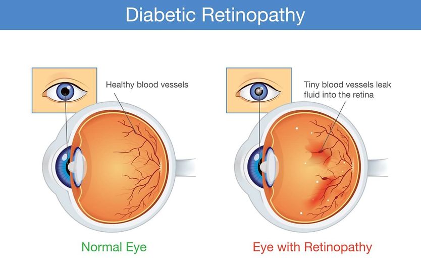
Diabetic retinopathy is a complication of diabetes that damages the small blood vessels in the retina, the light-sensitive tissue lining the back of the eye. Over time, high blood glucose can cause the retina’s blood vessels to weaken, leak or become blocked.
Recent estimates state that about 40 million U.S. adults, or roughly 12% of the population, have diabetes. About 1 in 4 people with diabetes in the U.S. has diabetic retinopathy, and about 1 in 20 has a form that threatens their vision.
Doctors divide diabetic retinopathy into two main types: non-proliferative diabetic retinopathy (NPDR), in which abnormal new blood vessels have not developed, and proliferative diabetic retinopathy (PDR), in which they have.
NPDR (stages 1-3) is the earlier form and is further divided into mild, moderate and severe. The earlier stages may develop before vision is affected.
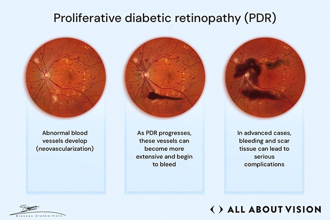
PDR (stage 4) is the advanced form in which new abnormal blood vessels begin to develop (also called neovascularization). This stage may lead to permanent vision loss.
When damaged blood vessels can no longer deliver enough oxygen, the retina releases a protein called vascular endothelial growth factor (VEGF). VEGF makes blood vessels leakier and triggers the growth of fragile new vessels.
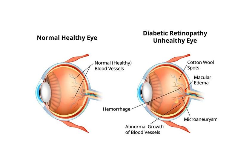
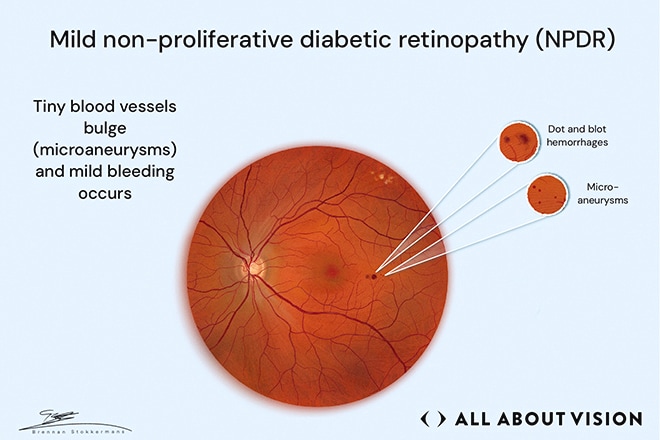
- Stage 1: Mild NPDR – Tiny bulges, called microaneurysms, are visible in retinal blood vessels.
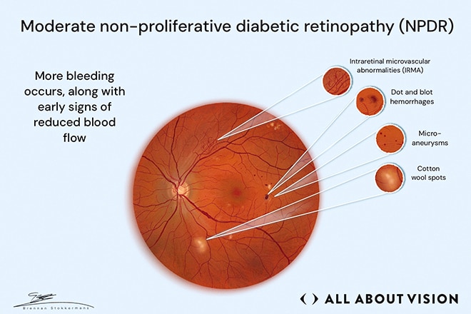
- Stage 2: Moderate NPDR – Additional damage has occurred, such as areas of reduced blood flow (cotton wool spots) and small amounts of bleeding (dot and blot hemorrhages).
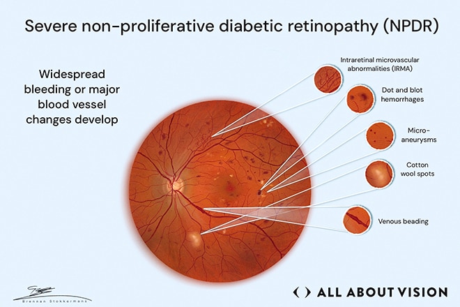
- Stage 3: Severe NPDR – Blood vessel damage is extensive, but abnormal new blood vessels have not started to grow.
- Stage 4: Proliferative diabetic retinopathy – PDR marks the transition to the most advanced stage. Abnormal new blood vessels begin to grow and can bleed inside the eye. Scar tissue may form and detach the retina.
What is diabetic macular edema and can it occur at any stage?
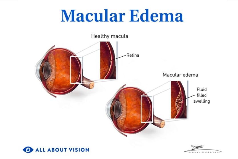
A complication that can occur at any stage is called diabetic macular edema (DME). It occurs when damaged retinal blood vessels leak fluid into the macula, the central part of the retina responsible for detailed vision, resulting in blurry central vision. It is the most common cause of vision loss in people with DR.
Another complication is diabetic macular ischemia (DMI). It occurs when the macula does not receive enough blood. Unlike DME, which involves fluid leakage and swelling, DMI results from reduced blood flow through the retina’s smallest blood vessels.
How quickly does diabetic retinopathy progress?
One long-term study found that most people with type 1 diabetes and more than half of those with type 2 diabetes had some signs of diabetic retinopathy after 20 years.
However, there is no set timeline for someone with diabetes to move from one stage to the next. Typically, the longer someone has diabetes, the more likely diabetic retinopathy becomes. As the retinopathy becomes more severe, it is more likely to progress. The risk is higher for people who:
- Have high blood sugar levels
- Have high blood pressure
- Have high cholesterol
- Smoke
- Are pregnant
Eye exams can detect retinal changes early, improving your chances of maintaining clear vision. As diabetic retinopathy progresses, you may need more frequent monitoring or treatment.
Tests that help determine the stage of diabetic retinopathy include:
- Dilated eye exam – Eye drops widen the pupils so the eye doctor can examine the retina for signs of diabetic retinopathy.
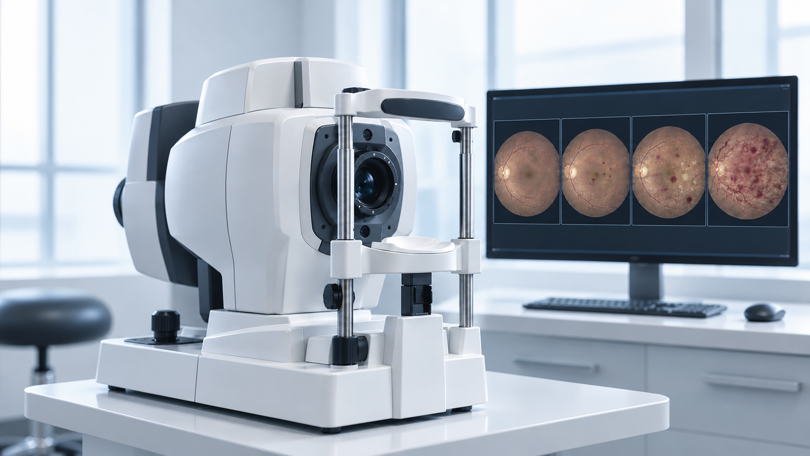
- Fundus photography – Photographs of the back of the eye can document retinal changes.
- Optical coherence tomography (OCT) – An imaging test that provides detailed images of the retina and can show and measure swelling when diabetic macular edema is suspected.
- Optical coherence tomography angiography (OCTA) – An imaging test that maps blood flow in the retina and shows areas of reduced blood flow and abnormal blood vessels.
- Fluorescein angiography – A dye is injected into a vein in the arm which travels to the retina’s blood vessels, showing leaks where present, areas without blood flow and abnormal new vessels.
What are the stages of diabetic retinopathy?
Diabetic retinopathy is generally described as mild, moderate or severe non-proliferative diabetic retinopathy, followed by proliferative diabetic retinopathy. The risk of progressing to more advanced stages of diabetic retinopathy increases the longer your blood sugar remains high.
Type: Non-proliferative diabetic retinopathy
The earlier stages of diabetic retinopathy are called non-proliferative diabetic retinopathy. Your eye doctor may detect signs of damage during an eye exam, even if vision seems normal early on. Early NPDR is often monitored rather than treated directly, but more severe disease or complications, such as DME, may require treatment.
Stage 1: Mild non-proliferative diabetic retinopathy
Mild NPDR is the earliest stage. Your vision may still seem normal, even though your eye doctor can see early changes in the retina. The main findings are microaneurysms (tiny bulges in the retina’s small blood vessels) and mild bleeding. They are usually the first visible sign of diabetic retinopathy. Early-stage findings can overlap, and leaking fluid may also result in deposits of fat and protein called hard exudates.
At this stage, your doctor may monitor your eyes for further changes. Managing blood sugar, blood pressure and cholesterol can reduce the risk of diabetic retinopathy or slow its progression.
Stage 2: Moderate non-proliferative diabetic retinopathy
In moderate NPDR, more changes appear in the retina’s blood vessels. Your vision may still seem normal, even though your eye doctor can see that the disease has progressed.
Your eye doctor may see:
- Dot and blot hemorrhages – Small, round areas of bleeding within the retina.
- Microaneurysms – Tiny bulges in weakened areas of blood vessels.
- Cotton wool spots – Small, whitish patches caused by temporarily reduced blood flow.
- Intraretinal microvascular abnormalities (IRMA) – Small blood vessels within the retina become more prominent in areas with poor blood flow.
Not everyone with moderate NPDR will have all of these findings, and signs such as hard exudates may be present as well. Moderate NPDR means there are changes beyond microaneurysms, but they have not reached the criteria for severe NPDR.
Stage 3: Severe non-proliferative diabetic retinopathy
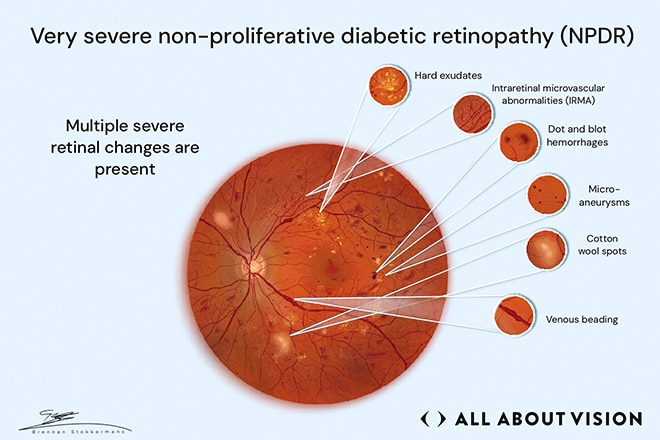
As NPDR becomes severe or very severe, the retinal damage becomes more widespread. Doctors look at how much of the retina is affected by bleeding and other blood vessel changes to determine the stage.
Key findings include those seen in moderate NPDR, with additional signs of damage, such as:
- Venous beading – Retinal veins that become irregular or look beaded.
- Hard exudates – Yellowish deposits left behind when damaged blood vessels leak.
- Prominent IRMA – Abnormal small blood vessels that are more widespread than in moderate NPDR.
Severe NPDR is diagnosed when any one of these is present:
- Bleeding in all four quadrants (sections) of the retina
- Beaded veins in at least two quadrants
- Prominent IRMA in at least one quadrant
Because severe and very severe NPDR have a greater risk of progressing to PDR, your doctor may recommend closer monitoring or treatment. Laser treatment may be considered in some cases.
In stage 3, new abnormal blood vessels have not begun to grow. Once they do, the disease is classified as stage 4 proliferative diabetic retinopathy.
Type: Proliferative diabetic retinopathy
This is stage 4, the most advanced form of diabetic retinopathy. Abnormal new blood vessel growth, called neovascularization, has developed, marking the transition from NPDR to PDR. These abnormal blood vessels can bleed and lead to serious complications, such as:
- Preretinal hemorrhage – Blood collects in front of the retina.
- Vitreous hemorrhage – Blood leaks into the vitreous, the clear gel inside the eye, and can cause new floaters, dark spots in vision or potential sudden loss of vision.
- Neovascular glaucoma – Abnormal new blood vessels interfere with fluid drainage from the eye, which can raise eye pressure along with causing eye pain, redness and blurred vision.
- Tractional retinal detachment – Scar tissue pulls the retina away from the back of the eye.
Complications of PDR may lead to vision loss. Seek immediate medical care (on the same day) from an eye doctor, retina specialist or emergency facility if you experience signs of a retinal detachment, such as:
- A sudden increase in floaters, which may look like small dark spots or squiggly lines drifting across your vision
- Flashes of light in one or both eyes
- A dark shadow or curtain moving across part of your vision
Treatment for PDR may include:
- Anti-VEGF treatment – Medicines injected into the eye that reduce abnormal blood vessel growth.
- Panretinal photocoagulation – Also called scatter laser surgery, this treatment uses a laser to decrease abnormal blood vessels.
- Vitrectomy – Surgery that removes most of the vitreous gel when there is significant bleeding or scar tissue inside the eye.
Treatment can stabilize diabetic retinopathy and lower the risk of severe vision loss, but it does not cure the disease. Many people need repeat injections or additional laser treatment. Vision that has already been lost may not fully return.
Other problems that can occur with diabetic retinopathy
Certain complications can occur at any stage of diabetic retinopathy, resulting in reduced vision.
Diabetic macular edema
Diabetic macular edema occurs when damaged blood vessels leak fluid into the macula, the part of the retina responsible for sharp central vision, causing it to swell. Vision may become:
- Blurry
- Warped or wavy
- Faded, especially colors
- Different in each eye, especially size of objects
- Worse over time
Treatment depends on where the swelling is and how it is affecting vision. Anti-VEGF injections are often the first step when DME affects the center of the macula and reduces vision. Laser treatments or steroid injections may also be needed in some cases.
Researchers are also testing longer-lasting approaches, such as standalone gene therapies designed to help the eye make its own anti-VEGF medicine, along with combination gene and anti-VEGF therapies, to reduce how often injections are needed. These treatments are still being studied and available only through clinical trials.
Diabetic macular ischemia
Diabetic macular ischemia (DMI) can occur when the macula does not receive enough blood supply. It can occur with diabetic retinopathy but is not another stage of the disease.
There is currently no approved treatment specifically for DMI. Researchers are studying treatments aimed at protecting retinal tissue or addressing impaired blood flow.
Protecting your vision
Diabetic retinopathy can progress before vision changes are noticeable. Eye exams and retinal screenings help doctors find and monitor changes that may not yet cause symptoms.
Protecting vision also involves managing the health factors that can affect diabetic retinopathy, like:
- Keeping up with recommended eye exams
- Maintaining glycemic control by keeping blood glucose in the target range
- Managing blood pressure and cholesterol
- Watching for sudden vision changes
- Scheduling an eye exam before pregnancy if you have diabetes
Managing diabetes can also help lower the risk of diabetes-related eye disease. This includes:
- Staying active
- Eating a healthy diet
- Taking medications as prescribed
The stage of diabetic retinopathy plays an important role in guiding your care, but it is not the only factor. DME or other complications can change over time. Any effect on vision can also determine how often you need follow-up and whether treatment is recommended.
Stay consistent with recommended eye exams so changes can be detected and treated early. If you notice sudden changes in your vision, contact your eye doctor promptly.