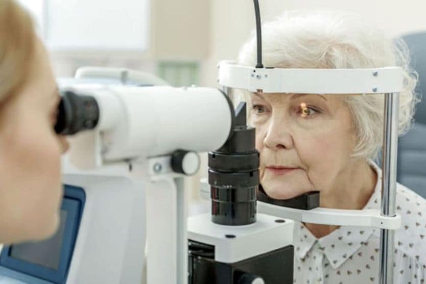
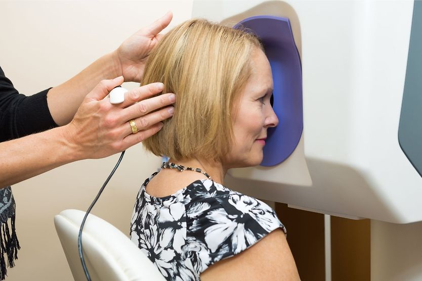
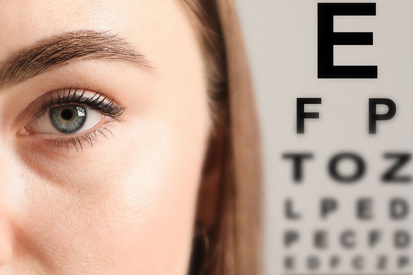
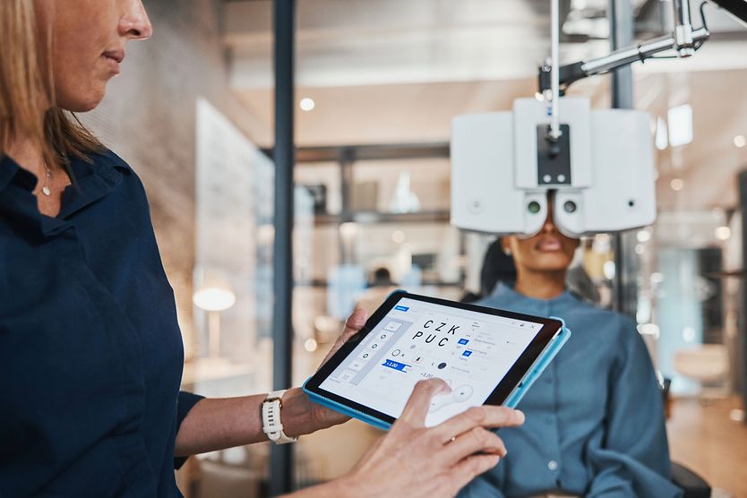
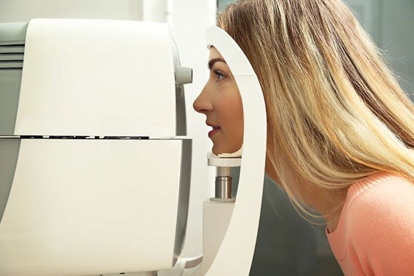
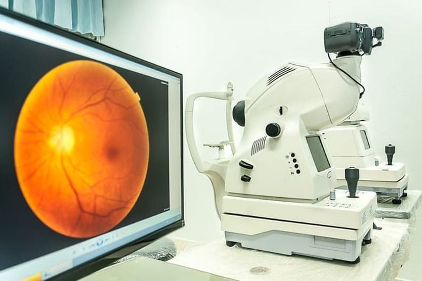
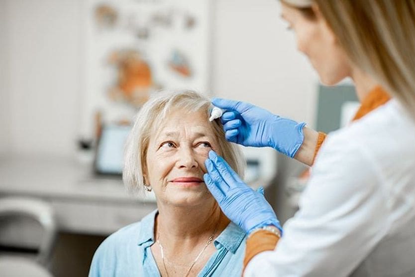
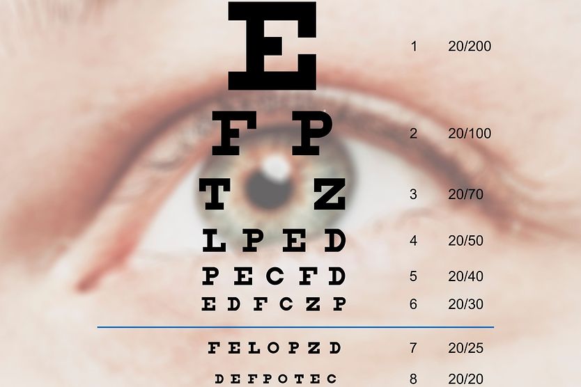
Home Eye Care Eye Exams | Low vision eye exams

How a low vision eye exam differs from a routine eye exam

Optometrist using phoropter to check eyesight value for a senior woman

What is a low vision exam? 

A low vision exam is a specialized exam for people with visual impairment, for whom glasses, contact lenses or surgery cannot fully correct vision. It includes additional tests and takes longer than a typical exam. A low vision specialist can provide recommendations for low vision aids and services.

Eye doctors specializing in low vision typically do an extra year of training. This gives them the skills to perform additional testing and assessments during a low vision exam.

A low vision doctor will begin the exam with a thorough eye health and overall medical history. They will discuss the individual’s regular activities and their specific visual needs for school, work and everyday life.

After a complete history, a low vision examination will include an assessment of:

  • Visual acuity – The ability to see fine details at distance and near

  • Contrast sensitivity – The ability to discern a figure from its background

  • Visual fields – The extent of your central and peripheral vision 

  • Glare sensitivity – A decrease in visual acuity due to bright lighting

  • Color vision – The ability to perceive different colors

  • Depth perception – The ability to judge the distance of an object (three-dimensional vision)

SEE RELATED: Cortical visual impairment

What is low vision?

Eye doctors refer to low vision as an uncorrectable, functional visual loss. This loss can make basic activities such as reading, driving, cooking and even walking a daily challenge. 

Low vision is more common in older individuals. In midlife, the likelihood of having low vision is less than 1%. By age 75, this increases to nearly 5%. By 85, about 15% of individuals are likely to have low vision. 

Low vision can be due to decreased vision or a reduced visual field. There are varying definitions of low vision. A common clinical definition of low vision is:

  • Decreased visual acuity – Visual acuity that is 20/70 or worse in the better eye with best correction. The American Optometric Association defines visual acuity that is worse than 20/200 (the largest letter on an eye chart) as “legally blind.” 

  • Visual field loss – Peripheral vision has been reduced, resulting in tunnel-like vision. The  American Optometric Association defines a visual field that is less than 20 degrees as “legally blind.” 

It is valuable to have clinical definitions of low vision as it is sometimes necessary to assess whether an individual qualifies for certain support services and assistance programs. However, a low vision exam may be recommended for any individual with decreased visual function.

Issues such as glare sensitivity, inability to see clearly in low light situations, blind spots and loss of depth or color perception can also result in a significant loss of visual function in individuals with low vision. 

READ MORE: Resources for the visually impaired

Causes of low vision

Low vision is typically a result of complications from eye disease or trauma to the eyes or brain. Some conditions that can lead to low vision include:

These conditions can result in different forms of visual impairment including:

SEE RELATED: Usher Syndrome

Visual acuity testing with a logMAR eye chart

An eye doctor specializing in low vision will perform a functional assessment of vision that is different from a routine comprehensive eye exam.  

One of the first steps of any eye exam is a visual acuity check at distance and near. Visual acuity tests measure how well a person sees fine details. The most commonly used visual acuity test during a routine eye exam is with a Snellen chart. This is the chart seen in all eye doctor offices, with rows of capital letters decreasing in size.

The logMAR eye chart is a type of eye test to further investigate visual acuity for cases of low vision.

During a low vision exam, a type of logMAReye chart (log of the Minimum Angle of Resolution) is commonly used instead of a Snellen chart. This chart is set up in incremental steps that allow the examiner to determine the degree of magnification an individual needs. 

Some individuals with low vision may not be able to see the largest line on an eye chart. Due to this, different testing methods such as finger counting may also be used to assess vision.

Contrast sensitivity testing

The Pelli Robson contrast sensitivity chart tests your ability to detect letters that are gradually less contrasted with the white background as your eyes move down the chart.

Contrast sensitivity is the ability to discern a figure from its background. It is not typically tested during a routine eye exam. When there is low contrast in a setting, such as a dimly lit room or a gray, rainy day, some individuals with visual impairment have a significant drop in their ability to see clearly. 

The most commonly used contrast sensitivity test is the Pelli-Robson chart. Testing contrast sensitivity allows a low vision specialist to recommend certain types of lighting and magnifiers.

Glare assessment at different light levels

Glare is caused by light scattering in the eye. It is common for individuals with low vision to experience significant glare that can lead to visual disability. When combined with decreased contrast sensitivity, glare can result in hazy vision. It can also cause discomfort and extreme sensitivity to light

Eyeglasses tints and filters or photochromic lenses are available to reduce the amount of light scatter and decrease the glare that an individual experiences at various light levels.

Depth perception evaluation

Depth perception allows you to see the world in three dimensions. It enhances your ability to judge distance and provides movement cues between objects and ourselves. It allows us to walk around without running into people and our surroundings. It also allows us to prepare meals safely, drive and walk up and down steps. 

Depth perception is a key aspect of our vision but can become compromised in people living with low vision. It is typically evaluated using tests such as:

Randot Stereotest – This test has a pattern made of black and white dots. When both eyes see clearly and work together, the image in each eye combines to form an identifiable shape.

Titmus circles – Also called the Wirt Stereo Fly Test, this requires proper depth perception to see the images of circles or fly wings that pop out. 

Color vision testing

Ishihara plates used to screen patients for color vision problems. Someone with red-green color blindness may not see the red number in this example.

A decrease or loss of color vision can be disorienting and troubling. Individuals who experience color vision loss may need to adjust some aspects of their life. Color vision can be tested during a low vision exam if such a loss is suspected. 

A color plate test is the most common type of color vision testing. A person must identify the number or shape inside an image made of multi-colored dots. An individual who is unable to see a shape in certain color plates may have color blindness. 

Prescription check with a trial frame

Most people are familiar with the device that eye doctors use for refraction. This device, called a phoropter, places different lenses in front of your eyes while the doctor asks, “Which is better? One or two?

During a low vision exam, refraction with a phoropter may be supplemented with other techniques. These techniques include retinoscopy, autorefraction and trial frame refraction. A retinoscope is a hand-held instrument that helps an eye doctor determine refractive error without needing patient feedback. Autorefraction, which determines a person’s prescription using an automated device, may also be used. 

A trial frame refraction is a traditional and often preferred method for checking prescriptions during a low vision exam. This type of refraction uses a pair of frames in which different power lenses can be inserted, allowing the prescription to be “trialed” in a more natural and functional way. 

This method takes much longer than other methods but provides a more functional assessment. Some low vision patients must use their side vision to see, and a trial frame refraction is essential. 

Low vision aids and services

It is critical for individuals with visual impairment to have routine comprehensive eye exams and follow the recommendations of their eye doctor. In addition, it is important to schedule an appointment with a low vision eye doctor who is specially trained to perform a low vision exam.

Many resources and technologies exist to help people who have low vision. After a thorough low vision exam — which includes an assessment of visual acuity, visual fields, contrast sensitivity, prescription, color vision, depth perception and glare sensitivity — low vision aids can be recommended. These aids can include magnifiers, special electronic devices and other assistive technology. A low vision specialist can also provide resources and recommendations for support organizations.

READ NEXT: Low vision: 10 coping strategies for people of all ages 

Blindness and vision impairment. World Health Organization. October 2021.

Low vision: What you need to know as you age. Johns Hopkins Medicine. Accessed October 2022.

Low vision and vision rehabilitation. American Optometric Association. Accessed October 2022.

Low vision: Causes, treatment, & prevention. Cleveland Clinic. Accessed October 2022.

Low vision. National Eye Institute. April 2022.

How to measure distance visual acuity. Community Eye Health. 2014.

Measuring contrast sensitivity. Vision Research. May 2013. 

Glare testing - an overview. The Ophthalmic Assistant. 2013.

Stereopsis: Are we assessing it in enough depth? Clinical and Experimental Optometry. July 2018.

Testing for color blindness. National Eye Institute. June 2019.

Trial frame refraction versus autorefraction among new patients in a low-vision clinic. Investigative Ophthalmology & Visual Science. January 2013.

Find Eye Doctor

Schedule an exam

Find Eye Doctor