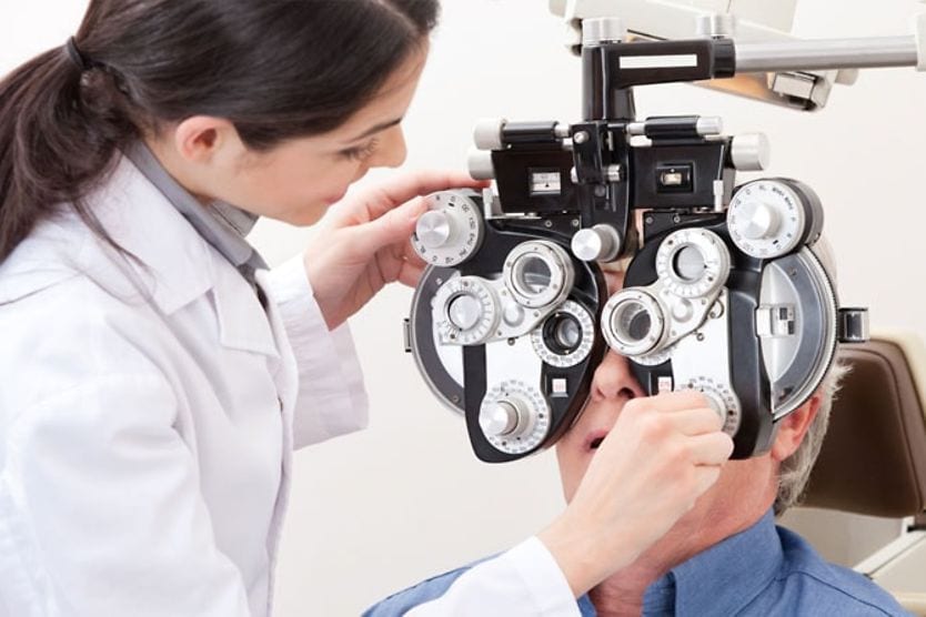
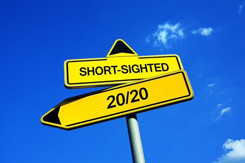
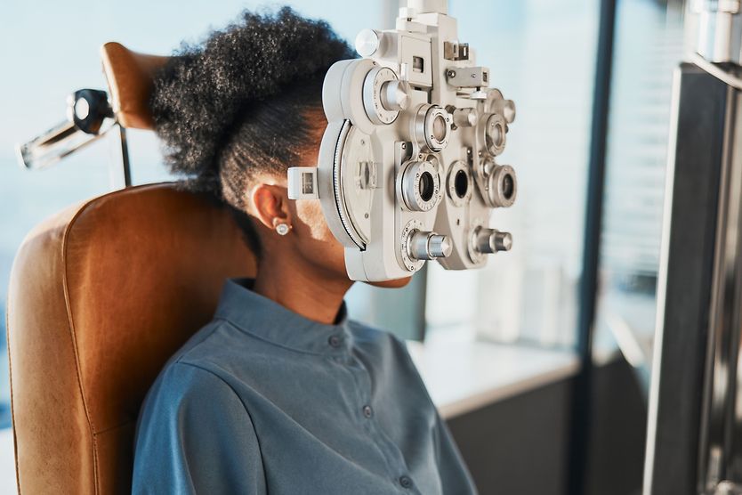
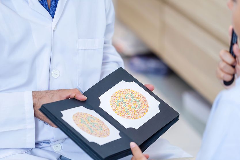
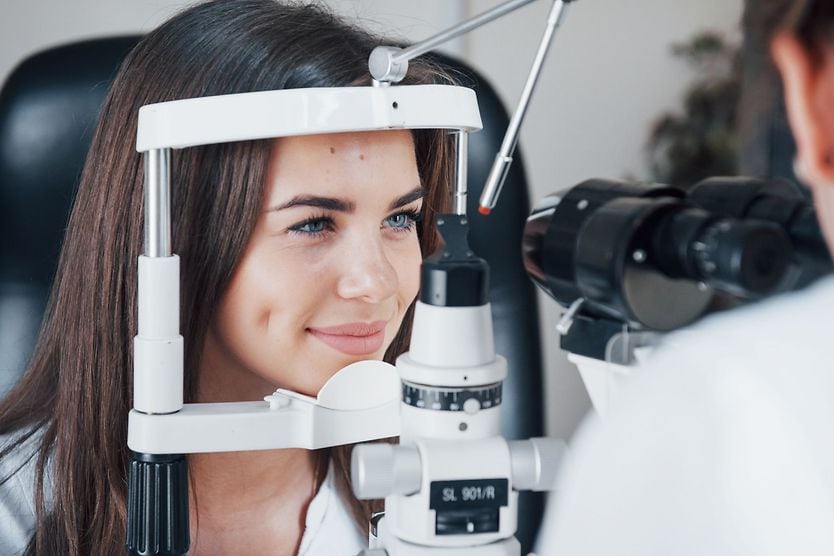
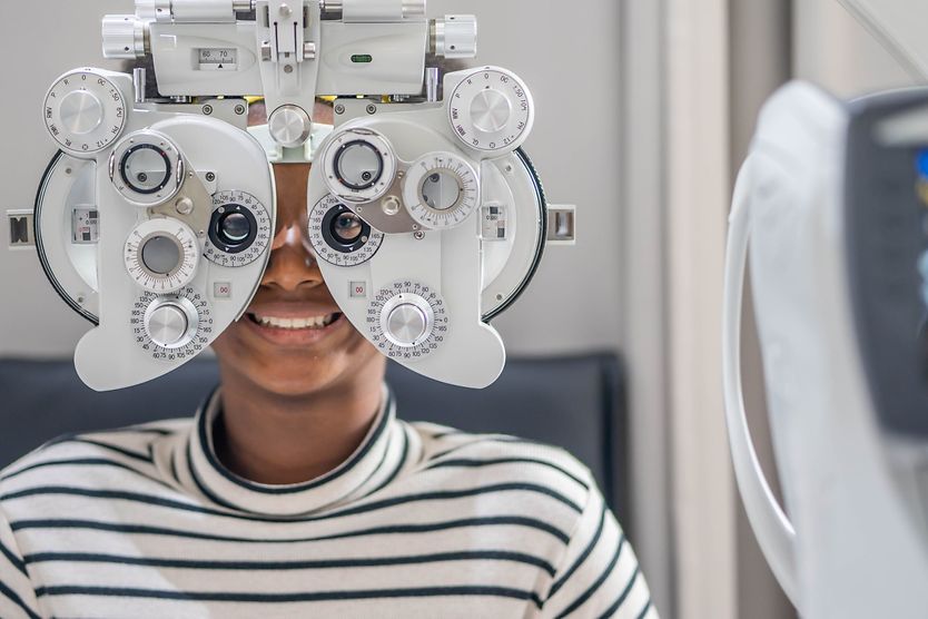
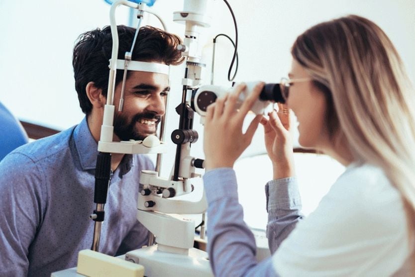
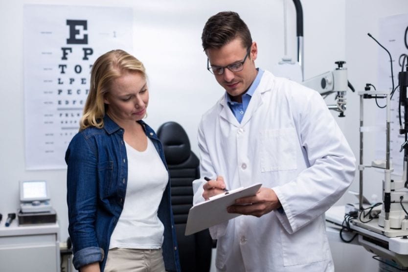
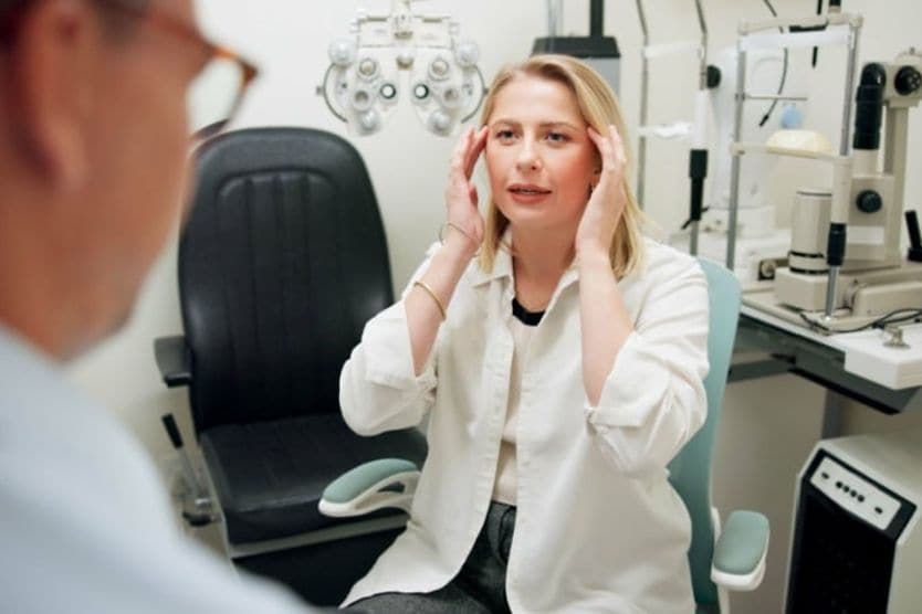
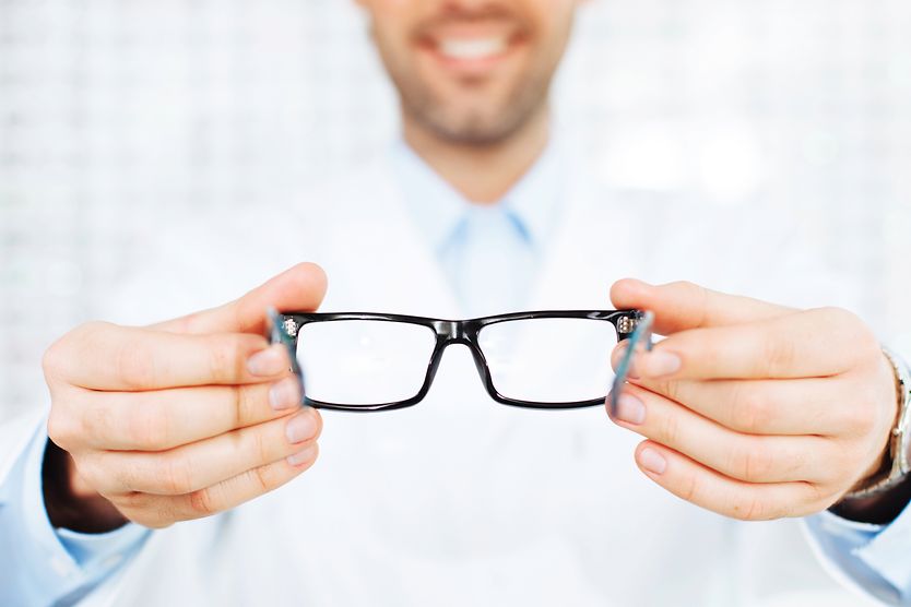
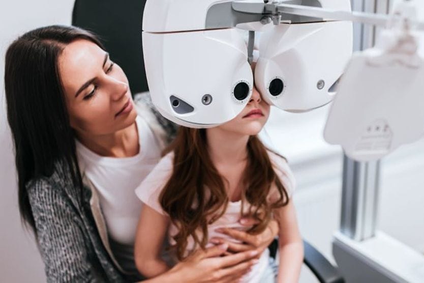
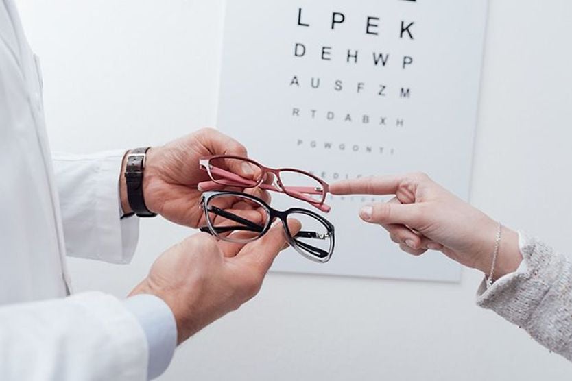
Home Eye Care Eye Exams | Ophthalmoscopy

Binocular indirect ophthalmoscope (BIO)

Ophthalmoscope lies on an eye test

What is a binocular indirect ophthalmoscope?

The binocular indirect ophthalmoscope (BIO) is a tool used by eye doctors to view the back of the eye, also called the fundus. It is a headband equipped with spectacles that sit in front of the doctor’s eyes with a light positioned just above the bridge of their nose. 

The BIO exam is conducted in a dark room in order for the physician to control the light entering the patient’s eyes. Plus, in order to get the most accurate results, the eye doctor will typically dilate the patient’s eyes. 

Dilation involves the use of eye drops that increase the size of the pupils, allowing more light to enter the eye. Dilation and the use of a BIO allow the physician to fully examine the retina — the layer of tissue that lines the back of the eye.

After light enters the patient’s eyes, it will bounce back out. The physician can hover a condensing lens over the patient’s eyes in order to gather and direct the exiting light. This light is what carries the image of the fundus back to the eye care provider. 

The BIO also has a triangular mirror. As the gathered light bounces off either side of the mirror, it is directed into both of the doctor’s eyes. 

Ophthalmoscopy

Binocular indirect ophthalmoscopy is only one way eye doctors can examine the fundus. There are two main kinds of ophthalmoscopes: direct and indirect. A direct ophthalmoscope produces a virtual, upright image of the fundus. An indirect ophthalmoscope allows the doctor to see the actual fundus, albeit upside down. 

Every ophthalmoscope requires three main parts: 

  • A light source

  • A reflective surface for directing the light

  • A way to focus the view of the fundus

Slit lamp exam of the front of the eye.

Physicians can also perform ophthalmoscopy using a tool called a slit-lamp. However, a slit lamp was designed to examine the front of the eye, rather than the fundus. With a condensing lens, a slit lamp exam can provide better magnification on the central retina than a BIO exam. But this enhanced visualization is limited to the central retina and does not extend as far into the periphery.

What is a binocular indirect ophthalmoscope used for?

The binocular indirect ophthalmoscope is commonly used in annual eye exams to check the retina. The retina contains cells called photoreceptors. There are two types of photoreceptors: rods and cones. They are responsible for converting light that enters the eyes into nerve signals. These nerve signals are sent through the optic nerve to the brain, where the signals are translated into images. 

The center of the retina, called the macula, is responsible for seeing images that are directly in front of the eye (central vision). This includes things like written text and faces. Getting these areas checked regularly is imperative for ensuring eye and vision health.

Ophthalmic surgeons may use BIOs to help them throughout their treatment of retinal disorders. It can also be a useful tool for eye doctors working with patients who have cataracts. The light can penetrate through the cataracts to give the physician a better view.

What can the binocular indirect ophthalmoscope help diagnose?

Regular exams with a BIO can help eye care providers diagnose and monitor many eye conditions. 

Cytomegalovirus (CMV) retinitis

CMV infections are common. In rare cases, CMV can cause inflammation in the retina. Some individuals with CMV retinitis may not notice any issues (asymptomatic). For others, the virus can lead to blind spots, blurred vision and floaters

If left untreated, CMV retinitis can eventually cause permanent vision loss within 4-6 months.

Diabetic retinopathy

While a BIO may not help a physician diagnose diabetes, it can help eye doctors monitor the potential impact that diabetes can have on the eyes. Diabetic retinopathy occurs when blood vessels in the eye leak, causing the retina to swell.

Diabetic retinopathy may not be noticeable to the patient at first. However, they may eventually develop blurred vision, floaters and blind spots. The patient may also have difficulty with night vision

The longer an individual has diabetes, the likelier they will develop diabetic retinopathy. The risk is further increased by unregulated blood sugar levels. But the earlier the disease is found, the better. And the BIO is the best instrument to detect diabetic retinopathy early.

Glaucoma

Glaucoma develops when the fluid of the eye (the aqueous humor) doesn’t properly drain, leading to pressure build-up. This pressure can eventually cause damage to the optic nerve, which attaches to the retina via the optic disc. 

Individuals with glaucoma may initially be asymptomatic. However, as the condition progresses, they may begin to notice blind spots, starting in their peripheral vision.

Damage caused by glaucoma cannot be repaired, so regular eye exams are an important preventative step.

Age-related macular degeneration (AMD)

Age-related macular degeneration, brought on by a thinning of the macula, is the main cause of vision loss in older adults. Over time, the thinning may worsen. Some may also develop abnormal blood vessels that can cause further damage to the macula.

AMD won’t lead to full blindness, but the associated loss of central vision can interfere with:

  • The ability to see faces

  • Driving

  • Reading

  • Cooking

  • Anything that requires close-up vision

Retinal detachment 

Retinal detachment occurs when the retina peels away from its usual position. It is often preceded by sudden floaters, flashes of light, a dark curtain in the peripheral vision or reduced vision. 

While retinal detachment isn’t painful, it does constitute an emergency situation. If not addressed quickly, it can lead to permanent vision loss.

Who invented the binocular indirect ophthalmoscope?

In 1851, German physicist Hermann von Helmholtz outlined the principles of direct ophthalmoscopy. Prior to this, physicians had no way to examine the fundus. In 1852, Christian Ruete introduced indirect ophthalmoscopy. 

Marc-Antoine Giraud-Teulon then created a binocular — or two-lensed — model. In 1944, Charles Schepens created what has become the modern BIO worn on the head. The BIO has continued to evolve as technology has advanced.

The importance of regular eye exams

Regular eye exams are important for maintaining healthy eyes and vision. The use of ophthalmoscopy in these exams can lead to early diagnosis of many retinal issues. 

Binocular indirect ophthalmoscopy. EyeWiki. American Academy of Ophthalmology. December 2021.

The dilated eye exam: Why it’s so important. National Glaucoma Research. Bright Focus Foundation. January 2023.

Retina. American Academy of Ophthalmology. September 2020.

Binocular indirect ophthalmoscopy: The beginner's guide. eOphtha. April 2021.

Ophthalmoscopy. MedLine Plus. March 2021.

CMV retinitis. Mount Sinai. November 2021.

Diabetic retinopathy. American Optometric Association. Accessed February 2023.

What is glaucoma? Symptoms, causes, diagnosis, treatment. American Academy of Ophthalmology. December 2022.

Age-related macular degeneration (AMD). National Eye Institute. June 2021.

Retinal detachment. Mayo Clinic. September 2022.

Slit-lamp exam. MedLine Plus. March 2021.

Find Eye Doctor

Schedule an exam

Find Eye Doctor