Fuchs' corneal dystrophy is a disorder of the front surface of the eye (cornea) that usually affects older adults. Here are seven key facts about Fuchs' dystrophy you should know:
1. What Is Fuchs' Dystrophy?
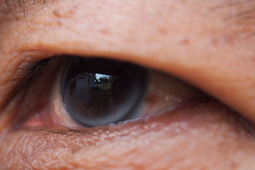
Fuchs' dystrophy (fooks DIS-truh-fee) is an eye disease in which the innermost layer of cells in the cornea undergoes degenerative changes. This cell layer, called the endothelium, is responsible for maintaining the proper amount of fluid in the cornea. The endothelium keeps the cornea clear for good vision by pumping out excess fluid that could cause corneal swelling.
Also called Fuchs' corneal dystrophy and Fuchs' endothelial dystrophy, the disease usually affects both eyes and causes a gradual decline in vision due to corneal edema (swelling) and clouding.
As the disorder progresses, swelling of the cornea can cause blisters on the front of the cornea known as epithelial bullae (BULL-eye). This condition is known as bullous keratopathy.
There is no known prevention for Fuchs' dystrophy.
2. What Causes It?
Fuchs' dystrophy can have a genetic cause, but it also can occur without a previous family history of the disease. In many cases, the cause is unknown.
3. What Are the Symptoms of Fuchs' Dystrophy?
Fuchs' dystrophy symptoms may include:
- Glare and sensitivity to light
- Eye pain
- Foggy or blurred vision
- Seeing colored halos around lights
- Difficulty seeing at night
- Poor vision upon awakening that may improve later in the day
- A feeling that something is in your eye (foreign body sensation)
4. Who Is Most At Risk For It?
Vision problems from Fuchs' corneal dystrophy usually affect people after age 50, though eye doctors can usually detect early signs of the disease in younger adults. It appears to be more common among women than men.
If your mother or father has Fuchs' dystrophy, you have roughly a 50 percent chance of getting the disease.
5. How Is It Detected?
A comprehensive eye exam by an optometrist or ophthalmologist is needed to detect Fuchs' corneal dystrophy.
During the exam, your eye doctor will use an instrument called a slit lamp to perform a detailed examination of the cornea. During this procedure, he or she will examine the cornea under high magnification to look for subtle changes in the appearance of cells in the endothelium that are characteristic of the disease.
The early clinical signs of Fuchs' dystrophy are typically a reduced number of endothelial cells and tiny drop-like lesions in the corneal endothelium called corneal guttata.
Another test your eye doctor might perform is a measurement of your corneal thickness (pachymetry), to detect increased corneal thickness that might indicate corneal swelling from the disease.
Also, visual acuity testing with an eye chart that is done during a comprehensive exam can reveal decreased vision due to corneal swelling.
6. What Treatment Is Available For Fuchs' Dystrophy?
Treatment for Fuchs' dystrophy depends on the stage of the disease. In the early cases, vision often can be improved by removing excess water from the cornea with 5% sodium chloride (hypertonic) eye drops.
If you have photophobia caused by corneal dystrophy, eyeglasses with photochromic lenses may be helpful to reduce your sensitivity to sunlight. Also, anti-reflective coating can also eliminate reflections in eyeglass lenses that may be particularly bothersome to someone with Fuch's dystrophy.
If you have endothelial dystrophy and ocular hypertension, your eye doctor might recommend glaucoma eye drops to reduce your intraocular pressure (IOP). High eye pressure can damage the corneal endothelium, worsening Fuchs' dystrophy.
As the disease progresses, the epithelial bullae can rupture, causing painful corneal abrasions and poor vision. If this occurs or if Fuchs' dystrophy progresses to the point of causing significant vision loss, a cornea transplant usually is needed.
An alternative to a full-thickness corneal transplant (also called penetrating keratoplasty, or PK) is deep lamellar endothelial keratoplasty (DLEK), which is a surgical method to replace the endothelium that leaves the upper layers of the cornea untouched. This procedure has shown success for the treatment of Fuchs' dystrophy with potentially fewer risks than penetrating keratoplasty.
In recent years, an advanced form of DLEK called femtosecond laser-assisted Descemet stripping endothelial keratoplasty (FS-DSEK) has shown results for treatment of the disease.
7. Precautions
If you have been diagnosed with Fuchs' corneal dystrophy, it is recommended to discuss this with your eye doctor if you are considering LASIK or other refractive surgery or if you have cataracts and need cataract surgery. These eye surgeries can worsen the condition, and corneal dystrophy often is considered a contraindication for elective refractive surgery.