Tarsorrhaphy

What is tarsorrhaphy?
Tarsorrhaphy, also known as blepharorrhaphy, is a procedure in which a surgeon joins the upper and lower eyelids together to cover the eye partially or completely. This procedure aids in protecting the corneal surface of the eye while promoting and accelerating the healing process.
Tarsorrhaphies are classified as either temporary or permanent. If the tarsorrhaphy is temporary, the cornea is expected to heal within two to eight weeks. If healing has not taken place within those weeks, the surgeon will perform a “permanent” tarsorrhaphy when eye closure is needed longer-term. The surgeon can then open the eye at a later time.
This procedure can also be performed on dogs and cats if their eye has been displaced following an injury.
Indications
Your eye doctor may recommend tarsorrhaphy when the cornea is unable to heal — due to an injury, surgery or the effects of disease — without additional protection. Conditions and situations that may benefit from tarsorrhaphy include:
- Dry eye that doesn’t respond to conventional treatment
- Cicatricial conjunctivitis, inflammation and scarring of the conjunctiva due to different underlying conditions
- Epithelial (outermost layer of cornea) defects that will not heal while the cornea is exposed
- Corneal ulceration associated with infectious keratitis, or infection of the cornea
- Progressive corneal thinning
- Stem cell deficiency in the limbus, which is the border between the cornea and the sclera (the white part of the eye)
The cornea may require protection from exposure in cases of:
- Lagophthalmos, a condition in which the eyelids do not fully close. This is often caused by facial paralysis
- Bulging eyes, which are sometimes seen with thyroid eye disease
- Eyelids that are no longer in the correct position because of trauma or surgery
- Floppy eyelid syndrome (FES), a condition where the upper eyelids are loose and may flip inside out
- Poor blinking rates related to certain neurological diseases or extensive sedation
A doctor may also recommend tarsorrhaphy following certain surgeries. This is done to prevent exposure and swelling. Some of those surgeries include, but are not limited to:
- Globe reconstruction surgery to correct globe luxation (an eye that pops out)
- Corneal transplant surgery
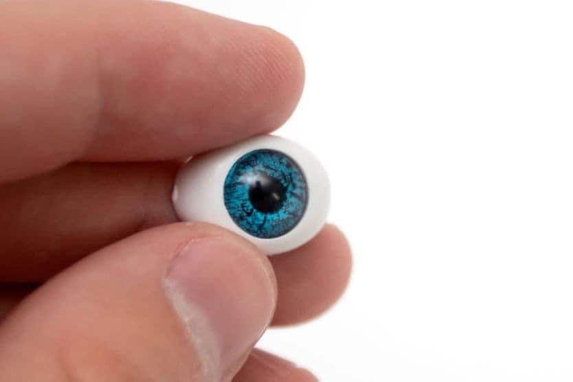
Tarsorrhaphy may also be done to help secure a prosthetic eye (also called an artificial eye).
Benefits
While eye patches can be used to protect the eye and promote healing, but your doctor may determine that tarsorrhaphy is a better option for you.
Tarsorrhaphy provides:
- More oxygen to the cornea, preventing bacterial growth
- Easier access to the eye for the doctor to examine and administer eye drops
- Partial vision for the patient if the eye is not completely sutured (or sewn) together
Temporary tarsorrhaphy vs. permanent tarsorrhaphy
There are two main classifications of tarsorrhaphy: temporary and permanent. Surgeons use different techniques and procedures within each category.
Temporary tarsorrhaphy
If the cornea is expected to heal in less than eight weeks, a temporary tarsorrhaphy may be appropriate. There are three types:
Temporary bolster tarsorrhaphy
In this procedure, the surgeon places bolsters made of plastic, cotton or other material on the eyelids to protect the skin from tight sutures. It also allows for better control of the closure of the eyelids.
If the surgeon needs to inspect the cornea throughout the healing process, they may use the drawstring technique. In this case, the surgeon adds another bolster. This extra bolster helps to slide the other bolsters apart and back together again. If the healing time is estimated to be less than two weeks, and there is no concern of skin damage, bolsters may not be necessary.
Cyanoacrylate glue tarsorrhaphy
This procedure can be done in an office setting. The surgeon applies glue gel to the outer part of the eyelids, which keeps them securely shut for about two weeks.
If needed, the doctor can reverse this process by cutting the eyelashes. This is appropriate for those who cannot have an invasive procedure.
Botulinum toxin temporary tarsorrhaphy
In this procedure, the doctor injects onabotulinumtoxinA into the upper eyelid to paralyze the muscle. Results can be unpredictable and more injections may be necessary.
Permanent tarsorrhaphy
Doctors typically recommend a permanent tarsorrhaphy if they don’t expect the cornea to heal within two to eight weeks. While labeled permanent, the surgeon can reverse it once the cornea heals. There are three types of permanent procedures:
Permanent lateral tarsorrhaphy
This is when the surgeon sutures the lateral, or outer, part of the upper and lower eyelids together. Bolsters are not used.
Permanent medial tarsorrhaphy
In this procedure, the surgeon sutures the medial, or inner, part of the upper and lower eyelids together. This allows the patient’s peripheral vision to remain accessible.
Pillar tarsorrhaphy
This is when the surgeon creates a pillar by suturing a flap of skin from the upper eyelid through the lower eyelid on either side of the pupil. The size and location of the pillar can be adjusted to account for the amount of exposure of the cornea.
This procedure is considered permanent, though it can be quickly reversed. It also tends to have better cosmetic results than other options.
Risks and complications
While few major risks or complications are associated with tarsorrhaphy, your doctor may prescribe antibiotics to prevent infection. They may also prescribe lubricating eye drops to preserve the cornea. In addition to following any post-operative instructions from your doctor, keep the affected area clean and avoid wearing makeup. Make sure you go to all follow-up appointments with your doctor.
Minor side effects such as redness, swelling and pain are common after surgery. If these symptoms occur, they typically resolve after a few days on their own. Acetaminophen and/or application of a cold compress may also help relieve discomfort. However, if symptoms persist or get worse, contact your doctor.
Because your peripheral vision can be affected by tarsorrhaphy, you may need to make adjustments to your daily activities. You should avoid driving until you get the go-ahead from your eye doctor.
Results
The success rate of tarsorrhaphy is very high, with 80% to 100% of procedures resulting in complete healing. Joining the eyelids together keeps the surface of your eye moist and protected, giving the cornea the time and space it needs to heal.