Corneal deposits


Understanding corneal deposits
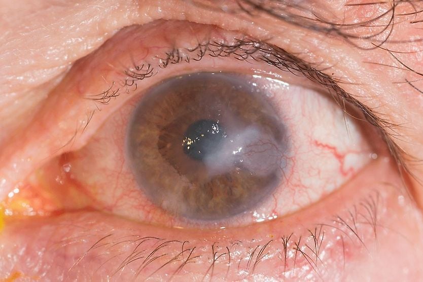
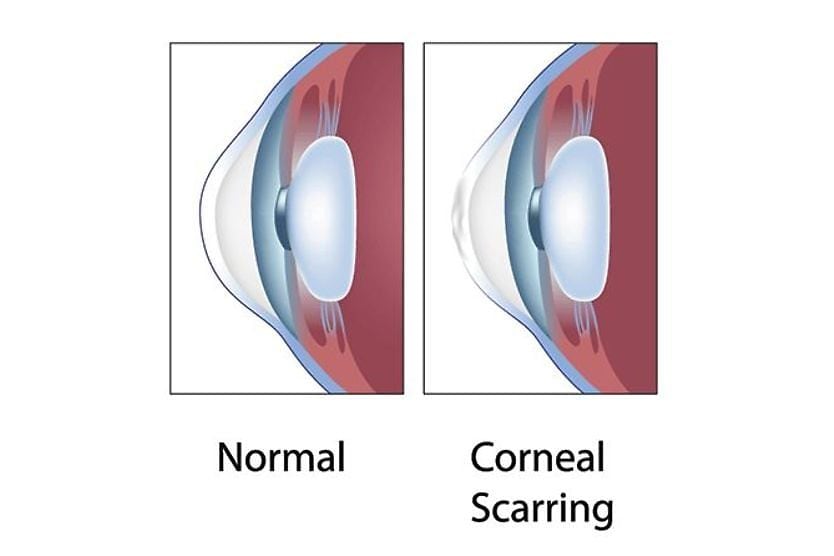
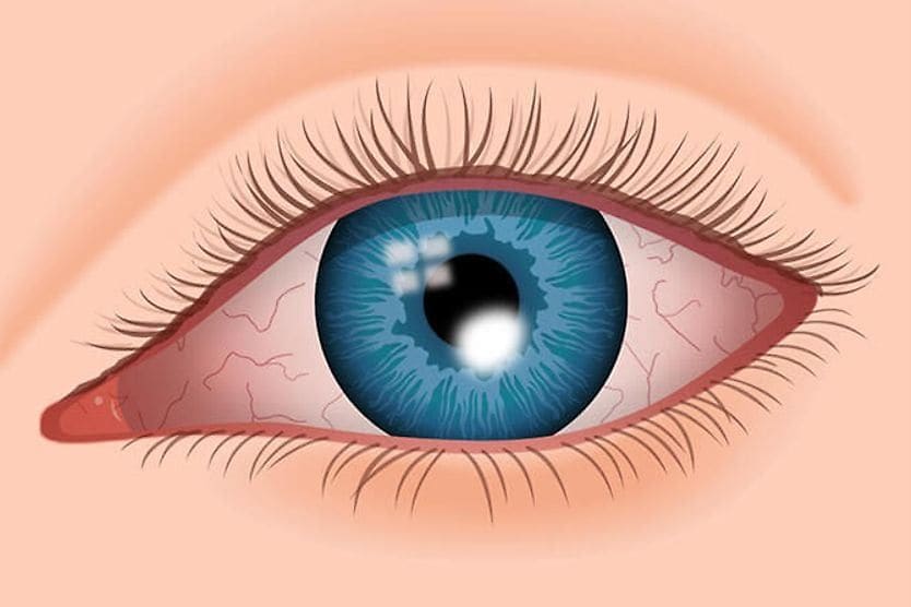
Corneal deposits occur when there is a buildup of material in the different layers of the cornea — the clear, dome-shaped outer layer of the eye. This buildup can be due to medications or a disease. The deposits may cause no symptoms or decrease vision due to clouding or scarring of the cornea.
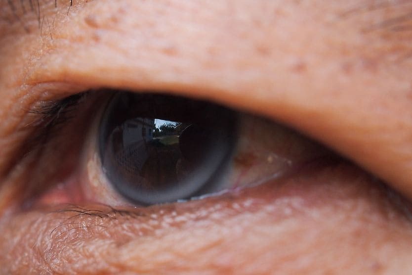
The most well-known pattern of corneal deposits is called cornea verticillata. It is also known as whorl keratopathy, vortex keratopathy or Fleischer vortex. The pattern, a whorl-like shape, is the accumulation of brown or gray deposits in the outer layer of the cornea. Cornea verticillata do not usually cause symptoms. These deposits can be caused by medications, products involved with the body's metabolism or diseases.
Some corneal dystrophies and degenerations can also be associated with deposits of material in the corneal layers. These abnormal deposits may accumulate in various patterns and layers of the cornea.
In what layer of the cornea do deposits accumulate?
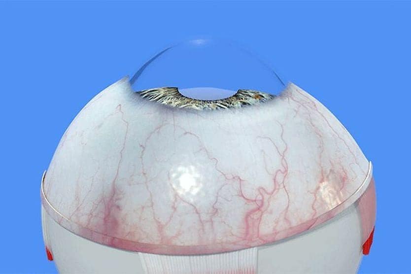
Corneal deposits can accumulate in different layers of the cornea depending on the underlying cause. The cornea provides much of the focusing power of the eye. It must remain clear so that light rays can enter without obstruction. A healthy cornea does not have blood vessels. Oxygen is diffused through tears, and the aqueous humor provides nutrients.
The five layers of the cornea provide structure and defense against particles and infection. Deposits that may accumulate in the layers of the cornea can potentially decrease and distort vision.
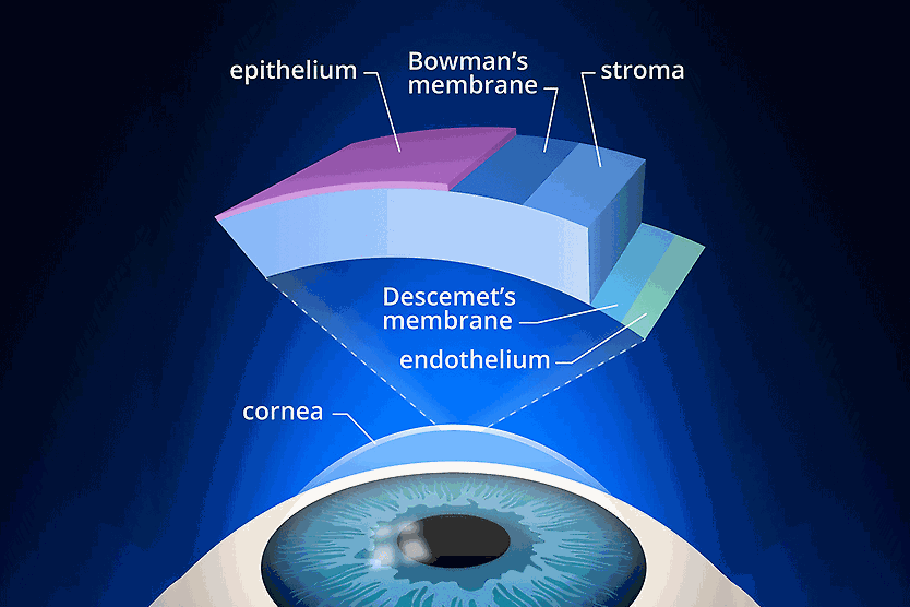
The layers of the cornea are:
- Epithelium – The nerve-rich outer layer that is the first barrier and absorbs nutrients and oxygen from tears.
- Bowman’s layer – A protective layer made of the protein collagen. It is transparent but can scar as it heals, impairing vision.
- Stroma – The middle layer made of collagen and water. It provides most of the cornea’s thickness, elasticity and structure, and plays a vital role in keeping the cornea transparent.
- Descemet’s membrane – A layer made of collagen fibers that provides a protective layer.
- Endothelium – A single layer of cells that keeps the stroma from absorbing too much water by pumping excess water out, keeping it from swelling and becoming hazy.
Medication and diseases that cause deposits to build up in the cornea typically do so in a unique pattern. If these deposits accumulate and damage the clear corneal tissue, scarring and haziness could result in vision loss.
Medications that can cause corneal deposits
A common cause of corneal verticillata is medications. Amiodarone, a heart medication for arrhythmia, and chloroquine, used to prevent and treat malaria, are two of the most common medications associated with a whorl-like pattern of corneal deposits. Additional drugs linked to the condition range from anti-inflammatory drugs and antibiotics to cancer treatment drugs, including:
- Indomethacin
- Phenothiazines
- Gentamicin
- Tamoxifen
- Meperidine
- Chlorpromazine
- Atovaquone
- Suramin
- Tilorone
- Perhexiline maleate
- Vandetanib
- Osimertinib
- Belantamab mafodotin
There is one topical eye drop that may cause a whorl-like pattern of corneal deposits called netarsudil.
Medical conditions that can cause corneal deposits
Several medical conditions cause deposits to build up in the cornea. Many of these cause the accumulation of material in other organs as well, which can create issues in multiple body systems.
Wilson's disease
Wilson’s disease is a rare disorder caused by copper accumulation in the cornea, brain and liver. It can lead to liver failure as well as behavioral and movement issues. The copper corneal deposits form dark circles around the iris (colored part) of the eye and are known as Kayser–Fleischer rings.
Fabry disease
Fabry disease is an inherited neurological disorder due to a deficiency in the enzyme alpha-galactosidase-A. With this condition, fatty substances in the body, called lipids, cannot be properly broken down into smaller substances that supply energy to the body. As a result, lipids build up in the cardiovascular and nervous systems, kidneys and eyes. The lipid buildup results in cornea verticillata.
Multiple myeloma
Multiple myeloma is a cancer involving plasma cells found in the bone marrow. Deposits of proteins — called immunoglobulins — made by malignant plasma cells can accumulate in the cornea and manifest as crystals. In some cases, this can cause the cornea to become cloudy. Symptoms such as dry eye and blurred vision can result.
Arcus senilis and arcus juvenilis
These ring-like deposits are composed of lipids, such as cholesterol, that form a circle or arc around the outer rims of the cornea. In older individuals with elevated cholesterol, the condition is known as arcus senilis. If the individual is younger than 40, then it’s called arcus juvenilis.
Corneal conditions that can cause corneal deposits
Corneal dystrophy or degeneration can result in deposits of material in different layers of the cornea.
Corneal degeneration
Corneal degeneration is the deterioration of the cornea’s tissue due to disease or aging. It usually begins on the edges of the cornea and occurs typically in one eye. Deposits, corneal thinning, corneal edema and abnormal blood vessel growth may occur. Corneal degenerations are not usually inherited.
Salzmann's nodular degeneration
This condition can be due to excess and irregularly arranged collagen deposits in Bowman’s layer. They are usually located at the edge of the cornea. These deposits do not usually cause symptoms if they are small and in the periphery. But, if they are large or occur more centrally, they can cause blurred vision and eye irritation.
Band keratopathy degeneration
This condition is due to calcium deposits in Bowman’s layer. It is usually due to an imbalance of calcium levels in the body. If the deposits are large enough, they can cause a scratchy feeling in the eye and decrease vision. Band keratopathy degeneration is usually treated with chelation, a treatment using eye drops to remove the calcium deposit.
Corneal dystrophies
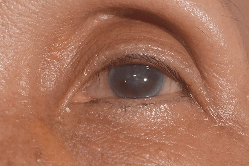
Corneal dystrophies are a group of disorders that are slow progressing, non-inflammatory and lead to a buildup of material in the cornea of both eyes. The dystrophy usually affects one layer of the cornea initially before progressing to the other layers. These conditions are typically inherited. Early on, people with corneal dystrophies typically have few, if any, symptoms. Over time, corneal deposits can accumulate, and the cornea may lose its transparency, resulting in progressive vision loss and other symptoms.
Macular corneal dystrophy
This condition is due to deposits of abnormal material in the stroma. It usually begins to cause clouding of the cornea after age 3, resulting in eye irritation and decreased visual acuity. Vision loss may be severe by age 20 or 30. Symptoms include painful recurrent erosion. This occurs when the corneal epithelium does not adhere to the eye properly and breaks down when the eyes are opened after sleeping. Decreased corneal sensation and light sensitivity are also common.
Schnyder crystalline corneal dystrophy
This condition is due to fat and cholesterol deposits progressively accumulating in the stroma, causing it to become opaque. Although it can occur in infancy, it usually begins in the early teenage years. Symptoms include blurry, hazy vision.
Lisch corneal dystrophy
This condition causes feathery microdeposits in a whorl or band pattern. Small cysts create a fuzzy appearance and can lead to blurry vision. It often appears in childhood and usually affects males.
Other conditions
Epidemic keratoconjunctivitis (EKC)
This is a very contagious inflammation of the conjunctiva (the clear tissue that covers the white of the eyes) that is caused by a virus. It also causes inflammation of the cornea, known as keratitis. EKC is caused by the same group of viruses that cause the common cold. It may cause temporary deposits of immune cells and collagen in the cornea. It results in red, irritated eyes and usually lasts two to three weeks.
Iron deposition (i.e., after radial keratotomy)
Radial keratotomy is an older refractive surgery that was used to correct refractive errors like nearsightedness. Lines composed of iron deposits that spoke out from the center of the cornea may appear sometime after the surgery in most people. These deposits do not usually impact vision.
Treatment for corneal deposits
There are many underlying causes for deposits in the cornea. Due to this, the treatment is very specific to the cause and severity of the disease. Some types of deposits, such as cornea verticillata, do not typically have any symptoms.
If symptoms are present, eye drops such as artificial tears and ointments can help relieve discomfort. Glasses and contact lenses can be prescribed to improve visual acuity. If corneal erosion occurs, contact lenses can be used as a bandage. Medical or surgical treatment may be necessary in cases of more severe underlying disease.
SEE RELATED: What is corneal ectasia?
When to see a doctor
If you are taking medication or have a condition known to cause deposits in the cornea, comprehensive eye exams are critical. Speak with your eye doctor and primary care physician regarding any medications or conditions that can lead to corneal deposits. It is important to follow their recommendation regarding how often you should be monitored. If you notice new, persistent eye pain or the sudden onset of blurry vision, contact an eye doctor as soon as possible.