Scotoma: Blind spot in vision

A scotoma is a blind spot in your vision. Depending on its size and severity, a scotoma also may look like a dark or blurry spot in your vision.
Most scotomas are permanent blind spots; but some can be temporary, depending on what causes them.
Scotoma is pronounced “skuh-TOE-muh.”
Scotoma definitions
There are different types of scotomas, based on appearance and location. Where a scotoma appears in your field of view and what it looks like can help your eye doctor determine its underlying cause.
It’s possible to have one or more types of scotomas, and blind spots can occur in one or both eyes. In many cases, a scotoma affecting one eye is only noticeable when you close your other eye
The term “scotomata” sometimes is used to describe multiple scotomas.
Central scotoma
A central scotoma is a blind spot directly in the center of your vision (directly in your line of sight). Central scotomas can make reading, driving and recognizing faces difficult or impossible.
Causes of central scotomata include macular degeneration and diabetic retinopathy or diabetic macular edema.
Eye infections and injuries that affect the macula of the retina also can cause a central scotoma.
SEE RELATED: Valsalva retinopathy
Paracentral scotoma
A paracentral scotoma is a blind or blurry spot in your vision that is slightly off-center (within 10 degrees of your line of sight). For example, if you have a paracentral scotoma and look at a road sign, you may be able to see the words on the sign clearly, but an area or spot very near the sign may be dark or blurry.
Glaucoma and diabetic retinopathy can cause paracentral scotomas. In the case of glaucoma, the scotoma may be arc-shaped and therefore called an arcuate scotoma.
A paracentral scotoma may occur by itself or there may be more than one of these blind spots in your vision.
In some cases, a paracentral scotoma or multiple scotomata are accompanied by peripheral vision loss as well, resulting in what’s commonly called “tunnel vision.”
SEE RELATED: Blurry vision during pregnancy
Scintillating scotoma
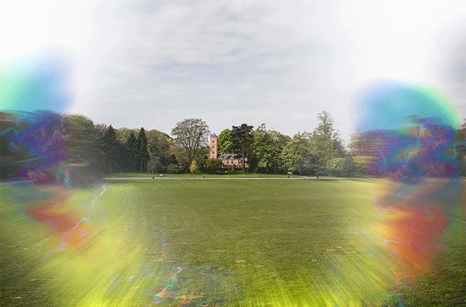
A scintillating scotoma is a visual disturbance that usually occurs on the paracentral or mid-peripheral portion of your visual field. However, this type of scotoma also may move across your field of view rather than staying in just one spot.
Because a scintillating scotoma is more of a visual disturbance (scintillating means “sparkling” or “shimmering”) than a true blind spot, it often is referred to as a visual aura.
Most scintillating scotomas are temporary and last anywhere from a few seconds to up to 30 minutes. Often, they are associated with migraine headaches (and therefore are sometimes called a migraine scotoma). But scintillating scotoma can occur without head pain as well.
Scintillating scotomas usually are arc- or ring-shaped, and may have wavy or jagged borders. They may be multi-colored or alternate between light and dark shades of gray.
In a addition to migraine, causes of scintillating scotomas range from benign to serious, including:
- Stress
- Hormonal changes
- High blood pressure
- Inflammation of the optic nerve
- Head injury
- Transient ischemic attack (TIA, or “mini-stroke”)
- Multiple sclerosis (MS)
Scintillating scotoma also can be a symptom of preeclampsia — a potentially dangerous complication of pregnancy characterized by high blood pressure, swelling of hands and feet, and possible liver or kidney damage.
SEE RELATED: Ocular and visual migraines: What's the difference?
Scotoma testing and diagnosis
A visit to an eye doctor is required to know for sure if you have a scotoma and to diagnose its underlying cause.
In most cases, testing and diagnosis of a scotoma requires an automated visual field test and a dilated eye exam.
Visual field test
Scotomas generally are detected and monitored using an automated visual field test (sometimes called a Humphrey visual field test). This test typically is supervised by a trained assistant in your eye doctor’s office.
During the visual field test, you will be seated in front of a large, bowl-shaped instrument. You will be positioned so you are looking into the opening of the bowl. Each eye is tested separately, and you will wear an eye patch to cover one eye at a time.
While you are looking at a target directly in front of you inside the instrument, tiny lights will flash (one at a time) from different points inside the bowl. Each time you see a light go on, you will click a hand-held device to indicate you saw it.
At the end of the test, the instrument prints out a map of the visual field of each of your eyes that your eye doctor will analyze to detect any scotomas. The data is stored in the instrument so the testing can be repeated in the future to detect any changes in your visual field over time.
Specific types of scotomas in specific areas of your visual field alert your eye doctor to look for health problems in specific parts of your eyes and optic nerve.
Dilated eye exam
In addition to analyzing the data from your automated visual field test, your eye doctor will closely examine the retina and optic nerve in the back of your eyes.
Typically, eye drops will be used to dilate the pupils of your eyes for this exam. However, in some cases, high-resolution images of your retina and optic nerve can be obtained with digital imaging devices that don’t require a dilated pupil.
The data from your visual field test and your dilated eye exam and/or imaging scans typically may give your eye doctor all the information needed to make an accurate diagnosis of the type of scotoma you have and what the underlying cause of the visual field defect is.
In some cases, however, referral to a neurologist or neuro-ophthalmologist and additional specialist testing may be needed to arrive at a definitive diagnosis.
SEE RELATED: Sudden blurry vision
Scotoma treatment
Scotoma treatment options depend on the cause and type of the blind or blurry spot in your vision.
Scotomas associated with migraine headaches typically are temporary and go away without treatment within 30 minutes or so.
If a scotoma appears to be caused by high blood pressure, stress, or other identifiable conditions, medications or other treatments for those conditions can help eliminate future blind or blurry spots in your vision.
Unfortunately, there is no successful treatment for many scotomas caused by glaucoma, diabetes, macular degeneration and certain neurological conditions. In such cases, however, your eye doctor may be able to prescribe specific low vision aids to help you cope with scotoma-related vision loss and use your remaining vision as effectively as possible.
When to see an eye doctor
If you suddenly notice a scotoma or other vision disturbances, see anoptometrist orophthalmologist immediately. Urgent medical care is especially needed if you experience any of following along with a scotoma:
- Confusion or disorientation
- Dizziness or nausea
- Muscle weakness
- Sudden, extreme headache
- Numbness in your limbs or face
- Slurred speech or difficulty speaking
- A head or eye injury before the scotoma appeared
Also, if you are at risk of diabetes, glaucoma, high blood pressure, cardiovascular disease or stroke, be sure to schedule an annual comprehensive eye exam to prevent scotomas and other vision problems before they begin.
READ NEXT: What is the blind spot in my eye?