Vitreous humor: Gel of the eye

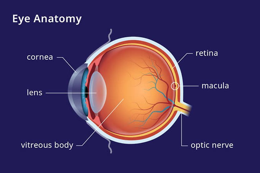
Vitreous humor, also called the vitreous body, is the transparent, watery, gel-like substance that fills the space in the center of the eye. Spanning from behind the lens of the eye to the retina, the vitreous body makes up the largest part of the eye.
What is the vitreous?
Vitreous is the fluid that fills a large portion of the inside part of the eye called the vitreous chamber. The vitreous chamber takes up approximately 80% of the eye, so the vitreous inside of it helps to give the eye its shape.
Fluid within the vitreous chamber is made up mostly of water, but it does contain small amounts of collagen, proteins, electrolytes and important sugars such as glycosaminoglycans. Vitreous fluid allows oxygen and nutrients to travel from the front of the eye to the back of the eye, while its antioxidants help safeguard the lens from cataract development.
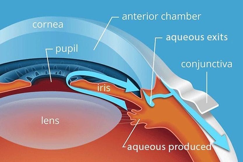
Unlike aqueous humor, the fluid located in the anterior chamber (front) of the eye, vitreous humor is a still fluid that does not constantly renew itself. However, a commonality between vitreous and aqueous humor is that neither rely on blood vessels to do their jobs.
Vitreous humor function
Vitreous humor’s function consists of several important jobs, including maintaining eye shape, keeping the eye clear and providing shock absorption.
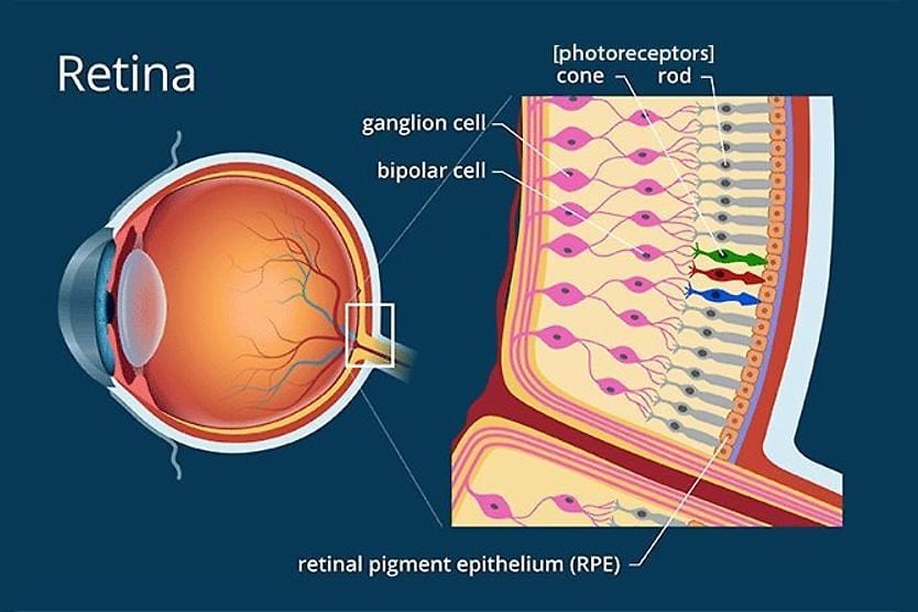
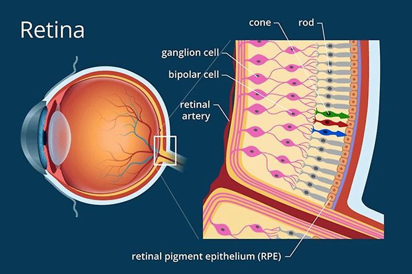
The most obvious function of the vitreous is to give the eye its spherical shape. When the vitreous chamber is properly filled, the vitreous humor stays attached to the retina (the light-sensitive tissue that lines the back of the eye).
In addition to maintaining eye shape, the vitreous body works to keep the center of the eye clear. For sharp vision to be possible, outside light must shine through the eye and focus directly on the middle part of the retina, called the macula. Because vitreous is clear and watery, light is more easily directed to where it needs to be for good vision.
Another important function of the vitreous is to help reduce any shock or disturbance that may affect the eye. For instance, when a person shakes or moves their head quickly, vitreous will absorb the shock from those movements.
This shock absorption also occurs during physical activities, such as running, or when someone sustains a head injury. The ability of the vitreous to lessen disturbances within the eyes helps prevent eye damage.
Vitreous conditions
As the eye ages, natural changes occur in the vitreous body. Such changes can increase the risk of age-related eye problems that vary from harmless to sight-threatening. Common vitreous-related eye conditions include vitreous detachment and retinal tearing.
Vitreous degeneration
Vitreous degeneration, also known as vitreous syneresis, is characterized by the thinning of the vitreous fluid. In a younger person’s eye, vitreous is thick and gel-like, which fills the vitreous chamber and keeps the vitreous humor plump.
As the eye matures, this gel-like substance begins to thin and liquify, making it difficult for the vitreous humor to fill the given space within the eye.
Vitreous syneresis can cause the vitreous humor to sag and pull away from the retina — a condition known as posterior vitreous detachment.
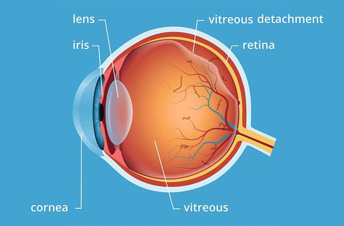
Posterior vitreous detachment
Posterior vitreous detachment (“PVD” or “vitreous detachment”) occurs when the vitreous humor begins to peel away from the connective tissue that attaches it to the retina. This typically happens because of vitreous degeneration, where the vitreous humor walls begin to sag as the gel-like substance liquifies.
As the vitreous detaches from the connective tissue, it’s common for vitreous floaters to appear (or worsen) in your vision. Eye floaters are tiny clumps or stands of collagen that break off into the vitreous as it pulls away from its connective tissue.
Shapes and types of eye floaters vary from person to person. While they can be distracting, they are typically not dangerous.
Though vitreous detachment is often part of the eye’s aging process and can occur with no issues, there’s a chance that more serious conditions can develop; these include retinal tears or retinal detachment.
Retinal tears and detachment
When the vitreous humor peels away from the tissue that connects it to the retina, it’s akin to removing a sticker from a sheet of paper: The sticker may peel away cleanly, or bits of paper may be torn off with the sticker.
It’s possible for bits of the retina to tear off with the vitreous humor when it detaches, resulting in a retinal tear. Because retinal tears can lead to retinal detachment, it’s important to have them examined by an eye doctor.
If severe, laser eye surgery may be required to seal the tear. However, it’s also possible that your retinal tear will be deemed low-risk and only require the monitoring of an eye doctor or retina specialist.
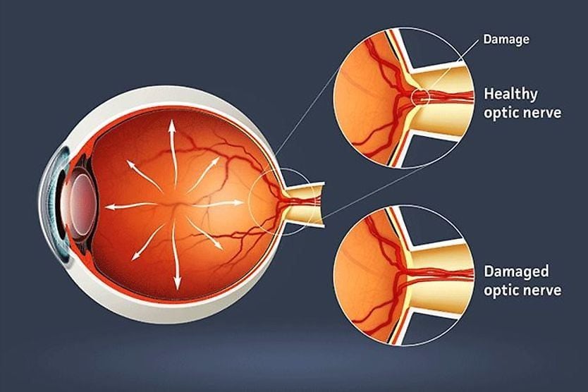
Retinal detachment occurs when the retina tears away from the back of the eye. This can happen following a retinal tear or from the vitreous humor tugging on the retina. Retinal detachment is a serious, sight-threatening condition that requires surgery to repair.
With retinal tears and detachment can come flashes of light (photopsia) in your peripheral vision. This is a result of the retina — a light-sensitive tissue — getting tugged on by the vitreous. The retinal stimulation triggers an appearance of light from inside the eye.
Vitreous hemorrhage
As mentioned earlier, the vitreous body is not composed of and doesn’t rely on blood vessels to work. However, there are many blood vessels surrounding the vitreous chamber.
In certain conditions, such as diabetic retinopathy or anemia, abnormal blood vessels can grow into the vitreous cavity. It’s possible for these blood vessels to rupture and bleed into or near the vitreous — this is known as a vitreous hemorrhage.
Vitreous hemorrhage is not painful and may appear as cloudiness or dark spots in your vision. Sight can be damaged or lost if vitreous hemorrhage is left untreated.
When to see an eye doctor
It’s important to see an eye doctor if you begin to experience symptoms associated with vitreous-related conditions, especially if you’re over 50 years of age.
See an eye doctor right away if you have any of these symptoms:
- Sudden development of or increase in eye floaters
- Flashes of light produced from within the eye rather than from an outside source
- Dark shadow or curtain closing in on the sides of your vision
- Sudden blurred or cloudy vision
These can be signs of retinal detachment or other urgent eye conditions that need to be treated right away. An eye doctor can examine the health of your vitreous and retina and administer treatment if needed.
In a less common procedure called avitrectomy, anophthalmologist drains the vitreous fluid from the eye and cleans the vitreous chamber. Once the chamber is cleaned, the doctor will replace the vitreous with a saline solution.
A vitrectomy is often used for people who have severe eye floaters or a vitreous hemorrhage that is affecting eyesight. The procedure can help repair structural damage to the vitreous and may restore clear vision to patients.
READ MORE: Eye floaters and flashes FAQ