What is detached retina surgery?
If the retina, the eye’s light-sensitive tissue, separates from the back of the eye, surgery for retinal detachment is typically needed to prevent vision loss. Depending on the type and area of detachment, one of the following procedures may be performed:
- Pneumatic retinopexy
- Laser or cryopexy
- Vitrectomy
- Scleral buckle
Retinal detachment requires immediate medical care. Without prompt treatment, the detachment may worsen, increasing the risk of vision loss. Although a minor retinal detachment may not produce any noticeable symptoms, a larger or central retinal detachment can cause sudden vision impairment.
Symptoms of retinal detachment include:
- A dark shadow that obscures part of your vision
- A flurry of small dark spots or lines in your vision
- Light flashing in one eye or both eyes
You should contact your eye doctor or go to the emergency room immediately if you experience these symptoms.
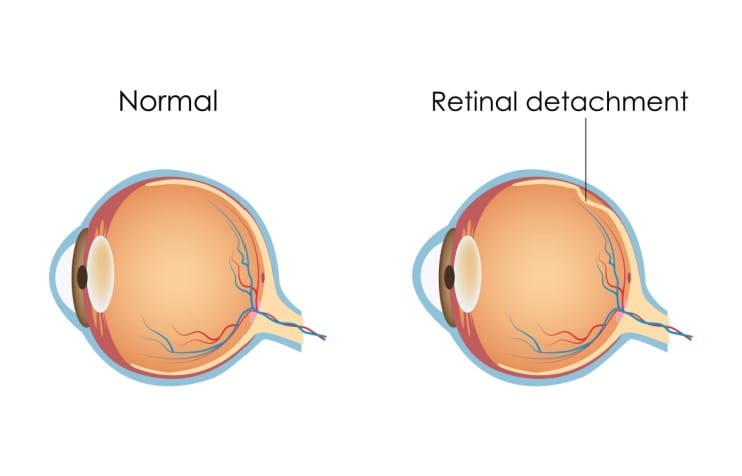
What is retinal detachment?
A retinal detachment occurs when the retina separates from the nourishing tissue beneath it. The retina is a thin layer of tissue at the back of the eye that contains specialized photoreceptor cells. These cells convert light into electrical signals that are sent to the brain and processed as images.
A network of blood vessels in the underlying tissue — called the choroid — supplies the retina with oxygen and nutrients. When the retina becomes detached from the choroid, it can no longer receive the necessary nourishment. This can lead to vision loss if not treated promptly.
Types of retinal detachment
Several types of retinal detachment can occur due to age, injuries or other conditions. As people get older, the most common type is a rhegmatogenous detachment. It can occur when the vitreous humor, a gel-type substance that fills the middle of the eye, shrinks and pulls on the retina, causing retinal tears. The vitreous can then seep under the retina, separating it from the choroid.
A tractional retinal detachment is caused by scar tissue forming on the retina, pulling it away from the back of the eye. Diabetes is a common cause of this type of retinal detachment.
Exudative retinal detachment occurs from fluid accumulation behind the retina, even without a retinal tear. The accumulating fluid separates the retina from the underlying choroid. Fluid buildup is mainly caused by leaky blood vessels or swelling behind the eye.
A detached retina is especially damaging if it is near or at the macula, the central part of the retina. The high concentration of photoreceptor cells there allows for detailed vision and a majority of our color perception. A detached retina at the macula often leads to serious vision problems.
Types of retinal detachment surgery
If performed promptly, retinal detachment surgery has a high success rate. However, more than one surgery is sometimes needed to reattach the retina. There are several types of retinal detachment surgeries:
Pneumatic retinopexy
Pneumatic retinopexy is a minimally invasive procedure appropriate for some types of detachments. It is typically performed in an office setting and can be done with anesthetic eye drops or local anesthesia.
During pneumatic retinopexy:
- The ophthalmologist will inject a gas bubble into your eye.
- The retina will be pushed back into place by the gas bubble.
- After the retina is positioned, your doctor will use a freezing probe (cryopexy), a laser (heat) or both to seal the tear and prevent further detachment.
- The gas bubble will disappear after about two to six weeks.
To ensure the retinal tear seals correctly, patients may have to hold their heads in a specific position for about a week after the procedure. The retina usually reattaches after a few days.
LEARN MORE about cryopexy (retinal cryotherapy)
Scleral buckling
Scleral buckling is a more invasive procedure performed in an operating room. It may be recommended for detachments that require external support from outside the eyeball.
During scleral buckle surgery:
- Your ophthalmologist will place a silicone band around the outside of your eye.
- The band will be stitched around your eyeball and attached to the white sclera that covers the outside of the eye, pushing the retina back into place.
- The tear will then be sealed with a laser or freezing treatment to prevent the retina from detaching again.
Vitrectomy
A vitrectomy may be recommended for complex retinal detachments, including those with significant scarring or large tears. It is performed in an operating room, and the vitreous gel inside the eye is removed and replaced with a solution, gas or oil bubble.
During a vitrectomy:
- Your ophthalmologist will remove the vitreous gel from the eye. The vitreous humor is the clear, gel-like substance that fills the inside of the eye.
- Once the vitreous gel is removed, your surgeon will use a laser or freezing treatment to seal the retinal tear.
- The eye is then filled with a gas bubble or silicone oil to help hold the retina in place.
Preparing for detached retina surgery
A thorough eye exam and medical history will be necessary before your surgery. The office staff will provide you with instructions on how to prepare for the surgery.
You will typically be asked to stop eating and drinking several hours before the surgery. You should also arrange for someone to drive you home afterward.
Examination before surgery
A comprehensive eye exam allows your eye doctor to evaluate your eye health and retina and determine the best surgical approach. This exam may include:
- Medical history and any medications you are taking
- Visual acuity to determine how well you are seeing
- Slit lamp examination to look at the front and inside of your eye
- Tonometry to check the pressure inside your eye
- Dilated retinal exam to examine your retina, including scleral depression (pushing on the outside of your eye to see the edges of the retina)
- Imaging tests such as an ultrasound, optical coherence tomography (OCT), or fundus imaging to help your doctor see the retina more clearly
Questions to ask your eye doctor
You will likely have many questions for your doctor. It is often helpful to write them down before your office visit to ensure you get answers for all of them. Some questions to ask before retinal detachment surgery include:
- What type of retinal detachment surgery am I getting and why?
- What are the risks and benefits of the surgery?
- What can I expect during and after the surgery?
- How long will it take to recover and return to my usual activities?
- What is the success rate of this type of surgery?
Retinal detachment surgery recovery
The recovery time after retinal detachment surgery varies depending on your retinal detachment, medical history and the type of surgery performed. Side effects depend on the specific surgery and individual patient. However, you can typically expect the following after the surgery:
Pneumatic retinopexy
After pneumatic retinopexy, your eye may be sore and red for a few days, and your vision may be blurry for a few weeks. To help your retina heal, you may need to keep your head in a specific position for up to a week after surgery. Most normal activities can typically be resumed within several weeks.
Scleral buckle surgery
After scleral buckle surgery, it is normal to experience some post-operative discomfort. You may require pain relief medication for several days. You may wear an eye patch for a while and should restrict your activities for a few weeks. You should be able to return to your regular routine within a few weeks or months.
Vitrectomy
After vitrectomy, some pain and discomfort are normal. You'll need to use pain medication, antibiotic drops and steroid eye drops for a few days or weeks. Maintaining a specific head position may be required for some time. Blurred vision could last for up to several months. Most normal activities can be resumed within several weeks to months. It is important to follow your surgeon's post-operative instructions and attend all follow-up appointments.
Potential risks and complications
Retinal detachment surgery is generally safe but carries some risks, just like any surgery. Possible complications include:
- Infection
- Bleeding
- High pressure inside the eye
- Cataract
- Redetachment of the retina
- Epiretinal membrane formation (a condition in which scar tissue forms on the retina)
You should call your doctor immediately or go to the emergency room if you experience any of the following after surgery:
- Increased pain
- Worsening redness, discharge
- Onset of blurry vision
- Signs of infection, such as fever and chills
If you've had an eye procedure with intraocular gas, your doctor will recommend precautions for the two to six weeks that the gas remains in your eye. For example, you should avoid air travel and nitrous oxide anesthesia due to the risk of expanding intraocular gas, which can damage your eyes.
Life after retinal detachment surgery
A majority of people have a good prognosis after retinal detachment surgery. Vision is usually improved and sometimes fully restored. Ninety percent of retinal detachment repairs are successful, although some patients may require more than one procedure.
Several factors influence how much vision is restored, including the severity and location of the retinal detachment and the length of time it remained detached before the operation.
Unfortunately, vision loss can occur in some patients even after successful retinal detachment surgery. Numerous resources are available to assist with adjusting to vision impairment and preserving independence, including:
- Low vision rehabilitation
- Support groups
- Assistive technology
If you have been diagnosed with a retinal detachment, it is important to discuss treatment options and post-surgical expectations with your doctor. Fortunately, prompt treatment frequently restores some or all vision.
After the retinal detachment or retinal tear surgery, you should continue to monitor your eye health with routine comprehensive eye exams and attend all follow-up appointments recommended by your eye doctor.
READ NEXT: Types of eye surgery and the conditions they treat


