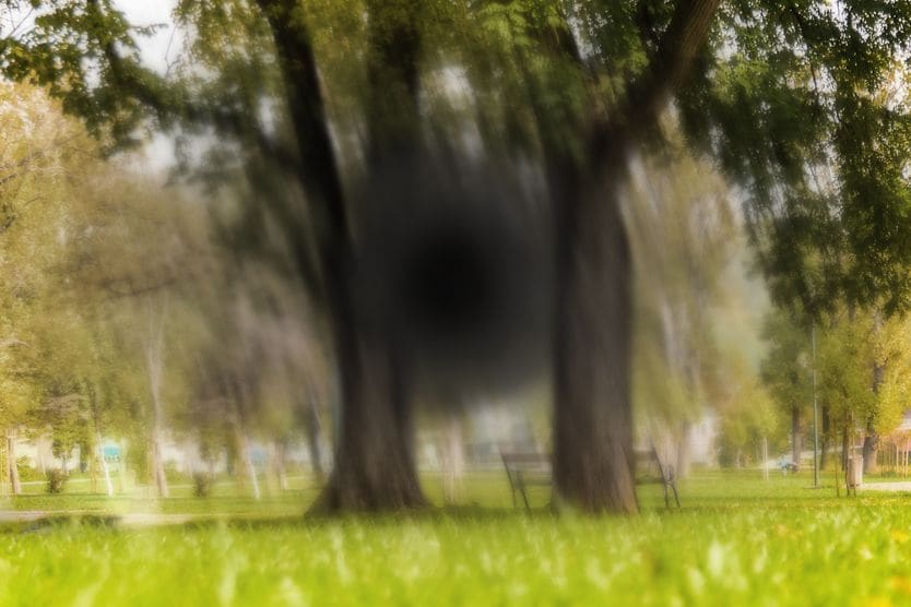
Home Conditions Refractive Errors | Myopic macular degeneration

Myopic macular degeneration: Causes, symptoms and prognosis

man getting an eye exam to check for myopic macular degeneration

What is myopic macular degeneration (MMD)?

Myopic macular degeneration (MMD) is damage to the macula that occurs in pathological myopia. Pathological myopia is due to progressive, abnormal elongation of the eyeball. This results in degenerative changes to the back of the eye. Myopic macular degeneration can lead to irreversible vision loss.

What are the symptoms of myopic macular degeneration?

The macula is located in the central part of the retina. It provides our sharpest central vision. Damage to the macula can cause individuals to experience symptoms such as:

https://cdn.allaboutvision.com/pathological-myopia-graphic.gif

Myopic maculopathy is a leading cause of vision impairment worldwide. It can result in permanent vision impairment and blindness. A recent study found that adults with high myopia and myopic macular degeneration may experience visual field loss as well. 

Myopic macular degeneration occurs in pathological myopia. This is myopia that is associated with degenerative changes in the back of the eye. These changes are due to elongation of the eyeball, which leads to overstretching and thinning of the retina, choroid and sclera.

Other degenerative changes to the back of the eye also occur with MMD. These include an increased risk of retinal detachment, retinal tears and posterior vitreous detachment. If a sudden onset of floaters or flashing lights or a dark curtain over your vision is noticed, contact a doctor immediately. This may be a sign of a retinal detachment and is an emergency condition

What causes myopic macular degeneration?

The overstretching and thinning of the retina that occurs in pathological myopia causes the cells of the retina to slowly die, or atrophy. When this occurs at the macula, it leads to myopic macular degeneration. 

Choroidal neovascularization can also occur at the macula. This is the formation of abnormal, leaky blood vessels as a result of thinning and atrophy. When deeper layers of the retina begin to crack and atrophy, it can lead to bleeding at the macula. 

Two important factors contribute to pathological myopia and can lead to MMD:

  • Elongation of the eyeball – An increase in the eyeball’s axial length (distance from the front of the eye to the back of the eye).

  • Presence of posterior staphyloma – When the sclera thins and bulges, it results in distortions in the overlying retina.

How do doctors diagnose and treat myopic macular degeneration?

An eye doctor will perform a dilated eye exam to view the back of the eye. This enables them to look for clinical signs of pathological myopia that often accompany MMD. These signs include:  

  • Posterior staphyloma – This can occur when the sclera thins and bulges. It causes distortions in the overlying retina. Posterior staphyloma is one of the major factors that can cause the development of MMD.

  • Peripapillary atrophy – This is deterioration and thinning around the optic nerve.

  • Chorioretinal atrophy –This is the result of a retina that has thinned to the degree that retinal cells have begun dying.

  • Choroidal neovascularization (CNV) – These are abnormal, leaky blood vessels that grow onto the retina from the choroid.

  • Lacquer cracks – These are breaks in the membrane between the choroid (blood vessel layer) and retina. They can result in retinal hemorrhage (bleeding).

  • Fuchs spots – This is scarring at the macula from CNV that causes missing spots in central vision.

The use of optical coherence tomography (OCT) allows eye doctors to identify structural changes, such as maculopathy, caused by pathological myopia. 

Current treatments focus on minimizing the growth of new leaky blood vessels. This can help to prevent irreversible vision loss. An individual may receive injections in the eye of a medicine that reduces the growth of abnormal blood vessels. This is known as anti-VEGF therapy. 

Photodynamic therapy is a procedure using a laser and a special medicine to seal off leaky, abnormal blood vessels. It may also be used to treat neovascularization of the retina. 

Vitreoretinal surgery is a surgical procedure. It may be performed to relieve traction at the macula.

I have myopia — will I get myopic macular degeneration?

It can be alarming to learn that myopia can lead to a sight-threatening condition such as myopic macular degeneration. It is important to understand that this condition is uncommon. It is also not due to the refractive error itself. It is a complication of a certain type of myopia. 

To gain a better grasp of which type of myopia increases the risk of myopic macular degeneration, a brief review of the types of myopia is helpful:

Severity of myopia

Myopia

In myopia, light rays entering the eyes are focused at a point in front of the retina. This results in blurry distance vision. 

Researchers have found that the risk of developing myopia increases with:

  • Less time spent outdoors

  • Continuous periods of near work activities at close viewing distance without a break

  • One or both parents having myopia

Most people’s myopia stabilizes by around age 20. Although some people's myopia continues to progress past this age. An alarming worldwide trend is an increasing prevalence of myopia. 

Scientists expect that in 30 years, about 50% of the world’s population will be myopic. According to the American Optometric Association, myopia currently affects one in three Americans.

Low myopia

Low myopia is considered to be a refractive error under –3.00 Diopters.

People with low myopia do not have a high risk of myopic macular degeneration. The prevalence of pathological myopia (which can lead to MMD) is between 1% and 19% for low myopes.

Moderate myopia

This generally refers to a refractive error that is between low and high myopia. In other words over –3.00 D but under –6.00 D. 

High myopia

When someone’s myopia progresses to the point that they require a prescription of –6.00 D or higher, they have developed high myopia. People with high myopia do not have a risk of visual impairment from the refractive error. 

High myopia is a concern because it leads to a higher chance of developing pathological myopia. This can result in conditions such as myopic macular degeneration. 

Typically, the younger someone is when they develop myopia, the more likely they are to have high myopia later in life. High myopia can also increase the risk of retinal detachment and retinal tears. Early onset of certain cataracts and certain types of glaucoma are also risk factors. Plus, the risk of posterior vitreous detachment and optic neuropathy is increased. 

Causes of myopia

Refractive myopia

Refractive myopia is due to a cornea or lens that is too steeply curved. It can also be due to the lens being too close to the cornea. 

Axial myopia

Axial myopia is the result of the eyeball being too long relative to the focusing power of the eye. It is the most common type of childhood myopia. As the eyeball grows longer, it stretches the tissue in the back of the eye. This type of myopia is associated with thinning and degeneration of the retina that leads to MMD. 

It is possible to have a combination of both types of myopia.

Progressive and pathological myopia

Progressive myopia

In some people, myopia continues to get higher (progress) each year. This is termed progressive myopia. It is important not to confuse progressive myopia with pathological myopia. 

Progressive myopia refers to an increase in refractive error over a relatively short period of time. 

Pathological myopia (also known as degenerative myopia)

Pathological myopia causes structural changes to the back of the eye that can lead to myopic macular degeneration. Excessive elongation of the eyeball also leads to other degenerative changes to the back of the eye. These include retinal detachment and retinal tears.

Pathological myopia is one of the leading causes of blindness in the world. It affects up to 3% of the world’s population. In the United States, pathological myopia is the seventh leading cause of legal blindness.

About 40% of individuals with pathologic myopia will experience chorioretinal thinning, atrophy and structural changes to existing scars. These changes to the back of the eye result in visual decline over time.

Scientists are investigating whether the genes that are responsible for pathological myopia are different from the ones responsible for myopia. According to the International Myopia Institute, environmental factors are most likely driving common childhood myopia. However, extreme myopia is likely driven by other factors. 

So how does myopia lead to myopic macular degeneration?

Someone who has axial myopia that continues to progress quickly over time has progressive myopia. A rapidly progressing refractive error can lead to high myopia

When someone has high myopia, their risk of pathological myopia, the development of degenerative changes in the back of the eye due to myopia, increases. Damage to the macula often seen in pathological myopia is myopic macular degeneration.

How common is myopic macular degeneration?

In general, someone’s risk of MMD is associated with their risk of pathological myopia. This risk increases as an individual’s myopia prescription increases. 

It is important to remember that not all people with high myopia will develop pathological myopia. It only increases their risk of developing it. Also, the complications associated with pathological myopia, such as myopic macular degeneration, can also occur in eyes that do not have high myopia. Although this is not common. 

According to the International Myopia Institute, the prevalence of pathological myopia in children and adolescents is low. This prevalence increases with age and degree of myopia. Children who develop pathological myopia will have a higher risk of vision threatening complications, such as MMD, as they get older. 

Researchers have found that about 1% of Caucasians have pathologic myopia. The prevalence of visual impairment caused by pathological myopia is 7%. The risk of pathologic myopia increases with higher age. High myopes aged 40 years and older have an increased prevalence and severity of changes to the back of the eye. 

Is age-related macular degeneration (AMD) the same as myopic macular degeneration (MMD)?

Age-related macular degeneration (AMD) is different from myopic macular degeneration. AMD is degeneration of the macula due to age. So it is seen in older people. Myopic macular degeneration is degeneration of the macula that is a complication of pathological myopia. MMD can occur in younger people.

The symptoms and appearance of myopic macular degeneration can be similar to those of AMD seen in older adults. But MMD starts at a much earlier age.

Can you prevent myopic macular degeneration?

High myopia can increase the risk of pathological myopia and, as a result, myopic macular degeneration. As mentioned earlier, low myopes have about 1% to 19% prevalence of pathological myopia. High myopes have a prevalence of 50% to 70%. 

This is why it is important to take steps to slow myopia as much as possible with myopia control strategies. According to a 2019 study, if myopia is slowed by 1 diopter in childhood, the risk of pathological myopia is lowered by 40%. 

Several strategies show promise in clinical trials for slowing myopia progression. These include:

  • Topical low dose atropine instilled at bed time

  • Myopia control contact lenses and glasses

  • Bifocal and progressive addition lenses

  • Overnight orthokeratology

An eye doctor can monitor myopia and implement strategies to slow down its progression.

Living with myopic macular degeneration

If you were given an amsler grid by your doctor to monitor your vision, check it on a daily basis. If any lines look wavy, immediately contact your doctor. 

Low vision aids

Low vision specialists are eye doctors who are trained to examine and manage the visual needs of individuals with decreased visual acuity. These doctors can provide devices and resources to help those affected by vision loss due to pathological myopia. 

Optical correction options

For those with very high myopia, high index lenses and smaller frames help to keep high prescription glasses lighter and more attractive. Contact lenses decrease distortions and improve peripheral vision. 

Lifestyle

Living a healthy lifestyle improves the health of not only your body, but also your eyes. Blood vessel damage due to underlying conditions, such as diabetes and high blood pressure, increases the risk of eye disease in general. 

Some simple lifestyle choices that help to maintain the health of your eyes are:

  1. Don’t smoke Smokers have a higher risk of macular degeneration.

  2. Eat a healthy, nutritious diet Leafy green vegetables, fruits and low-fat foods.

  3. Exercise – 30 to 60 minutes a day, for a total of at least 150 minutes a week.

  4. Watch your weight – Obesity greatly increases the risk of eye and heart disease.

  5. Wear sunglasses Make sure that the sunglasses are labeled with UV 400 protection. 

Some studies have found that nutritional supplements can help people with certain types of macular disease. Speak to your doctor about whether taking eye vitamins would benefit you.

And remember, routine, comprehensive eye exams are the first step to maintaining healthy eyes.

IMI pathologic myopia. Investigative Ophthalmology and Visual Science. May 2021.

Global epidemiology of myopia. Updates on Myopia. October 2019.

Visual field defects and myopic macular degeneration in Singapore adults with high myopia. British Journal of Ophthalmology. April 2021.

Age of onset of myopia predicts risk of high myopia in later childhood in myopic Singapore children. Ophthalmic & Physiological Optics: the Journal of the British College of Ophthalmic Opticians (Optometrists). June 2016.

Myopic macular degeneration. Macular Society. Accessed June 2022.

Posterior staphyloma in pathologic myopia. Progress in Retinal and Eye Research. Elsevier. June 2019.

Staphyloma. EyeWiki. American Academy of Ophthalmology. May 2022

Advances in diagnosis and treatment of pathologic myopia. Retina Today. July/August 2011.

Verteporfin photodynamic therapy in highly myopic subfoveal choroidal neovascularisation.

British Journal of Ophthalmology. February 2003.

IMI — Defining and classifying myopia: A proposed set of standards for clinical and epidemiologic studies. Investigative Ophthalmology and Visual Science. February 2019.

Prevalence of refractive error in Europe: the European eye epidemiology consortium. European Journal of Epidemiology. March 2015.

Global prevalence of myopia and high myopia and temporal trends from 2000 through 2050. Ophthalmology. American Academy of Ophthalmology. February 2016.

Myopia (nearsightedness). American Optometric Association. Accessed June 2022.

Epidemiology and disease burden of pathologic myopia and myopic choroidal neovascularization: An evidence-based systematic review. American Journal of Ophthalmology. October 2013.

Choroidal neovascular membrane in degenerative myopia. Medical Retina. Elsevier. 2006.

Pathologic myopia (myopic degeneration). EyeWiki. American Academy of Ophthalmology. March 2022.

How myopia develops. Community Eye Health. May 2019. 

Myopia control: Why each diopter matters. Optometry and Vision Science. June 2019.

Practical applications to modify and control the development of ametropia. Eye. December 2013.

Heart disease prevention: Strategies to keep your heart healthy. Mayo Clinic. January 2022.

Find Eye Doctor

Schedule an exam

Find Eye Doctor