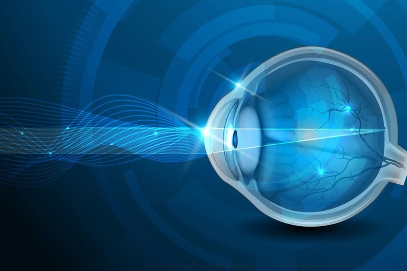
HomeEye careVision Resources

Eye anatomy: A closer look at the parts of the eye

eye diagram

When surveyed about the five senses — sight, hearing, taste, smell and touch — people consistently report that their eyesight is the mode of perception they value (and fear losing) most.

Despite this, many people don't have a good understanding of the anatomy of the eye, how vision works, and health problems that can affect the eye.

Read on for a basic description and explanation of the structure (anatomy) of your eyes and how they work (function) to help you see clearly and interact with your world.

How the eye works

In a number of ways, the human eye works much like a digital camera:

  1. Light is focused primarily by the cornea — the clear front surface of the eye, which acts like a camera lens.

  2. The iris of the eye functions like the diaphragm of a camera, controlling the amount of light reaching the back of the eye by automatically adjusting the size of the pupil (aperture).

  3. The eye's crystalline lens is located directly behind the pupil and further focuses light. Through a process called accommodation, this lens helps the eye automatically focus on near and approaching objects, like an autofocus camera lens.

  4. Light focused by the cornea and crystalline lens (and limited by the iris and pupil) then reaches the retina — the light-sensitive inner lining of the back of the eye. The retina acts like an electronic image sensor of a digital camera, converting optical images into electronic signals. The optic nerve then transmits these signals to the visual cortex — the part of the brain that controls our sense of sight.

Human Eye Anatomy (seen from above)

For more details about specific structures of the eye and how they function, visit these pages:

And for a description of common vision problems, see Refraction and Refractive Errors: How the Eye Sees.

FIND AN OPTICIAN: If you're concerned about your vision, visit an optician near you.

Find Eye Doctor

Find an optician near you

Find Eye Doctor