Optic neuritis and neuropathy: Symptoms, causes, treatment
The optic nerve transmits visual information from the retina to the brain to enable the sense of sight. Inflammation of this nerve is called optic neuritis.
What happens in optic neuritis?
In optic neuritis, inflammation can damage the optic nerve and the protective covering that surrounds it. This can affect one optic nerve or both optic nerves at the same time.
Vision symptoms from optic neuritis can include blurring, blind spots or complete loss of vision. You also may notice distorted vision, reduced color vision and pain when you move one or both eyes.
What causes it?
Optic neuritis can occur in anyone, but it usually occurs in adults younger than 45 and it affects more women than men. The condition is common in people who have multiple sclerosis, which occurs when the body's own immune system attacks and destroys protective nerve coverings.
Besides affecting eyesight, related nerve damage in multiple sclerosis can lead to loss of mobility and sensory functions, along with other debilitating conditions.
The term optic neuropathy more generally describes optic nerve abnormalities or damage. This damage could be from blocked blood flow, certain medical conditions or toxic exposure. Optic neuritis is one specific cause of an optic neuropathy. Other causes include:
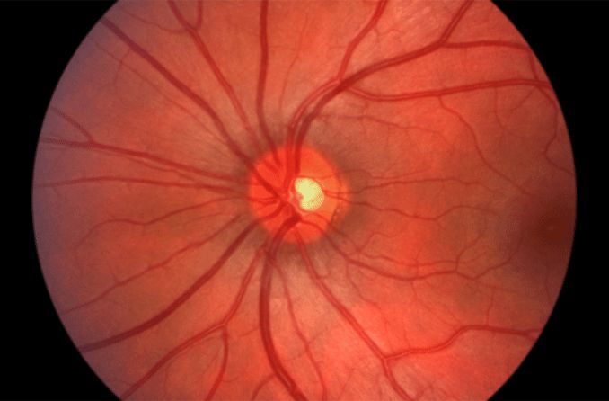
Image of the back of a healthy eye, where the optic nerve is located. When the optic nerve is damaged, poor vision results.
Infections such as herpes simplex
Other viral infections
Certain neurological disorders
Other signs and symptoms of optic neuritis
Optic neuritis also can affect pupil function, causing the pupil to get larger (dilate) instead of getting smaller (constricting) in the presence of bright light.
And depending on the severity of optic neuritis, the optic nerve may appear swollen.
During an eye exam, your eye care professional will look for signs of optic neuritis by directly examining the appearance of the head of your optic nerve in the back of your eye.
Other tests and measurements may include:
A visual field test to determine of you have an enlarged blind spot or other defects in your field of view
An imaging test called optical coherence tomography (OCT) to detect optic nerve damage
An eye pressure measurement to rule out glaucoma or ocular hypertension
You might also be referred for an MRI test of the optic nerves and the brain to detect possible underlying causes of optic nerve inflammation.
Treatments for optic neuritis
Eye care professionals usually treat optic neuritis with a combination of intravenous and oral steroids or monitor the condition without prescribing medical treatment.
For patients who are medically treated, the regimen typically includes three days of high-dose steroids followed by about 11 days of reduced doses.
Additionally, patients with optic neuritis may be offered treatment with a procedure called plasma exchange. This therapy requires the patient to be connected to a machine for one to two hours. During treatment, the patient’s blood is taken intravenously to the machine and certain plasma proteins are removed. The patient’s blood is then returned after being “cleaned” of some of the immune system components that promote inflammation.
Prognosis for those who have optic neuritis
Visual deficits caused by optic neuritis may worsen over a period of about seven days before vision typically stabilizes at that level for three to eight weeks. Gradual vision improvement may then occur.
About 95% of people with optic neuritis recover much of their vision within six months of onset.
However, about 19% will have a recurrence of optic neuritis in the affected eye, and 17% will develop optic neuritis in the other eye within 10 years. This is why careful testing to determine the cause is important. Recurrent events are preventable if an underlying disorder can be identified.
As mentioned above, sometimes optic neuritis is a precursor to the development of multiple sclerosis, so if you have optic neuritis, your eye care professional may recommend an MRI.
If imaging shows "white matter" lesions indicating damage to the protective covering of nerve fibres in the brain, there is an 80% to 90% chance of meeting criteria for a diagnosis of multiple sclerosis within five years. But even with normal results, a person with optic neuritis has a 22% chance of developing multiple sclerosis, making it very important to work with a specialist regarding treating and managing this condition.
Page published on Wednesday, 16 March 2022


